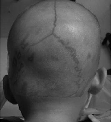
Fig. 5.1
Different shapes of the skull in lambdoidal synostosis (a, b, c on the left column) and in PPP (a, b, c on the right column), viewed from the top (a), from the back (b) and front (c). In lambdoidal synostosis, the head viewed from the top (a) has a trapezoidal shape (a-left), while in PPP a typical parallelogram shape (a-right)
Table 5.1
Comparison and differences in the shape of the skull in lambdoidal synostosis and in PPP
Lambdoidal synostosis | Posterior positional plagiocephaly | ||
|---|---|---|---|
Vertex view | Depressed right occiput | Vertex view | Depressed right occiput |
Prominent right forehead | The forehead is prominent on the left and severely flattened on the right side | ||
Parallelogram cranial shape | Trapezoidal cranial shape | ||
Anterior shift of the right ear | Anterior shift of the right ear | ||
Posterior view | Depressed right occiput | Posterior view | Depressed right occiput |
Compensatory increase in cranial height on the right side | Decreased cranial height on the right side | ||
Level ears | The right ear is positioned inferiorly relative to the left | ||
Frontal view | Forehead and cheek more prominent on the right than the left | Frontal view | Forehead prominent on the left and severely flattened on the right side |
Right eye appears more open than the left | Right eye appears more open than the left | ||
Nose is straight | The nose is slanted | ||
Chin point rotated to the left | Chin point deviation to the left | ||
In addition to the observational data, several anamnestic information can be helpful in supporting the different diagnostic hypotheses.
5.1.1 Risk Factors for PPP
It is known that posterior positional plagiocephaly (PPP) can be prevented, but it is also known that some individual or environmental factors, prenatal, neonatal, and postnatal, may give way to its onset [7–10].
The identification of these risk factors can be helpful in the diagnostic process and support the care management (Table 5.2).
Table 5.2
Risk factors for positional plagiocephaly
Obstetric factors |
Primigravidity |
Assisted delivery |
Low birth weight |
Preterm birth |
Infant factor |
Neck problems |
Infant difficulty turning head |
Decreased cervical rotation |
Limited passive cervical rotation |
Limited active cervical rotation |
Consistently sleeps with head turned to one side |
Male sex |
Larger cerebrospinal fluid spaces |
Infant care factors |
Cumulative exposure to the supine position |
>20 h per day in supine position |
<1 h per day upright |
Placed in prone <3 times per day |
Position of head not varied when infant is put to sleep |
Firmer mattress |
Position of infant when fed |
Only bottle fed |
With reference to Table 5.2 information about the prenatal, neonatal, and postnatal period, let make us presume the positional nature of the disorder.
Observational data and medical history will be sufficient to enable us to orient ourselves in the differential diagnosis between postural plagiocephaly and synostotic plagiocephaly.
If we are oriented towards the diagnosis of positional plagiocephaly and we have checked the absence of postural head anomalies of clinical relevance (head tilted to the contralateral side of rotation, deficit of contralateral rotation), we will not need to conduct further diagnostic exams, and we can explain to the parents the developmental dimension of the clinical features addressing the case to a pediatric physiotherapeutic treatment.
5.1.2 Developmental Risks of PPP
PPP has a prognosis of spontaneous improvement, but in the most “serious” cases, a residual deformity can still be observed at the end of preschool age [11]. The possibility that PPP may represent a clinical problem not only concerning “aesthetics” has been studied by evaluating the quality of case studies of neurobehavioral development in children.
It has been reported that children with PPP get different achievements in neurobehavioral development at 18 months and at 36 months (according to the Bayley Scales of Infant and Toddlers Development – III Edition): the cognitive and the linguistic areas seem to be those which mostly show recognizable immaturity [12, 13].
It has been reported that PPP can alter the development of the visual field [14] and determine disorders of the auditory processing [15].
It has been observed that, when beginning primary school, 39.7 % of children diagnosed as having PPP at 6 months of age required special care and educational needs in comparison to only 7.7 % in the control group [16].
5.2 Synostotic Plagiocephaly
The observational diagnosis of synostotic plagiocephaly requires confirmation by neuroimaging studies. The three-dimensional computerized tomography (3D CT) of the head will be the investigation of choice to confirm the clinical hypothesis of synostosis (Fig. 5.2).


Fig. 5.2
A 3D CT image showing a synostosis of the suture was superimposed to the baby’s head photography in which the anterior and inferior position of the left ear clinically suggests a left lambdoid suture synostosis. The image overlapping led to diagnostic confirmation
The 3D CT study will help us to verify the presence of other cranial suture synostosis (differential diagnosis between single synostosis and multiple synostoses).
Magnetic resonance imaging (MRI) of the brain is not universally recommended for routine examinations in isolated craniosynostosis as it usually does not add any information that may affect treatment decisions [17]. MRI of the brain may, however be useful, even in isolated forms, to assess the presence of brain malformations (i.e., agenesis of the corpus callosum, Chiari malformation type 1, or conditions of intracranial hypertension) [18].
Neuroimaging has otherwise a specific indication when neurobehavioral delays, dysmorphisms, or malformations of other organs and systems are detected during the clinical observation of the child. In this diagnostic context, a consultation by a geneticist pediatrician becomes very useful.
The diagnosis of a specific syndrome may provide prognostic factors of great importance in order to define a correct therapeutic approach to the cranial shape disorder. Some genetic syndromes that include the presence of cranial synostosis are listed below (Table 5.3).
Table 5.3
Syndromic forms of craniosynostosis, with involvement of different sutures (metopic, coronal, sagittal, lambdoidal)
Pfeiffer syndrome |
Crouzon syndrome |
Apert syndrome |
Jackson-Weiss syndrome |
Muenke syndrome |
Saethre-Chotzen syndrome |
5.2.1 Developmental Risks of Synostotic Plagiocephaly
Several longitudinal observations have investigated whether isolated craniosynostosis may involve risks of neurodevelopmental delay or worse outcomes.
Stay updated, free articles. Join our Telegram channel

Full access? Get Clinical Tree


