Fig. 76.1
Chest wall hamartoma in a newly born male patient. (a) Radiography shows a large expansile rib lesion with areas of mineralization and soft tissue expansion. (b) CT scan shows a large heterogeneous lesion with irregular areas of mineral deposition
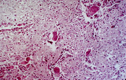
Fig. 76.2
Medium-power magnification microphotography. Highly cellular cartilage nodule next to aneurysmal cyst-like cavity (Courtesy of Prof. Eliane Ingrid Amstalden, Campinas, SP, Brazil)
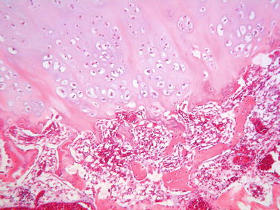
Fig. 76.3
Medium-power magnification microphotography. Cartilage nodule undergoing enchondral ossification, a common finding in chest wall hamartoma (Courtesy of Prof. Eliane Ingrid Amstalden, Campinas, SP, Brazil)