53
Carpometacarpal Joint Injuries: Bennett’s Fractures (Wire Technique)
Michael Jablon
History and Clinical Presentation
A 35-year-old, right-handed dentist injured his left, nondominant thumb in a bicycle accident. His injury occurred when he ran into a cement post. He fractured his right clavicle as well as his left thumb. The thumb fracture was not initially recognized by his surgeon, although initial radiograph reports indicated a “marginated bony fragment adjacent to the medial aspect of the base of the first metacarpal.”
Physical Examination
The left thumb had tenderness at the carpometacarpal joint, with a marked prominence of the first metacarpal base. Skin, two-point discrimination, and Allen’s test were intact.
Diagnostic Studies
Radiographs of the left hand and thumb carpometacarpal joint indicated the presence of a fracture dislocation. The base of the first metacarpal was fractured intraarticularly and the metacarpal itself was radially and proximally subluxed (Fig. 53–1).
PEARLS
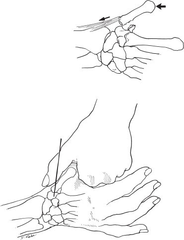
- Once traction, pronation, and abduction have been applied to the thumb for reduction, a K wire may be placed overlying the first metacarpal trapezial level and a fluoroscopic image obtained.
- If the wire is in the appropriate position, draw a line with a marking pen to help as a guide when placing the K wire.
- The transfixing K wire need not cross the fracture site. Strive for perfect anatomic reduction, but a minor step-off may be acceptable.
PITFALLS
- Poor radiographs may delay correct diagnosis. Accurate imaging requires a pronated thumb view to profile the base of the first metacarpal.
- During the surgical approach, care to avoid injury to the superficial radial nerve branches is necessary.
- Prolonged immobilization may lead to loss of mobility and circumduction.
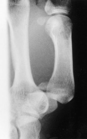
Figure 53–1. Radiograph of patient’s thumb demonstrates small fracture fragment and dorsal radial subluxation of the trapezial metacarpal joint.
Differential Diagnoses
Bennett’s fracture dislocation
Rolando’s fracture dislocation
Metacarpal shaft fractures
Trapezium fractures
Thumb dislocation
Carpometacarpal joint osteoarthritis
Thumb sprain
Pathologic fracture
de Quervain’s tenosynovitis
Diagnosis
Bennett’s Fracture Dislocation
The diagnosis was a Bennett’s fracture and intraarticular fracture of the first metacarpal bone with two fragments; the larger fragment shows subluxation from the deforming pull of the abductor pollicis longus tendon.
The Bennett’s fracture is the most common intraarticular injury involving the carpometacarpal joint of the thumb. Because the abductor pollicis longus tendon acts as a deforming force, stabilization by fixation is necessary. An attempt at closed reduction is recommended by applying traction to the thumb metacarpal with radial abduction and a pronation rotatory torque. A wire is percutaneously driven while holding the thumb in palmar abduction and opposition if reduction is achieved. The pin may be driven into the index metacarpal or trapezium. Always strive for anatomic reduction. However, acceptance of a 2-mm or less articular step-off does not biomechanically predispose a patient to posttraumatic osteoarthritis if the fragments heal in close apposition. The priority of treatment should be reduction of the metacarpal shaft relative to the trapezium and small fragment.
Surgical Management
Six weeks after injury, the patient was referred with the established diagnosis of a Bennett’s fracture. Informed consent was obtained, including discussion of the risks and benefits of the various treatment options. Problems discussed included stiffness, loss of motion, pain syndromes, infection, and possible need for future surgical removal of hardware—all concepts important to the patient’s profession. The patient agreed to surgical treatment.
Surgical management consisted of an attempted but unsuccessful closed reduction and percutaneous pinning. Open reduction and internal fixation with 1.0-mm Kirschner wires (K wires) was performed. Axillary block anesthesia and intravenous prophylactic antibiotics were administered. The left hand and forearm were prepped and draped, and a well-padded tourniquet was applied to the upper arm. Fluoroscopy was utilized during the procedure.
Axial traction was placed on the left thumb while simultaneously pronating, abducting, and applying pressure at the base of the first metacarpal to reduce it (Fig. 53–2). If closed reduction had been successful, a single K wire may be driven while holding the reduction from the first metacarpal into the trapezium (Fig. 53–3). Due to the elapsed time from injury, closed reduction was not successful, and therefore a lateral approach to the thumb carpometacarpal joint was performed. The incision was marked with a sterile marking pen along the dorsal radial border of the first metacarpal extending ulnarward at the base, exposing the thenar muscles along the radial border of the metacarpal shaft. Sensory branches of the radial nerve were identified and carefully protected (vessel loops were used for retraction). The thenar musculature was then elevated subperiosteally from the radial aspect of the left thumb metacarpal, exposing the carpometacarpal joint. The capsulotomy was performed, revealing the joint surfaces and showing a step-off and early healing. The abductor pollicis longus tendon was markedly scarred down at the capsular level, necessitating tenolysis to allow access to the carpal metacarpal joint. Capsular release was required for joint reduction. The step-off of the articular surface of the first metacarpal was identified and osteotomized through the fracture site with a small osteotome. The medial fragment was deemed too small to adequately accept a screw and also had a central area of fibrous healing.