52
Carpometacarpal Joint Injuries: Bennett’s Fractures (Arthroscopic and Percutaneous Screw Technique)
Joseph F. Slade III and John D. Mahoney
History and Clinical Presentation
An 18-year-old varsity field hockey player presented with pain in the base of her right thumb. She was injured in the course of a game.
Physical Examination
Inspection of the right hand revealed a swollen, obviously deformed thumb. The base of the thumb was tender to palpation. The skin was intact, with normal sensation over the surface of the thumb. Palpation of the carpometacarpal (CMC) joint did not reveal a step-off, but passive movement of the thumb elicited pain.
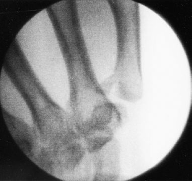
Diagnostic Studies
Radiographs demonstrated an oblique, intraarticular, displaced fracture of the base of the thumb metacarpal. To obtain a true lateral view of the joint, the hand was placed flat on the cassette and then pronated 15 to 20 degrees. The tube was directed obliquely 15 degrees distal to proximal, centering over the CMC joint.
Differential Diagnosis
Bennett’s fracture
Pure dislocation of the carpal metacarpal joint
Rolando’s fracture
Metacarpal shaft fracture
Carpal fracture
PEARLS
- Malunion is present in a small minority of Bennett’s fractures managed surgically. These patients may be candidates for osteotomy or arthrodesis.
- Nonunion is practically non-existent.
- Reduction maneuvers may change the type of fracture; that is, a type I may become a type III on manipulation.
PITFALLS
- The majority of patients treated conservatively will develop subluxation of the trapeziometa-carpal joint.
- The superficial branch of the radial nerve may be injured in Bennett’s fracture. Careful neurological exam is warranted.
- For fractures managed with K wires, use of a single K wire is often associated with K-wire migration, displacement, and pin tract infection.
Diagnosis
Bennett’s Fracture: Type III
The diagnosis in this case was Bennett’s fracture, type III, an oblique intraarticular fracture dislocation of the basal joint of the thumb metacarpal. The most common mode of injury is a motorcycle or bicycle accident, forcing the handlebar into the web space between the thumb and index finger. Other common causes include fights, sporting accidents, and falls on an outstretched hand. Generally, any trauma causing abduction and hyperextension of the thumb can cause a Bennett’s fracture. Most of the injuries (70–90%) occur in men, and the peak age incidence is 35 to 45 years. A classification scheme for Bennett’s fracture has been devised (Table 52–1).
| Fracture Type | Description |
| Type I | Distal fragment separation with preservation of continuity of the proximal joint surface |
| Type II | The opposite of type I, with proximal fragment separation, joint surface disruption, and preservation of the distal cortex |
| Type III | Most common (67% incidence), with total separation of the fragment |
Surgical Management
We performed an arthroscopically assisted closed reduction and fixation with percutaneously placed cannulated screws. Four days after referral from the emergency department, the patient was taken to the 1-day surgery center for definitive management. The patient was placed supine, using general anesthesia, exsanguination of the arm, and tourniquet control. To begin, longitudinal traction was applied through a single finger trap to the thumb with 10–12 pounds of traction. A mini-fluoroscopy unit placed horizontally around the hand assisted in placement of percutaneous guidewires (Fig. 52–1). Nineteen-gauge needles were used to provide portals to the dorsal CMC joint. A skin incision was made over the needles. Blunt dissection to the thumb CMC joint protected the superficial branch of the radial nerve. A blunt trocar was used to enter the joint, and a small-joint, 2.7-mm, 30-degree, angled scope was introduced. Through the second portal, a small-joint shaver was used to remove clots and perform a limited synovectomy. The fracture was now observed. The displaced fracture was reduced using longitudinal traction. Percutaneously, a guidewire was placed in the metacarpal and directed toward the proximal avulsed fragment. With longitudinal traction, pressure was applied to the base of the thumb. This maneuver levered the displaced proximal fragment to a reduced state countering the distracting force of the abductor pollicis longus. With reduction confirmed by minifluoroscopy, the guidewire in the distal metacarpal was driven into the proximal fracture fragment, securing the reduction (Fig. 52–2). A depth gauge was used to select the appropriate screw length. The guide wire was next overdriven into the adjacent trapezoid bone, which permitted a cannulated drill to be used in preparing for implantation of the screw without loss of reduction. A countersink was used in the distal metacarpal segment to permit placement of a buried washer (Fig. 52–3). The cannulated washer was driven until it was firmly seated beneath the bone cortex. Next, a cannulated screw was placed (Figs. 52–4 and 52–5). Occasionally, a second screw is required for rigid fixation, and the above process is repeated. Arthroscopy confirmed articular alignment of the fracture, and minifluoroscopy confirmed architectural restoration of the metacarpal base and good position of the implant. Skin incisions were closed with 4–0 nylon suture, and a thumb-spica splint was applied.