Fig. 26.1
Elongation of the anterolateral (AL) and posteromedial (PM) bundles of the PCL with weight-bearing knee flexion from 0° to 135°. PCL—posterior cruciate ligament. ([8] Reprinted with permission from SAGE Publications)
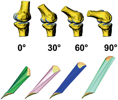
Fig. 26.2
Schematic representation of the relative length and orientation of the PCL with varying degrees of knee flexion. PCL—posterior cruciate ligament ([7] Reprinted with permission from SAGE Publications)
The dysfunction and instability seen in the PCL -deficient knee reflect this characteristic of varying force through the arc of motion. When the PCL is sectioned in cadaveric knees, posterior translation increases from 2.4 mm in full extension to 10.1 mm in 90° of knee flexion [9]. For most athletic activities, knee flexion is less than 60°; however, posterior translation still averages 9 mm at 60° of flexion [9]. Indeed, posterior translation typically exceeds 5 mm once the knee flexes to 20–30°, so instability may be experienced even in terminal stance with normal gait [5, 9]. Posterior sag of the tibia in a PCL-deficient knee (Fig. 26.3) also affects the mechanical advantage of the extensor mechanism and may lead to anterior (patellofemoral) knee pain in patients with PCL insufficiency [5].
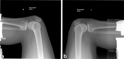
Fig. 26.3
Posterior sag of the right tibia a compared to the normal left knee b as seen on stress X-ray with a 20 lb. weight placed over the patient’s anterior tibia while the hip and knee are each flexed to 90°
The ideal brace for PCL -deficient knees would thus provide an anterior force on the proximal tibia that increases in magnitude as the knee flexes to 90°. Similarly, postoperative braces following PCL injury and reconstruction should increase support with increasing knee flexion to relieve strain on the reconstructed ligament as it heals. Braces should be worn for ambulation as well as rehabilitative exercises until the early stages of healing are complete, so comfort and ease of use are also important considerations in brace design.
Indications for Bracing
For all types of orthopedic injuries, bracing falls into different categories. Braces may be rehabilitative, functional, or prophylactic. Rehabilitative braces for PCL injuries are intended to protect the surgically reconstructed ligament or to provide a stable environment for the native torn ligament to heal by limiting tibial translation and rotation. Functional braces may provide external stability in the setting of ligamentous insufficiency allowing patients to complete daily activities and progress to higher-level athletic pursuits. The goal of prophylactic braces is to prevent or limit the severity of future injuries, particularly in knees that have been injured or experience excessive forces with certain activities [2, 5, 10]. Unfortunately, this is still just a theoretical benefit as we have no evidence that bracing for PCL insufficiency reduces the development of osteoarthritis. Osteoarthritis following nonoperative treatment of PCL insufficiency occurs in as much as 78 % of patients, with the medial and patellofemoral compartments most susceptible [2].
Most braces will be prescribed for rehabilitative purposes, for either initial nonoperative treatment or in the postoperative period. These braces should reduce the posterior translation of the tibia, which is created by the pull of the hamstrings and by gravity in the supine position, so that the healing ligament or graft does not elongate [2]. Different methods of bracing and immobilization have been used, but the principle of applying an anteriorly directed force to the proximal tibia remains consistent.
In theory, braces can control anterior–posterior tibial translation and even varus–valgus angulation relatively well as long as the braces are adequately rigid [10]. Internal and external tibial rotation, on the contrary, will not be well controlled without the hip and ankle included in the brace [10]. Therefore, standard PCL braces may not be sufficient when the PCL has associated injuries to the posterolateral corner or other rotational stabilizers of the knee. There is a lack of biomechanical research specifically on PCL braces to support this theory, however. Biomechanical testing of the Lenox Hill brace placed on cadaveric knees with sectioning of the ACL and medial collateral ligament (MCL) demonstrated a 20 % reduction in anterior–posterior translation [11]. An in vivo study by Jonsson and Kӓrrholm [12] also found a reduction in anterior–posterior translation by approximately one third using the Lenox Hill and Ecko braces for ACL-deficient knees. The Lenox Hill brace also controlled external rotatory laxity, but not internal rotatory laxity [12]. Despite these encouraging studies, extrapolating the biomechanical data from ACL bracing studies is inadequate for directing PCL management. Biomechanical testing of PCL braces is needed to understand if the current braces can achieve comparable reductions in abnormal tibial translation and rotation.
Brace Specifications
Selecting the proper brace for a patient depends on matching various brace specifications to both the injury and the individual. This thoughtful attention will increase the likelihood of the brace achieving its desired goals.
The first decision is choosing between static and dynamic PCL braces. Static braces rest passively on the leg in a position that resists pathologic motion. Their countering force is only applied when the pathologic motion is encountered [5, 10]. An example of a static brace would be a device that has additional padding between the calf and the posterior tibial support to counteract the posterior translation of the proximal tibia in the supine position (Fig. 26.4). In contrast, dynamic braces are constantly applying a force or preload that resists the undesired motion [5, 10]. This may be accomplished with springs, as with the PCL Jack brace (Albrecht, Stephanskirchen, Germany; Fig. 26.5) [2]. While dynamic braces are considered superior in their ability to resist tibial translation, they may create abnormal forces on the knee [10]. As discussed above, the anterior–posterior translational forces in the knee vary with range of motion. Similar to the PCL, the in situ force on the ACL changes throughout the arc of motion. The ACL experiences its peak force at low flexion angles between 15° and 30°, with a significant drop in force by 45° of flexion [13, 14]. Therefore, a constant anterior force on the proximal tibia from a dynamic PCL brace may increase strain on the intact ACL in a non-physiologic pattern. Whether or not the force generated by the brace is enough to damage the ACL remains to be seen.
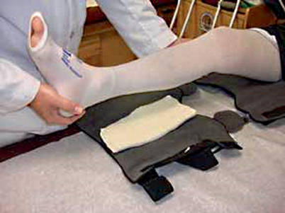
Fig. 26.4
Padding added to the posterior tibial support of a static knee brace. ([21], reprinted with permission from Elsevier Limited)
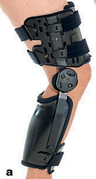
Fig. 26.5
The PCL Jack brace. PCL— posterior cruciate ligament. ([2], Reprinted with permission from Springer)
The strength and rigidity of the brace will determine the degree of unintended motion and the resistance to high loads, as with a fall. Straps should interlock with the brace struts to provide the best support. Braces with bilateral hinges and hard-shell supports are more rigid than those with unilateral hinges and soft-shell supports [10]. Condylar pads (Fig. 26.6) that keep the hinges centered over the joint line enhance the ability of the hinges to control motion [10]. Hinge mechanisms with a shear pin stop may limit unintended motion [5]. Straps and components may be attached with Velcro, rivets, stitching, or glue, and the quality of these attachments should be inspected prior to use of a particular brace. As PCL braces will typically be worn for several weeks, normal wear and tear of the brace should be expected and monitored so that the brace can be replaced as needed [5].
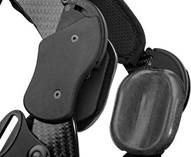
Fig. 26.6
A knee brace with condylar pads. (Image courtesy of Össur, Inc.)
Regardless of the strength and sophistication of the brace, an improperly fitted brace will not adequately protect the patient’s knee. The tightness of fit must be balanced to prevent slippage without compromising circulation and lymphatic drainage. The brace should also be appropriately padded over bony prominences to prevent irritation to the underlying skin and soft tissues [5]. Even with a secure fit, braces tend to allow more motion than indicated by the hinge stops. Cawley et al. [15] found that during ambulation, patients could achieve 15–20° more extension than the amount set by the extension stop. The amount of adipose tissue between the bone and the brace will also affect how well the brace can limit motion [5]. Similarly, as the patient regains muscle girth during rehabilitation, the fit of the brace will need to be adjusted [10].
Finally, comfort and ease of use are important factors to the patient, who will ultimately determine if the brace is to be worn as prescribed. Custom braces may provide a more comfortable fit since they can be specifically contoured to the patient’s anatomy. However, they are more expensive and may become loose as swelling subsides or tight as the muscles regain their normal size. Longer braces will provide more leverage for applied forces, but are often less tolerable to the patient and more difficult to achieve a snug fit with rigid struts [10]. Dynamic braces tend to be bulkier and may become too restrictive as the patient progresses in activity level. For example, the PCL Jack brace limits knee flexion from 0° to 90–110°, and hinge mechanisms at both the knee and the ankle make it cumbersome for athletic participation [2].
A single type of rehabilitative brace may not be satisfactory for all patients with PCL injuries, and individual patients may need more than one type of brace over the course of recovery and rehabilitation. However, braces are costly, and more complex designs are not always better. Attention to the needs of the patient and the brace specifications can help to provide optimal chances for successful treatment without incurring unnecessary costs.
Duration of Bracing
There is no clear evidence supporting a particular duration for bracing knees with PCL injuries as part of nonoperative management. Studies on PCL bracing report various length of time in a brace, ranging from 4 weeks to 6 months [2–4]. Some authors report that their selected duration for bracing was chosen somewhat “arbitrarily” [3], though it is based on the current understanding of ligament and soft tissue healing. During the first 2–3 weeks following injury, fibroblasts enter the zone of injury and collagen fibers proliferate [4]. Protection of the healing ligament during this time is critical, so it is necessary to brace or even immobilize the knee in a cast or splint.
Stay updated, free articles. Join our Telegram channel

Full access? Get Clinical Tree


