Fig. 58.1
Dinamic slink for anterior instability
Moving on to the acromio-clavicular joint (ACJ), clavicle fractures make up 5 % of all fractures; thus, they are not uncommon. It is a common injury in contact sports (rugby, martial arts) and impact sports (such as horse, motor and bike racing). Most clavicle fractures are mainly treated in a sling for about 4–6 weeks, which may provide more comfort if applied correctly and align some fractures in a more stable position. However, complete healing can be slow and may take up to 3–6 months. The conservative treatment of mid-shaft clavicle fractures with simple slings vs figure-of-eight braces has been studied to examine whether one method is better than the other.
Concerning trauma, the ACJ in many contact sports, such as rugby, American football, ice hockey, basketball etc., is susceptible to frequent injury. Before the brace is applied, in terms of prevention, the foam pad is important in avoiding the impact on this small joint. Also, taping has been used for first- and second-degree sprains and ACJ separations are one of the most common injuries seen in orthopaedic and sports medicine practices, accounting for 9 % of all injuries to the shoulder girdle. Various operative and non-operative treatment schemes have been described for the management of ACJ injuries.
The ACJ consists of the articulation between the distal end of the clavicle and the acromion, functioning to anchor the clavicle to the scapula and shoulder girdle.
Early classification of AC joint injuries by Tossy and colleagues [2] and Allman [3] were based on radiographic displacement and the degree of ligamentous damage. They were initially graded I through III, and Rockwood and his group [4] later added types IV through VI to the classification system.
Non-operative treatment is typically reserved for types I, II and acute type III injuries. This involves a 7- to 10-day period of rest, immobilisation and anti-inflammatory medications. Historically, braces, such as the Kenny–Howard brace, were used to treat these injuries. It acted to reduce the ACJ by applying a downward force on the clavicle and an upward force on the humerus. It was worn for 6–8 weeks and was associated with recurrence of deformity. Fourth-generation bracing was made for the non-operative treatments of the type I, II and for some type III ACJ dislocations for 4–6 weeks, with good compliance of the patients (Fig. 58.2).
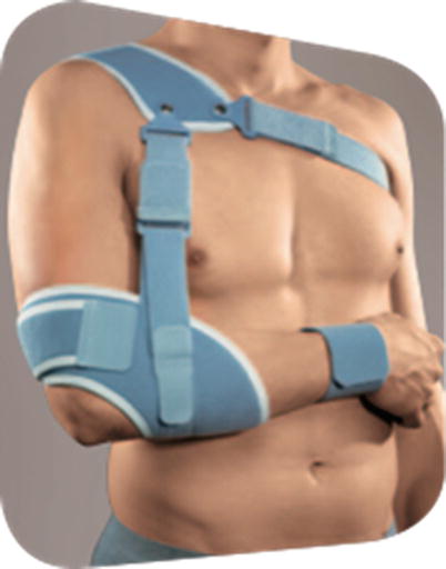
Fig. 58.2
AC joint stabilizer for acute dislocations
58.3 Elbow
The most frequent injury is over-use syndrome and its effect on basic mechanics. The stress on the extensor muscles of the wrist (due to the frequent pronation and supination/extension movements against moderate or high resistance) cause microtrauma at the proximal osteotendinous insertion.
Painful symptoms play a role in the primary lesions of the extensor carpi radialis brevis, which is involved in 100 % of cases, but in 35 % of cases the extensor digitorum communis is also involved. This is termed epicondylitis and the aim of the therapy is to reduce or to eliminate the painful symptoms and recover the functionality of the arm. To enhance the individual effects, a route is created that applies more methods of treatment: pressure, cold, to the rest of the muscle at night-time (Fig. 58.3).
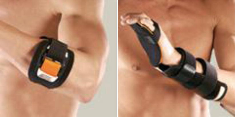
Fig. 58.3
Supports for the treatment of the epicondilytis for day and night time
Overuse of the wrist extensors is mentioned by many authors as a major factor in the development of lateral epicondylitis [5–9]. In tennis it has been shown that force and flexibility deficiencies in the forearm muscles and lack of movement accuracy lead to increased load at the lateral epicondyle [10]. These factors also correlate with the incidence of lateral epicondylitis.
Most braces on the market for the treatment of patients with epicondylitis claim to reduce the load at the lateral epicondyle. One type is clasp-based braces, which compress locally at the insertion of the wrist extensor tendons. In acute epicondylitis in particular, these braces are often not tolerated by the patient, because local compression at the insertion can be painful. Another type of brace applies compression over an area of several square centimetres with a silicone pad. A third type of brace was investigated in which a high-viscosity fluid pad is placed at the forearm over the extensor muscles. Conservative treatment of patients with lateral epicondylitis requires the limitation of repetitive stress to the origins of the two common extensors. It seems reasonable to consider the effectiveness of a brace in reducing the load at the lateral epicondyle as one of its quality criteria. The data show that the influence of a brace on the load at the lateral epicondyle depends on the characteristics of the product. Pad-based braces result in a much higher reduction of the load at the lateral epicondyle than braces with the a clasp. Placing a pad at the forearm, distal to the lateral epicondyle, seems to be superior to placing pads directly on the lateral epicondyle.
Hinged elbow braces may be used to provide medio-lateral support for elbow dislocations and collateral ligament sprains, while allowing a full range of motion. Postoperatively, a hinged elbow brace allows for a gradual increase in the range of motion. A dial on the hinge may be set to limit motion to a specific range, which can then be increased gradually during the healing process. The brace should run from the mid-bicep to the wrist. Straps should be secured from distal to proximal.
58.4 Wrist
A cock-up wrist brace is one of the most frequently used braces. It is appropriate for the treatment of wrist sprains and contusions. It may also be used for buckle (torus) fractures of the distal radius. Appropriate fitting requires the distal end of the splint to be just below the palmar crease so as not to restrict the movement of the meta-carpo-phalangeal joints.
For the treatment of distal radius buckle fractures, level I evidence studies demonstrate that a wrist splint is as good as a cast for the prevention of refracture and/or loss of alignment [11]. There was no difference in pain with use of a splint compared with a cast. Patients treated using the splint found it easier to bathe, had better function and did not need to return for cast removal.
A thumb spica splint or brace is used for the treatment of scaphoid injuries, gamekeeper’s thumb (ulnar collateral ligament sprain), De Quervain’s tenosynovitis, and wrist and thumb sprains. The brace may be soft or rigid, depending on how much support is needed.
Appropriate fitting is for the distal end of the splint to sit just below the palmar crease and for the thumb portion to extend to the distal phalanx of the thumb to immobilise the interphalangeal joint.
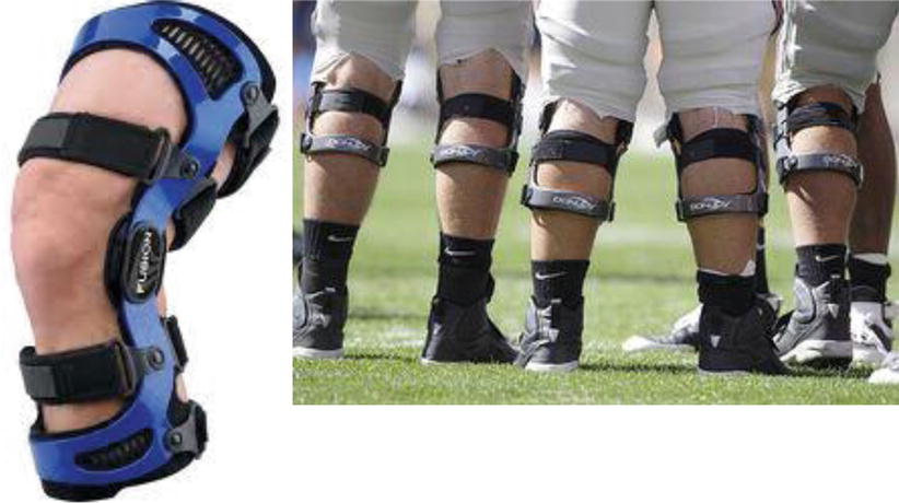
58.5 Knee Braces
Controversy has long surrounded the use of knee braces in the practice of sports medicine. From their use as a prophylactic measure in high-risk sports to their prescription for ligamentous instabilities, knee braces have always been viewed with widely varying amounts of acceptance and scepticism. This has ranged from enthusiastic endorsement to outright condemnation. Braces for patellar-femoral disorders, osteoarthritis and postoperative rehabilitation have come to abound in the practice of sports medicine.
The idea of wearing a brace to prevent sports-induced injuries of the knee was first proposed in 1979 by Anderson and colleagues [12], who used a lateral, upright, dual-hinged device on National Football League players.
After it had been tested on nine players for short periods (one to nine games) without reinjury, this brace was proposed as a means of preventing ligamentous injury to the medial side of the knee. The authors believed that in addition to preventing a significant valgus stress to the knee, their brace also helped to restrict antero-posterior displacement. This report led to an explosion in the demand for prophylactic knee braces (PKBs) in football and other demanding sports, and a multitude of manufacturers sold off-the-shelf PKBs to meet the demand.
These can be classified into two basic types.
Numerous laboratory studies investigating the effects of knee bracing have been performed. Some studies were designed to study PKBs, which are used to prevent knee injuries, whereas others looked at functional knee braces, which are structured to compensate for torn ligaments. Most studies are useful, but the reader must carefully consider the biomechanical parameter reproduced in the study and its subsequent effect on the ligaments of interest.
Related posts:

Stay updated, free articles. Join our Telegram channel

Full access? Get Clinical Tree


