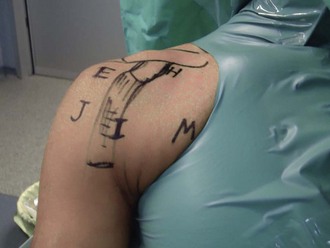
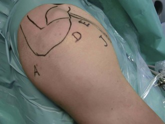
Chapter 19 Several options exist for the surgical treatment of anterior glenohumeral instability. Although the Bankart procedure has been shown to have reliably favorable results, there exist certain conditions (e.g., glenoid fracture, engaging Hill-Sachs lesion, inferior ligament hyperlaxity) that can signal a potentially suboptimal outcome.1,2 The open Latarjet procedure, transfer of the coracoid such that its inferior surface abuts the anterior glenoid, has been reported by several authors to return a high rate of good to excellent results in long-term follow-up studies.3–9 Mid-range stability is increased by the additional bone stock increasing the effective surface area of the glenoid. End-range stability is aided by the sling effect of the transferred short head of the biceps; as the arm is brought into increasing abduction, this tendon is brought under increasing tension, resisting anterior translation of the humeral head. The goal of this procedure is to recreate the open Latarjet procedure through an all-arthroscopic approach. This allows not only the reproduction of a reliable procedure, but also enhanced ability to address concomitant pathology, as well as accelerating patient mobility. • Instability with glenoid, humeral, or combined bone loss • Complex soft tissue injury, humeral avulsion of the glenohumeral ligaments (HAGL lesions) • High-risk activity—for example, in throwing athletes, contact athletes (American football, rugby), overhead athletes (rock climbing, swimming), manual laborers General anesthesia with a regional nerve block is the preferred technique. The patient is prepared as for a normal shoulder arthroscopic procedure, with care taken to have a wider area exposed medially for the most medial portal (Fig. 19-1). The patient is placed in the beach chair position. The following technical tips are important and specific for this procedure: • The arm should be free (without any traction). • The medial border of the scapula should be supported by a back support. This helps to act as a hinge on which the scapula can be rotated posteriorly with the help of switching sticks (as described later) to make the glenoid face more laterally. Supporting the shoulder on its back at the level of the spinal edge of the scapula maintains it in an external rotation position and facilitates the fixation of the bone graft to the glenoid. Seven portals are used, denoted by the letters A, D, E, H, M, I, and J (Figs. 19-1 and 19-2). Portals A and E are the starting portals, and the rest are introduced as the surgery proceeds. The three portals that are used specifically in this surgery are as follows: Specific steps for this procedure are outlined in Box 19-1. Box 19-1 Surgical Steps 1. Intra-articular preparation
Arthroscopic Latarjet Procedure
Preoperative Considerations
Indications
Surgical Technique for Arthroscopic Latarjet Procedure after a Failed Bankart Procedure
Surgical Landmarks, Incisions, and Portals
Specific Steps
Related posts:
 Knot-Tying and Suture-Passing Techniques
Arthroscopic Remplissage for Management of Engaging and Deep Hill-Sachs Lesions
Open Repair of Multidirectional Instability
Arthroscopic Meniscus Repair: Inside-Out Technique
Tendon Augmentation Devices in Rotator Cuff Repair
Arthroscopic Lateral Retinacular Release and Lateral Retinacular Lengthening
Knot-Tying and Suture-Passing Techniques
Arthroscopic Remplissage for Management of Engaging and Deep Hill-Sachs Lesions
Open Repair of Multidirectional Instability
Arthroscopic Meniscus Repair: Inside-Out Technique
Tendon Augmentation Devices in Rotator Cuff Repair
Arthroscopic Lateral Retinacular Release and Lateral Retinacular Lengthening
![]()
Stay updated, free articles. Join our Telegram channel

Full access? Get Clinical Tree