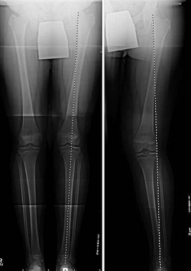
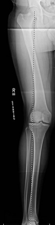
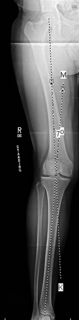
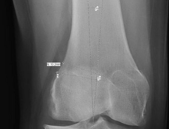
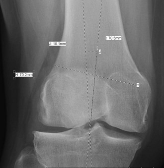
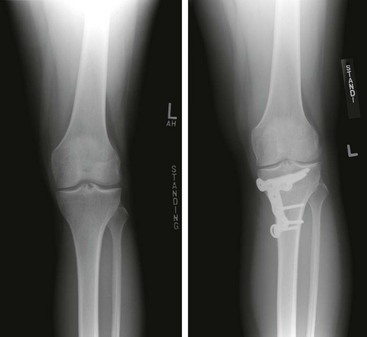
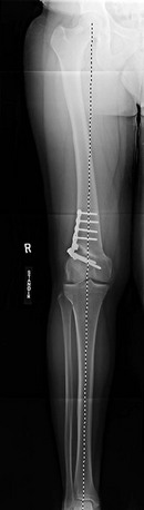
Chapter 71 Several studies have investigated the association between valgus limb alignment and the development and progression of lateral compartment osteoarthritis (OA). Brouwer and colleagues1 found that valgus alignment was associated with a borderline significant increase in the development of knee OA (odds ratio [OR] 1.54; 95% confidence interval [95% CI] 0.97–2.44). Other studies have found a strong correlation between valgus alignment and the progression of knee OA.2,3 A cross-sectional study by Issa and colleagues4 showed that the presence of cartilage defects in the lateral compartment increased with greater valgus malalignment. Lateral unicompartmental knee arthroplasty (UKA) is performed much less commonly than medial UKA, and therefore the available data on long-term outcomes are limited. In general, proximal tibial osteotomy is used to correct varus limb malalignment, and distal femoral osteotomy is used to correct valgus limb malalignment. A proximal tibial osteotomy can be used to correct valgus malalignment, but this usually leads to a change in the joint line orientation (Fig. 71-1).5 Perhaps, small corrections can be tolerated in the proximal tibia. Figure 71-1 A valgus-producing high tibial osteotomy results in obliquity of the tibiofemoral joint line. It is well known that proximal tibial osteotomy alters the tibial slope, with opening wedge having the tendency to increase the slope6 and closing wedge having the tendency to decrease the slope. This change in the slope has an effect on knee kinematics and stability. In addition, the tibial osteotomy will alter the contact mechanics through a full range of motion, whereas the femoral osteotomy will change the contact forces mainly near extension.5 Although there are complications that may accompany any osteotomy including femoral osteotomy, the alignment changes with distal femoral osteotomy involve only the coronal plane. In the varus knee, slight overcorrection into valgus is encouraged, but in the valgus knee the most common complication is overcorrection into varus, and this must be avoided. Patellar height changes after distal femoral osteotomy are also minimal and largely ignored7 compared with proximal tibial osteotomy. Sometimes a single standing long-leg view reveals an obscure deformity if clinical findings do not correlate with the initial long-leg view (Figs. 71-2), especially if there is a valgus thrust. The mechanical axis, which is a line drawn from the center of the femoral head to the center of the talus (Fig. 71-3), should normally pass through the center of the knee joint between the two tibial spines or just lateral to the medial spine. A line is drawn from the center of the femoral head to the medial tibial spine. Another line is drawn from the center of the talus to the same point (medial tibial spine), and the angle between these two lines is the angle of correction (Fig. 71-4). Rather than calculating the correction angle in degrees, calculating the actual correction in millimeters is more practical and avoids an extra step. This conversion is made by drawing the planned osteotomy wedge or triangle (planned level, length, angle of inclination, and angle of correction) on the distal femur and measuring the width of the base of this wedge at the lateral femoral cortex (Figs. 71-5 to 71-7). Figure 71-7 Follow-up long-leg radiograph of the patient shown in Figs. 71-3 to 71-6 3 months after the distal femoral lateral opening wedge osteotomy with a 10-mm plate showing the mechanical axis passing between the two tibial spines as planned before surgery. • Lateral compartment gonarthrosis with valgus limb alignment (mechanical axis passes through the lateral compartment) • Lateral femoral condyle osteochondritis dissecans (OCD) lesion with valgus limb alignment in isolation or in addition to other surgical procedures such as autograft or allograft osteochondral resurfacing • Deficient lateral meniscus with valgus limb alignment when lateral meniscal transplantation is considered (mechanical axis passes through or beyond the lateral tibial spine) • Valgus deformity with or without valgus thrust in chronic medial collateral ligament (MCL) or MCL and cruciate instabilities (mechanical axis passes beyond the lateral tibial spine) Indications for medial closing wedge distal femoral osteotomy are as follows: • Angle of correction greater than 17.5 degrees • Limb length discrepancy in favor of the operated limb • Presence of risk factors that delay healing—for example, smoking, neuropathy, poor bone quality, obesity
Distal Femoral Osteotomy
Preoperative Considerations
Imaging
Indications
Related posts:
 Knot-Tying and Suture-Passing Techniques
Arthroscopic Remplissage for Management of Engaging and Deep Hill-Sachs Lesions
Open Repair of Multidirectional Instability
Arthroscopic Meniscus Repair: Inside-Out Technique
Tendon Augmentation Devices in Rotator Cuff Repair
Arthroscopic Lateral Retinacular Release and Lateral Retinacular Lengthening
Knot-Tying and Suture-Passing Techniques
Arthroscopic Remplissage for Management of Engaging and Deep Hill-Sachs Lesions
Open Repair of Multidirectional Instability
Arthroscopic Meniscus Repair: Inside-Out Technique
Tendon Augmentation Devices in Rotator Cuff Repair
Arthroscopic Lateral Retinacular Release and Lateral Retinacular Lengthening
![]()
Stay updated, free articles. Join our Telegram channel

Full access? Get Clinical Tree


Distal Femoral Osteotomy