The purpose of rotator cuff repair is to restore a biomechanically sound, anatomically accurate, and durable connection between the torn cuff tendons and the humeral head. The surgeon has limited abilities to achieve these objectives due to various factors, many of which are out of his/her direct control. These factors include the preexisting state of the muscle-tendon unit (including tendon retraction and muscle atrophy, fatty infiltration, and connective tissue changes), the biomechanical stability of the repair construct, the healing potential of the bone-tendon interface, and the rehabilitation protocol and postoperative activities/compliance of the patient. The complex interplay between these factors makes objective interpretation of surgical repair strategies very difficult and probably explains the confusing discrepancies between laboratory biomechanical experiments and clinical outcome studies when we consider double-row rotator cuff repair.
Normal tendons rarely tear in the first place. Therefore, it seems logical that unraveling the underlying cause(s) of tendonopathy will provide important clues about possible solutions to this ubiquitous and challenging clinical problem. The biology of tendonopathy is germane to tendon disorders throughout the body. However, we should be very careful not to directly extrapolate findings from one tendon site to another because there may be subtle differences in cellular composition, gene expression, and local factors (such as blood supply or biomechanical factors) that would make therapeutic interventions highly variable in terms of regional efficacy. It is likely that in the future we will be better able to manipulate the biologic elements of the healing process, with a higher degree of clinical success. Nonetheless, a fundamental requirement will continue to be creation of a repair construct that is stable throughout the healing interval (even if that interval becomes shorter), so we will continue to strive for surgical strategies that are reproducible, reliable, and durable.
Arthroscopic rotator cuff repair has evolved to the point where patients can expect clinical outcomes that are similar to open or mini-open methods, and these results have been reviewed previously within this book. Arthroscopic outcomes are in part related to the surgical skill and experience of the operating surgeon, and there is a significant learning curve associated with these techniques. This field is rapidly advancing in terms of the surgical tools and implants at our disposal. We have almost reached a point where reports of clinical results are outdated by the time articles achieve publication because the surgical techniques are evolving so rapidly.
Advances in surgical technique, implants, and equipment have substantially improved our ability to tackle larger and more complex tears while simultaneously extending the general capabilities of surgeons to achieve good clinical outcomes (in other words, arthroscopic rotator cuff repair is no longer limited to the domain of “master arthroscopists”). It is critical to understand that the clinical outcome of rotator cuff repair depends upon surgical creation of a stable tendon-bone construct: regardless of whether that is achieved through arthroscopic or open methods (these principles are presented in Table 10.1). The surgical approach should therefore be dictated by the skill and preference of the operating surgeon in concert with informed consent regarding the pros and cons of the specific approach with the patient. At the current time, there is no clear evidence that long-term clinical outcome is markedly affected by the surgical approach with rotator cuff repair. However, patients clearly appreciate the advantages of less early postoperative pain and improved cosmetics associated with minimally invasive techniques, so it is likely that the indications for arthroscopic cuff repair will continue to expand (assuming at least long-term clinical equivalence).
HISTORY AND RATIONALE FOR THE DEVELOPMENT OF DOUBLE-ROW REPAIR
It is fascinating and humbling to look back at the history of surgery and to appreciate the amazing intuition of the early pioneers of orthopedic surgery. Early methods of rotator cuff repair involved wide open exposure of the rotator cuff, which was achieved by deltoid takedown and subsequent repair. Before the advent of suture anchors, the cuff was repaired to the bone with sutures that were passed through small holes in the greater tuberosity, the so-called transosseous repair. When the sutures were tied circumferentially around the tendon and the bone, the surgeon essentially achieved a footprint repair, with a point of medial fixation adjacent to the articular surface and a zone of more lateral compression of the tendon toward the tuberosity. Early strategies focused upon lateralizing the torn cuff to achieve a water tight repair if possible, and it is likely that the various patterns of cuff tearing and reduction were under-appreciated (with associated tension mismatch as attempts were made to overlateralize the cuff). The era of arthroscopy has clearly augmented our understanding of tear pattern (e.g., U-shaped vs L-shaped tears). Proper tendon reduction leads to better soft tissue balance, decreased tension overload, and a lower propensity for retear during the rehabilitation period.
Table 10.1 Basic principles of rotator cuff repair, regardless of surgical approach
Identification of tear pattern and extent, which usually requires bursectomy
Removal of offending bone and soft tissue impingement (smoothing)
Mental visualization of the repair strategy
Mobilization of the tendon
Stimulation of a biologic healing response
Stable fixation of rotator cuff to bone
Solid repair of structures violated by the approach, particularly the deltoid
The advent of suture anchors and early arthroscopic methods led to new surgical alternatives, including arthroscopic subacromial decompression and mini-open cuff repair. This method decreased the morbidity associated with deltoid takedown and repair. Deltoid splitting methods could also utilize transosseous cuff repair with sutures if desired; however, exposure of the greater tuberosity was somewhat limited by the acceptable extent of distal deltoid split (due to risk to the axillary nerve).
As arthroscopic skill and experience advanced, rotator cuff repair focused upon our surgical ability to place suture anchors in bone, pass sutures through cuff, manage multiple subacromial sutures, and tie reliable arthroscopic knots. In the 1990s, various tools and obturated cannulas were developed to assist with arthroscopic suture passage, and these tools continue to evolve today. Antegrade methods, retrograde methods, and shuttling techniques allowed for accurate passage of sutures through the tendon, which was further facilitated by various safe portals in the region to create the proper trajectories for arthroscopic instruments. Subsequently, so-called super-sutures were introduced that markedly decreased the tendency for suture breakage during knot tying. These sutures increase the durability of the repair construct, assuming the suture anchors remain in their original position and solid knots that reliably resist suture slippage are created.
In the first decade of the new millennium, appreciation of the anatomy of the rotator cuff “footprint” reemerged. Although the medial-lateral extent of anatomic cuff attachment has been understood for many decades, arthroscopic application of this information became a topic of strong interest upon the presentation of compelling visual examples of normal footprint anatomy (Fig. 10.1). The theoretical and practical pros and cons of arthroscopic footprint repair are presented in Table 10.2. The rest of this chapter essentially deals with our current knowledge regarding these advantages and disadvantages. Suffice it to say, the jury is still out when it comes to double-row repair, and it is likely that our knowledge, experience, and clinical algorithms will mature substantially over the next 5 to 10 years.
FOOTPRINT RESTORATION AND BIOMECHANICAL STABILITY
The published data in regard to the biomechanics of double-row rotator cuff repair are relatively confusing (Table 10.3). To date, laboratory data are relatively compelling in terms of improved contact area and contact force between tendon and tuberosity, when comparing single-row against double-row constructs. It should be emphasized that most laboratory experiments use animal or human cadaveric tissue and involve creation of cuff defects that are repaired immediately, thus these models minimize the adverse but clinically relevant impact of excessive cuff tension (due to chronic retraction and/or tension mismatch). Nonetheless, laboratory data do support the notion that the area of tendon-bone apposition is augmented by double row and therefore it is logical to attempt footprint restoration if possible (assuming this theoretical advantage leads to better clinical results and the method is not outweighed by the “cons” noted in Table 10.2).
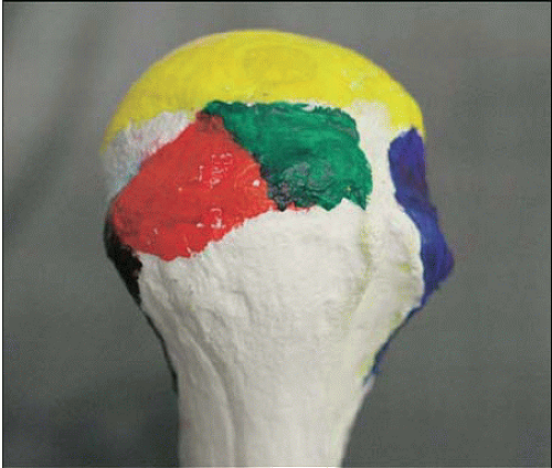
FIGURE 10.1. The rotator cuff has a broad medial-lateral insertional footprint. The supraspinatus (shown in green) extends from the articular surface of the humerus laterally approximately 15 mm. Note the oblique anterior extension of the infraspinatus (shown in red) whose fibers sweep forward and blend with the posterior fibers of the supraspinatus tendon. This architecture correlates with common L-shaped tears that propagate medially between the supraspinatus and the infraspinatus muscles and tend to have horizontal split components as well. (Reproduced with permission from Curtis AS, Burbank KM, Tierney JJ, et al. The insertional footprint of the rotator cuff: an anatomic study. Arthroscopy 2006;22(6):603-609.e1.)
Table 10.2 Theoretical and practical pros and cons of arthroscopic footprint repair
Pros
Cons
Increased contact area between tuberosity and cuff should augment bone-tendon healing
Overtension of a chronically retracted cuff could lead to tension overload/mismatch and early construct failure
Greater cuff apposition and more anchors and sutures should improve biomechanical stability
More suture anchors and longer surgical time may increase cost compared with simpler arthroscopic repair methods
Anatomic restoration of the footprint should lead to better long-term rotator cuff function and durability
Increased technical complexity may lead to an unacceptably long learning curve for the typical arthroscopist
Some double-row methods avoid arthroscopic knot tying, which should simplify the surgical technique
More implants and sutures could interfere with revascularization of the cuff, with a negative impact on healing
In contrast to the relatively consistent data regarding footprint restoration, laboratory studies of biomechanical stability/durability with double-row repair are more inconsistent and harder to interpret (Table 10.3). This is probably due to the wide variety of experimental paradigms and loading protocols that are applied in these studies. A thoughtful review by Reardon and Maffulli (6) provides a good overview of the background and scientific basis for double-row repair. This literature review suggests that there is good evidence for increased footprint coverage with double-row techniques and more inconsistent but generally positive biomechanical rationale. The authors conclude that we do not yet have strong clinical evidence of a difference in clinical outcome between single and double-row techniques.
Most experimental studies utilize a static position of the glenohumeral joint with direct medial pull of the supraspinatus to analyze cuff repair construct stability. However, recent evidence strongly suggests that the tendon-bone interface is asymmetrically loaded as a function of humeral rotation and abduction (13). Although this is a completely intuitive concept, relatively a few data are currently available in regard to the stability of various cuff repair constructs under these more physiologic conditions. A critical implication of this concept is that it may be advantageous to avoid some rotational positions during the early postoperative period to minimize extremes of cuff tension: in other words, it may be better to limit passive motion until moderate healing and bone-tendon stability are achieved after rotator cuff repair. Solid clinical outcome data are not available to answer this important question, and a potentially adverse impact upon long-term shoulder range of motion must be considered as well.
Some of the footprint restoration constructs involve passage of sutures from medial, periarticular suture anchors, superiorly through the cuff, with lateral fixation of the sutures either by a direct lateral transfer or by a crossing configuration (see below). These methods are convenient because they do not require arthroscopic suture tying. However, from a biomechanical perspective, these techniques probably transfer a large part of the suture load to the lateral anchors when medial tension is applied to the construct. Unfortunately, bone quality is sometimes poor in the greater tuberosity (especially in the setting of a chronic cuff tear) and many of the current surgical methods involve a lateral “punch-in” anchor in order to fix the sutures in position. Punch-in anchors tend to be biomechanically weaker than similarly sized screw in anchors, especially in osteoporotic bone. In addition, simple passage of the sutures through the tendon (without a knot) is likely to increase the propensity of the sutures to cut through the tendon itself (compared with mattress sutures with bursal-sided knots or some kind of tendon augmentation with a suture loop just lateral to the point of suture egress). Unfortunately these methods require operative time and arthroscopic dexterity. Nonetheless, it is probably mechanically advantageous to stabilize the medial points of fixation prior to lateral suture bridging. In a recent study, Busfield and coworkers et al. (7) demonstrated the biomechanical advantage of medial knots in a double-row repair in human cadaveric shoulders, as opposed to simple transtendon sutures that were “bridged” laterally. We demonstrated very similar findings in our laboratory using a bovine cuff repair model utilizing suture anchors Leek et al. (26).
Table 10.3 Biomechanical studies of double-row rotator cuff repair
 Labral (Including Slap) Lesions: Classification and Repair Techniques
 Periarticular Ganglion Cysts of The Shoulder
 The Stiff Elbow: Degenerative Joint Disease and Arthrofibrosis
 Arthroscopic Treatment of Anterior Glenoid Bone Loss: Latarjet Techniques
 Double-Bundle Acl Reconstruction
 Arthroscopy and Management of Ankle Fractures


