Fig. 30.1
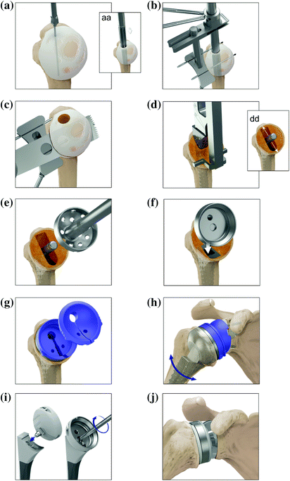
Operative steps for the Arthrex Univers Revers™ Glenoid Preparation

Fig. 30.2
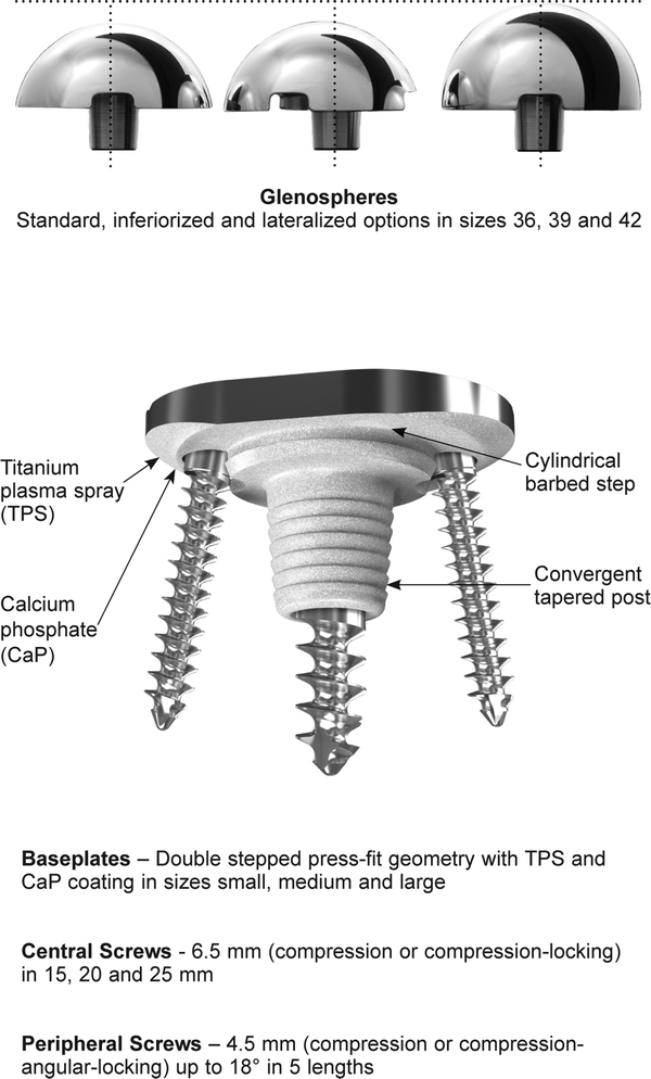
Arthrex Univers Revers™ glenoid baseplate, screw and glenosphere feature overview
2.
3.
Once completed, the reamed surface should be evaluated to ascertain if remnants of sclerotic subchondral bone remain, in which case a second corrective reaming step should be performed. Additional reaming is performed in a control stepped manner to remove 1 mm “slithers” from the central post junction until a suitable fresh bleeding bone surface is exposed, preserving a maximum amount of valuable subchondral bone stock (see Fig. 30.1d, dd).
Glenoid Baseplate Implantation Steps:
1.
The appropriate sized baseplate (see Section “Recommended Surgical Steps to Prepare the Glenoid for Improved Fixation and Outcome” and Fig. 30.3a) for the native glenoid size is impacted into position (see Fig. 30.1e). In complex bone loss cases, such as revision after prior total shoulder arthroplasty, glenoid bone grafting may be required to achieve proper glenoid position and fixation, although the goal remains of primary fixation to the native glenoid bone through the central post and screws.
2.
The Arthrex baseplate is fixed by three cortical bone screws including one 6.5 mm central and two 4.5 mm peripheral screws (see Section “Recommended Surgical Steps to Prepare the Glenoid for Improved Fixation and Outcome”).
3.
The optimal trajectory for the inferior 4.5 mm angular locking screw is drilled into the scapular neck [26, 27] (see Fig. 30.1ff) and the inferior screw is seated (see Fig. 30.1f) to maximize inferior tilt and compression. The central screw pilot hole is then tapped with a 6.5 mm tap to penetrate the dense cortical bone of the scapula (see Fig. 30.1g), followed by the insertion and seating of the central screw (see Fig. 30.1h). This compresses the titanium plasma spray (TPS) and calcium phosphate (CaP) coatings against the freshly reamed bone to further encourage osseointegration (see Section “Recommended Surgical Steps to Prepare the Glenoid for Improved Fixation and Outcome”). The length of the central screw is determined by the initial guide pin depth which is laser marked at 15, 20 and 25 mm (see Fig. 30.1a). Finally, the superior screw trajectory is angled toward the base of the coracoid and seated into the baseplate [26, 27] (see Fig. 30.1i, ii), completing baseplate fixation. A peripheral trench is then reamed to accommodate the glenosphere flange (see Fig. 30.1j). Attention is then directed toward humeral preparation before trial reduction can begin (see Sections “Recommended Surgical Steps to Prepare the Humerus for Maximum Function” and “Trial Reduction to Maximize and Balance Range of Motion and Stability”).
Arthrex Univers Revers™ Baseplate Design Rationale and Glenoid Fixation Methodology
The primary purpose of baseplate screws is to apply and maintain compression between the baseplate and glenoid interface to avoid micromotion and resultant fibrous tissue formation. Compression provides immediate and short-term baseplate stability while creating an environment for solid osseous formation to the native glenoid for long-term fixation [26–29]. The time frame for this has been reported by Cameron et al. to be as short as four months for porous-coated endoprostheses with initial mechanical fixation [30].
Many rTSA baseplates typically offer four peripheral screws: superior (S), inferior (I), anterior (A), and posterior (P), but no central screw. It is widely accepted that only two or three of these peripheral screws are beneficial for fixation. Specifically, the benefit of the A and P screws has been questioned by James et al. [31] A or P screws are rarely both engaged in the glenoid bone [22, 25] but even when engaged, they have been reported to provide far less fixation strength than S or I screws [32]. A central baseplate screw has been reported to be the most effective screw for fixation strength [5]. The Arthrex Univers Revers™ design offers a comparably large 6.5 mm central screw (available in 15, 20 and 25 mm lengths as compression or compression-locking), along with superior and inferior 4.5 mm variable angle screws (available in 24, 30, 36, 42 and 48 mm lengths as variable angle compression or variable angle compression-locking screws). These three screws are aligned vertically in the central plane of the baseplate to enable optimal position, length and trajectory into the best bone stock in the glenoid vault as recommended by Humphrey et al. [22], DiStefano et al. [26], and Parsons et al. [27] (see Section “Recommended Surgical Steps to Prepare the Glenoid for Improved Fixation and Outcome”).
The Arthrex Univers Revers™ baseplate test results were comparable to systems with four screws when tested according to ASTM F2028 [33, 34] guidelines (Standard Test Methods for the Dynamic Evaluation of Glenoid Loosening or Disassociation), suggesting comparable short-term fixation. Systems with four or more screws have reported good clinical short-term fixation [35]. However, filling the glenoid with an overabundance of screws may lead to stress shielding, adversely affecting long-term implant fixation. Stress shielding is the degeneration of native bone due to reliance on the rigid metal prosthesis for load transfer, discouraging the remodeling of new bone [12]. Stress shielding at the glenoid interface is believed to contribute to suboptimal osseointegration and loosening or worse glenoid neck fracture resulting in challenging revision surgeries with limited options.
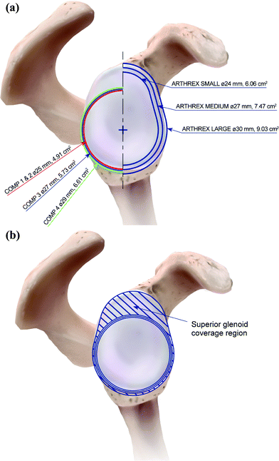
Three Arthrex baseplate sizes have been designed to maximize contact area by closely matching the native glenoid’s “pear”-shaped surface (see Fig. 30.3). This presents several potential benefits over typically “round” baseplates, commonly offered in one size only. First, by maximizing the surface area, size for size, the highly localized pressure regions can be distributed more evenly across a larger implant-to-bone surface area (see Fig. 30.3a). Second, the Arthrex baseplates extend vertically to match the superior region of the native glenoid anatomy (see Fig. 30.3b). Standard round baseplates fail to utilize this superior region, which not only enables additional surface area contact for improved long-term fixation but also acts as a buttress, helping to counteract and distribute the torsional forces imposed at the implant-to-bone interface. An even distribution of forces has been reported to encourage optimal conditions for scapular remodeling [12]. Furthermore, in the event of glenoid bone loss, which commonly occurs in the region of the superior glenoid with rotator cuff deficiency [2], the additional surface area of the implant provides the opportunity to better capture bone graft under the implant, encouraging osseointegration of the graft.
Anglin et al. reported that glenoid component loosening generates the greatest concern among surgeons performing rTSA. The authors devised a specific test to analyze different baseplate designs and geometries, concluding that a roughened fixation surface far outperformed a smooth fixation surface and a curved backed surface showed almost half the distraction of a flat backed surface [36]. Many current baseplate designs are smooth and flat or just slightly curved with no protruding or press-fit geometry, subjecting the screws to shear forces applied to the baseplate bone interface. These designs have reported failure rates between 11.7 % and 40 % [37], confirming that bony ingrowth is an important factor for avoiding baseplate failure.
The underside of all Arthrex Univers Revers™ baseplates includes a unique double stepped press-fit geometry, roughened to 0.30 µm with a TPS coating, to minimize micromotion and encourage bone ingrowth [38, 39], and a slow release CaP coating on the outer surface to encourage bone ongrowth [40]. A roughened titanium substrate of higher than 0.10 µm improves anchorage for the CaP and enables increased surface area for CaP and resultant bone coverage [41]. CaP coatings induce more rapid bone apposition onto prostheses, optimizing osseointegration for long-term fixation and increasing bone quality and strength [42, 43]. The critical micromotion threshold has been disputed. However, what is evident is without stable fixation, the formation of fibrous membrane is more prevalent, predisposing shoulders to early loosening and poor clinical outcomes [28, 29]. The Arthrex Univers Revers™ press-fit baseplate geometry has been specifically designed for maximum stability to encourage bone ongrowth in a reduced micromotion environment. The Arthrex baseplate design has been tested in-line with load protocols specified in ASTM F2028 [34] and subsequently verified by results with significantly lower micromotion values than the threshold limits suggested by Cameron et al. of to 150 µm [44].
The Arthrex press-fit baseplate is specifically designed to match the patient’s anatomy and help to maximize the surface area in contact with bone (see Fig. 30.3). The three centrally aligned screws offer optimal trajectories into the native glenoid bone stock to support a stable compressive healing environment. This combination of a macro-press-fit geometry and micro-roughened TPS substrate with a CaP coating encourages stable osseointegration of the prostheses, optimizing the chances for long-term fixation.
Recommended Surgical Steps to Prepare the Humerus for Maximum Function
1.
The humeral osteotomy is performed either with the supplied extra- or intra-medullary cutting jig (see Fig. 30.4b, c) or by using a free-hand technique, after the subscapularis has been released. Repair of the subscapularis tendon has been shown to improve postoperative stability [14, 15], therefore the intact portion of the subscapularis is released directly from its lesser tuberosity insertion and preserved during the procedure. The goal of the humeral osteotomy is 20° of retroversion to match normal humeral anatomy. This angle facilitates the ideal position for activities of daily living (ADL) [13]. With the Arthrex Univers Revers™, the resection angle and depth are non-critical, as the definitive implant seats within the metaphysis to dissipate loads directly to the proximal humerus and calcar. Many commercially available rTSA designs force the user to respect the resection surface, which the implant sits on top of, or risk overstuffing the joint [14, 15]. Removal of greater tuberosity deformity should be undertaken to avoid impingement with the acromion, which has been reported in some incidences to inhibit abduction ROM (see Section “Recommended Surgical Steps to Prepare the Glenoid for Improved Fixation and Outcome” for glenoid preparation) [45].


Fig. 30.4
Operative steps for the Arthrex Univers Revers™ humeral preparation
2.
The intramedullary canal is then step reamed until a desired diameter match has been achieved. Aggressive reaming is unnecessary and potentially increases the risk of humeral fracture. The purpose of humeral reaming is to identify the endosteal bone diameter.
3.
The humeral canal is then progressively broached to desired fit (see Fig. 30.4d). The broach depth mark (135° and 155°) represents the minimum impaction line. A large range of stems which increase incrementally in size and offset enable an optimal press-fit in the native humerus (see Section “Arthrex Univers Revers™ Humeral Stem, Metaphyseal Cup and Liner Articulation Design Rationale”). For rare situations where cemented application is indicated with the Arthrex Univers Revers™ due to bone deficiency, a stem, one size smaller than that broached should be used to ensure adequate cement mantle thickness.
4.
Once the desired broach size and height has been attained, disconnect the handle, leaving the broach in the humeral canal. Due to the variability of the osteotomy level, it may be necessary to impact the broach further to ensure the post is equidistant between the medial (M) and lateral (L) aspects of the osteotomy (see Fig. 30.4dd).
5.
The A and P position of the broach is then evaluated to determine whether a central or an offset reamer guide post is required (see Fig. 30.4e). The Arthrex Univers Revers™ offers the ability to match the typically posterior metaphyseal offset [46–48]. The proximal humerus reamer guide post is available in centralized, 2 mm offset left or 2 mm offset right, enabling the surgeon to match the cup position to the native metaphyseal/shaft offset.
6.
Following metaphyseal reaming, the selected trial cup size is fixed to the broach (see Fig. 30.4f) (which is still in the humeral canal) and trial reduction is performed with trial spacer and liner components (see Section “Trial Reduction to Maximize and Balance Range of Motion and Stability” and Fig. 30.4g, h).
Arthrex Univers Revers™ Humeral Stem, Metaphyseal Cup and Liner Articulation Design Rationale
The Arthrex Univers Revers™ consists of 11 humeral stem sizes ranging from 6 to 15 in lengths of 111 to 146 mm, respectively. Three additional extra-long stems are available for revision purposes in size 6, 9 and 12 at lengths of 180 mm. An extra small size 5 mono block stem is also available in both 135° and 155° inclination angles. All stems offer a rectangular proximal geometry to ensure optimum rotational stability in the proximal humerus. The proximal portion of all stems is CaP coated with a press-fit design. Rotationally, stable and compressive proximal load bearing environments have been reported to encourage solid osseous formation for long-term diaphyseal fixation [28, 29]. All these stems are also used for anatomic shoulder arthroplasty configurations, so if well-fixed during revision surgery, the stems can be left in situ and converted to the Arthrex Univers Revers™.
Compatible with all humeral stem sizes and types, the Arthrex Univers Revers™ system includes three metaphyseal cup sizes in 36, 39 and 42 to match the patient’s metaphyseal anatomy. Each size is offered in central, offset left, and offset right to best match the patient’s native metaphyseal offset. All cups can be positioned in either a varus 135° or valgus 155° neck-shaft angle to support the surgeon’s decision to individualize each case and optimize their patient outcomes. The underside of the cups, which transfers the load of the joint to the proximal humerus, is also CaP coated to encourage solid osseous formation for long-term metaphyseal fixation.
UHMWPE liners that match the cup sizes are available in both standard and constrained designs. Constrained liners, with deeper socket depths, are utilized in specific situations to increase biomechanical stability. However, due to the increased rim height of constraint liners, ROM is reduced in favor of increased stability [37, 49]. The articular constraint of the Arthrex Univers Revers™ liners upholds the principles outlined by Gutiérrez et al. of humerosocket depth “d” normalized by radius “r” [50]. The resultant articular constraint ratio closely matches the mean value for commercially available standard liners of 0.56 and a ratio of 0.66 for constrained liners, across all sizes 36, 39 and 42.
Stay updated, free articles. Join our Telegram channel

Full access? Get Clinical Tree


