Fig. 9.1
This arthroscopic view from a posterior portal demonstrates thickening of the upper rolled border of the subscapularis with synovitis and inflammation of the adjacent anterior capsule
9.3 Capsular Ligamentous Complex
The glenohumeral joint capsule, coracohumeral ligament, and glenohumeral ligaments (superior, middle, and inferior) make up the capsular ligamentous complex. These structures connect the humerus and glenoid and also attach to the glenoid rim via the labrum [4].
9.3.1 Anterior Superior (The Rotator Interval)
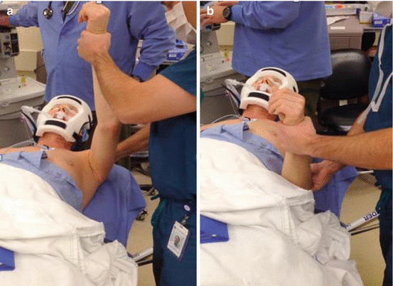
The rotator interval is a confluence of tissue in a triangular shape located between the anterior supraspinatus leading edge and the upper border of the subscapularis tendon. The apex of the rotator cuff interval is located at the margin of the transitional ligament where the biceps sulcus is located. The rotator interval includes the coracohumeral ligament, the superior glenohumeral ligament, and the anterior-superior capsule. Contracture of these structures, the most commonly affected in frozen shoulder, results clinically in loss of flexion and external rotation in adduction [6, 10, 14, 17, 18] (Fig. 9.2).


Fig. 9.2
Examination under anesthesia in a patient with frozen shoulder. (a) Forward elevation with the scapula stabilized is limited to 90°. (b) External rotation with the arm at the side (0° of abduction) is limited to 0° (The authors would like to acknowledge Dr. Bryce Wolf for his assistance with these images)
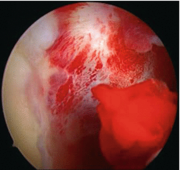
Clinically, the rotator interval may be obliterated by fronding synovitis or inflammation, making initial appreciation difficult during arthroscopy (Fig. 9.3).


Fig. 9.3
In this arthroscopic view from a posterior portal, there is complete obliteration of the rotator interval by proliferative synovitis and capsular contracture
9.3.2 Anterior
The anterior-inferior quadrant of the glenohumeral joint contains the anterior-inferior capsule and the middle glenohumeral ligament (MGHL). These structures are also almost invariably involved in frozen shoulder. The anterior capsule is frequently scarred to the middle glenohumeral ligament and the subscapularis. This results in further limitations of external rotation at the side and with increasing degrees of shoulder abduction [6, 11, 16].
9.3.3 Inferior
In Neer’s original description based upon open surgical dissection, he described scarring of the axillary fold to itself and to the anatomic neck of the humerus [15]. However, newer research based on arthroscopic findings suggests that no such scarring of the axillary recess exists and that instead there is contracture of the inferior capsular tissues [25].
Nonetheless, this results in significant losses of abduction, forward flexion, and both external and internal rotation due to involvement of the anterior and posterior bands of the inferior glenohumeral ligaments (AIGHL/PIGHL) as well as the remainder of the capsular tissue [6].
9.3.4 Posterior
Clinically, internal rotation is less affected in frozen shoulder. When involved, the posterior capsule results in loss of internal rotation of the adducted arm. Because this tissue frequently appears to be less involved, the need for surgical release of the posterior capsule during surgical treatment has been debated [2, 6, 24].
9.3.5 Posterior-Superior
There has been recent evidence to suggest the existence of a posterior-superior glenohumeral ligament (PSGHL) [19]. Whether this represents a discrete structure or simply a thickening of the capsule is unclear [3, 20]. The relevance of this finding and its relevance to frozen shoulder are unknown. The expected effect of contracture would be a loss of internal rotation in adduction – an uncommon clinical finding.
Conclusions
While frozen shoulder has been shown to involve all portions of the capsule, it affects certain quadrants more predictably and severely. Additionally, capsular contracture has effects on the surrounding periscapular musculature. Surgeons treating this difficult problem need to consider not only the intraarticular pathology but its associated effects on the entire shoulder girdle.
References
1.
Boyle-Walker KL, Gabard DL, Bietsch E, Masek-VanArsdale DM, Robinson BL. A profile of patients with adhesive capsulitis. J Hand Ther. 1997;10(3):222–8.CrossRefPubMed
Stay updated, free articles. Join our Telegram channel

Full access? Get Clinical Tree


