Fig. 15.1
Knees: varus position in both knees, subchondral sclerotisation and asymmetrical narrowing of tibiofemoral articular slit. Changes are accentuated in the lateral part of articular slit of the left knee with its narrowing to the minimum. Mild impaction and fragmentation of medial condyle of the right tibia. Osteophytes of articular surface margins, accentuated in patellofemoral area on the right side. Hyperostose rim on patella anterior surface
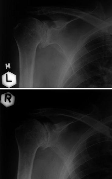
In shoulder joint, ochronosis is manifested most frequently as typical osteoarthrosis sometimes with atypical secondary finding. Numerous fibro-ostoses on the tuberculum majus or acromioclavicular and ochronotic foci localised in deeper structure of the head and neck of humerus and neck of scapula in supraglenoid area can be the atypical findings. Insignificant manifestations of osteoarthrosis are represented by spike-forming of the inferior margin of glenoid fossa and a small osteophyte on the lower pole of humeral head. This finding rapidly progresses to advanced stage characterised by considerable narrowing of glenohumeral articular slit, significant osteoplastic changes as well as possible mushroom-like flattening and fragmentation of humerus head (Fig. 15.2).


Fig. 15.2


Shoulders: Position of heads of both humeri in subacromial retraction, with sclerotisation and enthesophytes in the area of major tubercle – periarthropathia humeroscapularis (PHS) bilaterally. Narrowing of articular slit in humeroscapular joint to the minimum with slight incongruence and subchondral sclerotisation of articular surfaces, with osteoplastic rim of their margins that creates exostotic formation at the caudal margin of glenoid fossa. The humeral head is mildly flattened on the right side, with irregular contours, shallow usurations and cystoid translucencies in deeper structure
Stay updated, free articles. Join our Telegram channel

Full access? Get Clinical Tree


