Achilles Tendon Repair
James D. Granata
Jason Calhoun
The Achilles tendon is the most commonly ruptured tendon of the lower extremity (5,7), with an overall incidence of 18 per 100,000 people (1,11,18). Middle-aged recreational athletes are particularly susceptible, accounting for 40% of all surgical repairs (4,18). The majority of ruptures are indirect injuries that occur during sports and recreational activities involving rapid acceleration and deceleration movements at the ankle.
The tendons of the gastrocnemius and soleus muscles, collectively known as the triceps surae, combine to form the Achilles tendon. The combined tendon rotates 90 degrees from proximal to distal, with the medial fibers rotating posteriorly, and the posterior fibers rotating laterally (16). The tendon is extrasynovial and receives its blood supply from three different sources: the paratenon, the musculotendinous junction proximally, and the osseotendinous junction distally. This leaves the central portion of the tendon the least vascularized. The majority of Achilles tendon ruptures are midsubstance tears within this relatively avascular zone, 2 to 6 cm proximal to the calcaneal insertion (13).
Although the Achilles tendon is the strongest tendon in the body, a number of factors make it susceptible to complete rupture. Most patients with acute ruptures have no preceding Achilles tendon symptoms, yet histopathology of ruptured tendons shows the presence of degenerative tissue in the vast majority of cases (4,5,8,13). Rapid loading of the degenerated tendon can then lead to a complete tear, with running and jumping transmitting forces up to eight times body weight through the Achilles tendon. Other risk factors for acute ruptures include previous corticosteroid injections, use of fluoroquinolone antibiotics, and high longitudinal arches with a decrease in the pronation range of the foot and ankle (16).
INDICATIONS/CONTRAINDICATIONS
The choice between operative and nonoperative management of an Achilles tendon rupture remains controversial. The best form of treatment depends on patient characteristics and expectations. Age, activity level, and medical comorbidities help guide treatment recommendations. Patients should be educated about the risks and benefits of both treatment options so that an informed mutual agreement can be reached.
Nonsurgical management offers the advantages of decreased cost and the absence of surgical complications, although the overall complication rate may be higher (4). Traditional treatment includes immobilization in a plaster cast for 6 to 8 weeks. Nonsurgical management is recommended for patients with sedentary lifestyles, heavy tobacco use, poor skin quality, and medical comorbidities, which increase the risk of wound healing problems.
Operative management offers the potential benefit of a decreased rerupture rate, restores the length and tension of the muscle-tendon unit, and may result in earlier return to sport as well as increased strength and endurance (1,4,5,7,11,13,18). We recommend surgery for active patients with minimal risk factors for wound healing complications. Surgery may also benefit chronic Achilles tendon tears that have failed conservative management and remain symptomatic.
PATIENT EVALUATION AND PREOPERATIVE PLANNING
The diagnosis of acute Achilles tendon ruptures can usually be made with a thorough history and physical exam. Patients often describe acute pain in the back of the calf while participating in an athletic activity, followed by an inability to fully bear weight. A subjective feeling of being kicked in the back of the leg and hearing a loud pop is common. Physical exam findings include soft tissue swelling and ecchymosis around
the distal posterior leg and ankle. A tender palpable defect is often appreciable along the course of the tendon (Fig. 53.1). Active plantar flexion may still be intact due to the long flexors, but strength is decreased compared to the contralateral side.
the distal posterior leg and ankle. A tender palpable defect is often appreciable along the course of the tendon (Fig. 53.1). Active plantar flexion may still be intact due to the long flexors, but strength is decreased compared to the contralateral side.
The Thompson test, also known as the calf squeeze test, is the most reliable part of the physical exam used to assess the continuity of the Achilles tendon (12). The calf musculature is squeezed, and in a normal individual the ankle will plantar flex (Fig. 53.2A). In Achilles tendon tears, plantar flexion is absent or significantly decreased compared to the normal side (Fig. 53.2B). The squeeze test can be quite painful and patients may not allow it.
Uncommon injury patterns and chronic tears may be difficult to diagnose with history and physical exam alone. Imaging studies can be helpful when the physical exam is equivocal. Avulsion injuries may include a portion of the calcaneal tuberosity and are well visualized with lateral plain radiographs (Fig. 53.3). MRI can be helpful in detecting partial tears, proximal tears, and direct avulsions of the tendon off the calcaneus
(sleeve avulsion). MRI can confirm the diagnosis in delayed presentations or chronic tears and provide relevant information for preoperative planning, including the length of tendon involved.
(sleeve avulsion). MRI can confirm the diagnosis in delayed presentations or chronic tears and provide relevant information for preoperative planning, including the length of tendon involved.
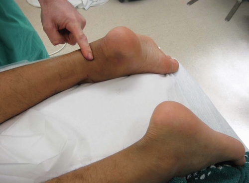 FIGURE 53.1 A palpable defect is noted along the middle portion of the Achilles tendon. |
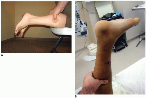 FIGURE 53.2 A: Negative Thompson test: intact plantar flexion with calf squeeze. B: Positive Thompson test: absent plantar flexion with calf squeeze. |
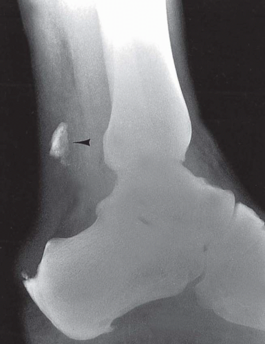 FIGURE 53.3 Lateral radiograph of the calcaneus demonstrating a displaced calcaneal tuberosity fracture. |
After the initial evaluation, the ankle should be immobilized in plantar flexion with a well-padded posterior splint. Patients are encouraged to ice and elevate the affected extremity to minimize swelling. In order to minimize scar formation and contracture of the tendon, surgery should be performed within 2 weeks of the initial injury. Delaying surgery beyond the acute period may decrease the chance of a direct end-to-end repair.
Related posts:
 Arthroscopic Management of Elbow Osteochondritis Dissecans Lesions
HAGL: Arthroscopic/Open
Meniscal Débridement
Aperture Fixation in Primary Arthroscopic Double-Bundle, Single-Bundle, and Single-Bundle-Augmented ACL Reconstruction
LCL/PLC Reconstruction
Cartilage Transplantation: Fresh Osteochondral Allograft
Arthroscopic Management of Elbow Osteochondritis Dissecans Lesions
HAGL: Arthroscopic/Open
Meniscal Débridement
Aperture Fixation in Primary Arthroscopic Double-Bundle, Single-Bundle, and Single-Bundle-Augmented ACL Reconstruction
LCL/PLC Reconstruction
Cartilage Transplantation: Fresh Osteochondral Allograft
Stay updated, free articles. Join our Telegram channel

Full access? Get Clinical Tree


