Baseline values
Mini incision
Standard incision
p-value
N = 103
N = 88
Gender (M/F)
49/54
50/38
0.205*
Age
67.0 ± 10.9
67.3 ± 12.6
0.838**
Primary OA (%)
85.4 %
78.4 %
0.928**
BMI (kg/m2)
27.2 ± 4.1
27.9 ± 4.4
0.272**
Preop PMA score
10.0 ± 1.4
9.6 ± 1.6
0.206***
Statistical Analysis
Data were evaluated with Statistica 6.1 (StatSoft Inc., Tulsa, OK, USA). Alpha was chosen at 0.05. Between-group comparisons were performed with the Student’s t-test or the Mann-Whitney test for continuous variables, the Mann-Whitney test for ordinary scaled variables, and the chi-square and Fisher exact test for nominal scaled variables.
Surgical Technique
Patient Positioning
The patient is placed on the operating table in the lateral decubitus position with the pelvis locked perpendicular to the table. The entire leg and hip are prepared and draped. A supplementary sterile pouch is dressed in front of the operating table in order to place the leg in a vertical position at the femoral preparation step.
Incision
The skin incision is made longitudinally in a straight line over the greater trochanter from 3 cm above the tip to 5 cm below (Fig. 8.1). The fascia lata is divided in a straight line and the gluteus maximus is splitted in line upwards. This division is extended 3 cm proximal and distal beyond the limits of the skin incision; the incision of the trochanteric bursa reveals the anterior and posterior borders of the great trochanter and its attaches. Using cutting diathermy, a longitudinal incision is made to divide the tendinous periosteum over the great trochanter centered midway between the anterior and posterior margins and extended distally in the middle of vastus lateralis tendon to a point 1 cm beyond the vastus ridge. The incision extends proximally to divide, in an anterior curved direction, 1/3 anterior of the gluteus medius muscle in direction of the fibers and not more than 2 cm above the tip of the great trochanter (Fig. 8.2).
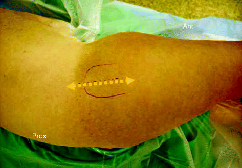
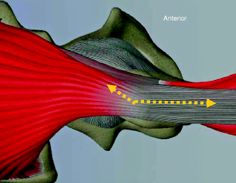

Fig. 8.1
Skin incision

Fig. 8.2
1/3 gluteus medius-1/2 vastus lateralis digastric anterior flap developed with bony intermediate junction created by osteotomy of the lateral facet of the greater trochanter
Approach
With use of an oscillating saw, an osteotomy of the lateral aspect of the great trochanter is performed in an upward direction from the vastus ridge in order to preserve the transverse branch of the lateral circumflex artery (Fig. 8.3). The trochanteric fragment is vertical, linear, about 5–8 mm thick and carries with it the continuation of the anterior part of the gluteus medius and the vastus lateralis. It is attached proximally to the anterior part of the gluteus medius and distally to the anterior half of the vastus lateralis. Rotating the extremity laterally achieves a medial slide of the fragment which is then mobilized anteriorly to expose the gluteus minimus and the capsule which are incised in the same line. The distal part of the gluteus minimus is detached jointly from the capsule and from its femur insertion. The proximal part of the incision is extended along the femoral neck in an anterior direction toward the superior acetabular rim (Figs. 8.4 and 8.5). The femoral neck is transected in situ or after dislocation; then, the femoral head is excised.
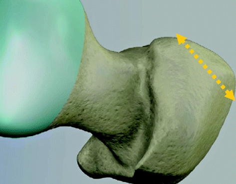
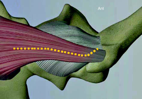
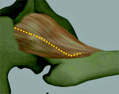

Fig. 8.3
Osteotomy of the lateral facet of the greater trochanter

Fig. 8.4
Split of the gluteus minimus and incision of the capsule in the same line

Fig. 8.5
Line drawing of the capsule incision
Acetabular Exposure
After removal of the femoral head, the position of the leg is adjusted to give the exposure of the acetabulum. In most cases, lateral rotation and slight flexion of the hip give the best access. After excision of the labrum, two spiked Hohmann retractors are inserted over the anterior and posterior edges of the acetabulum (at 4 and 8 o’clock). Then the capsule can be released if necessary to the medial border of the femur. A Steinman pin or a self-retaining retractor is placed proximally to retract the capsule and the gluteus muscles (Fig. 8.6). The entire acetabular cavity can now be seen and remnants of the labrum are excised.
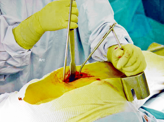

Fig. 8.6
Acetabular exposure is provided by two spiked Hohmann retractors and a Steinman pin
The acetabular bony preparation is performed with an angled reamer handle designed for use in minimally invasive surgery of the hip. Either a curved impactor through the incision directly or a straight impactor through a separate percutaneous incision is used to insert the cup in proper position.
Femoral Exposure
The femoral preparation is made with the foot placed vertically. The exposure is provided by two spiked Hohmann retractors, one placed on the medial and the other posterolateral side of the femur. A third spiked Hohmann can be placed advantageously under the posterior femoral neck anterior to the ventral gluteus medius part to prevent muscle damage possibly encountered by the femoral rasps. Sharp-cutting femoral rasps of rectangular cross section and increasing size are used with a pneumatic hammer to achieve direct anchorage by press fit. After satisfactory trial reduction with a trial device the definitive prosthesis is inserted and the hip is reduced.
Closure
The closure is made in layers. The capsule and the gluteus minimus are jointly sutured and can be reattached to the femoral bone (Fig. 8.7). Then the trochanteric slide fragment is reattached to the proximal femur by a single cerclage wire (monofilament 1.2 mm steel) passed anteriorly to the stem of the prosthesis through drill holes. The twist of the metal knot is placed under the vastus ridge to prevent trochanteric bursitis related to the cerclage wire (Fig. 8.8). The fascia lata, gluteus fascia, subcutaneous tissues, and skin are closed in usual fashion.
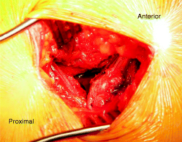
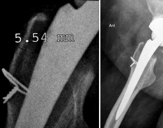

Fig. 8.7
Operative aspect before the closure (right hip)

Fig. 8.8
The lateral radiograph and focus show the reattachment of the trochanteric fragment with a wire
Clinical Results
Average time for surgery was 62 min for study group and 63 min for the control group (p = 0.51). No decreased time related to the learning curve was observed between mid-practice in the mini-incision group. Postoperative day 1 after surgery, the hemoglobin level was 11.8 g/l for the study group and 11.6 g/l for the control group (p = 0.42). However, fewer patients in the study group received blood autotransfusion with hemocare (16 % vs. 49 %, p < 0.001), and the amount of blood transfused was less for the study group (119 ml vs. 130 ml, p < 0.001).
Stay updated, free articles. Join our Telegram channel

Full access? Get Clinical Tree


