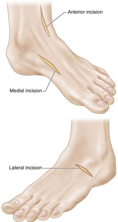
Split Transfer of the Tibialis Anterior Tendon
Indications
 Spastic, flexible equinovarus-cavovarus foot deformity in the ambulatory child or adult with cerebral palsy or brain injury
 Recurrent flexible clubfoot cavovarus deformity
 Weak peroneal muscles and relative “over-pull” of the tibialis anterior leading to foot supination and cavus deformity
Examination/Imaging
 The peroneus longus may be relatively weaker, allowing the tibialis anterior to “over-pull” the midfoot into supination and cavus, especially during early stance and during the swing phase of gait.
 The gastrocnemius-soleus and tibialis posterior must also be assessed for contracture.
 The goal is a balanced foot in both stance and gait.
 Hindfoot varus flexibility may be tested with the Coleman block test (Coleman and Chestnut, 1977).
 Preoperative plain films, including standing anteroposterior, lateral, and oblique views of both feet, should be obtained to confirm no fixed bony deformity.
Surgical Anatomy
 The tibialis anterior has a broad insertion and is best found proximal to the first ray through the medial incision.
 When transferring to the cuboid, care should be taken to protect sensory nerve braches from the superficial peroneal nerve.
 The surgeon should try to avoid injury to the short toe extensor muscle belly when exposing the cuboid.
52: Split Transfer of the Tibialis Anterior Tendon


