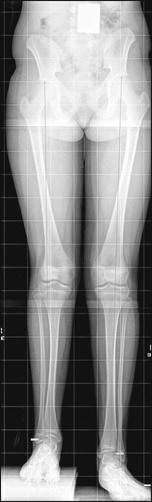
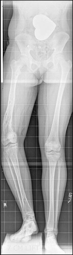
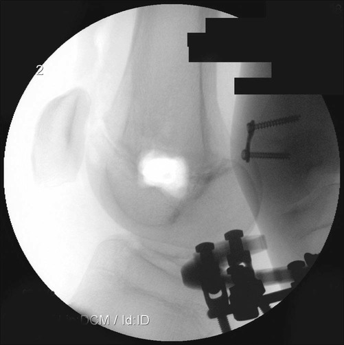
Figures 9–14 modified with permission from Springer-Verlag (Paley D. Principles of Deformity Correction. Berlin: Springer-Verlag, 2002). Figures 1–3, 4–7, and 15 Copyright 2010, Rubin Institute for Advanced Orthopedics, Sinai Hospital, Baltimore, Maryland. • Deformity correction can be performed simultaneously with gradual distraction for lengthening at the same osteotomy site, provided the osteotomy is at or near the apex of the deformity. • Double-level tibial osteotomies can be performed in older children, especially if there are multiple apices of deformity. • Possible valgus procurvatum of the diaphysis—Requires simultaneous angular correction with slight overcorrection of valgus • Instability of the knee—May need crossing of the knee with the fixator for stabilization (fixation to femur with external fixation or cast brace linked to tibial fixator) • Fixed equinovalgus of the ankle—Peroneal/Achilles tendon lengthening plus supramalleolar osteotomy or subtalar osteotomy to realign the hindfoot/ankle (can be staged or simultaneous) • Intrinsic soft tissue contractures—Vulpian lengthening of the gastrocnemius-soleus at the time of tibial lengthening • Concurrent instability of the ankle joint • The knee is often unstable, requiring the external fixator frame to be extended to the femur with hinges. • Foot equinovarus needs simultaneous correction (supramalleolar osteotomy for ankle diastasis type). • Typically has proximal varus and distal valgus/recurvatum; check for compensatory subtalar varus contracture • It is difficult to predict the final limb length inequality during growth. • The surgeon should wait until skeletal maturity to equalize limb lengths. • Type V growth pattern with upward slope–plateau–downward slope with improvement without intervention. • Final LLD should be assessed at skeletal maturity and then corrected. • Limb lengthening at ages less than 8 years can result in growth inhibition. • Staged lengthenings should be planned to gain functional height. • Delayed regenerate bone formation and healing—Plan decreased rate of distraction. • Slight ankle equinus and residual LLD may be desireable to aid knee extension in the face of weak quadriceps. • May need medial hemiplateau elevation as preliminary staged procedure, followed by metaphyseal osteotomy for lengthening and residual varus correction • Ollier’s disease always has angular deformities. • Angular deformities recur with subsequent growth until skeletal maturity. • The leg may be lengthened through the edge of the Ollier lesion. • Bone healing may be premature, requiring faster rate of lengthening. Controversies • Severe deformities requiring extensive lengthening • LLD less than 5 cm in the skeletally immature patient • LLD less than 5 cm in the skeletally mature patient • Contractures or joint/bony abnormalities must be documented. • Concurrent joint instabilities and ligamentous deficiencies must be documented. • Malalignment and malorientation tests are performed to determine concurrent deformity and location of deformity. • Limb lengths are assessed (the pelvis should be leveled with blocks and the number of blocks noted on the radiograph). • The radiographs are checked for fixed flexion deformity of the knee. • A malorientation test is performed to determine the presence and location of sagittal plane deformity. • The radiographs are checked for joint subluxation (significant anterior-posterior instability). • Figure 1 shows a preoperative radiograph of an 11-year-old girl with FH and valgus knee, with 5-cm LLD. • Figure 2 shows a preoperative radiograph of a 14-year-old girl with Ollier’s disease, demonstrating shortening of the femur and tibia, with angular deformity in both. Treatment Options • LLD less than 5 cm (see Fig. 2): shoe lift or epiphysiodesis • The surgical approach for osteotomy is through the internervous interval (peroneals/soleus). • It is safer to perform the fibular osteotomy in the distal half to avoid traction injury to the nerve to the extensor hallucis longus. • A prophylactic anterior compartment fasciotomy is safe, and can give a larger diameter appearance to an atrophic leg. • Prophylactic Vulpian gastrocnemius-soleus recession helps the Achilles tendon to stretch to accommodate the tibial lengthening. • This nerve is at risk during the external fixation application if wires are placed near the fibular neck or slightly below, or if the fibular osteotomy is high. • Rapid lengthening or concurrent deformity correction can place tension on the peroneal nerve. • The superficial peroneal nerve may be entrapped by distal tibial wires. • This nerve is at risk during the external fixation application if wires are passed posteromedially in the distal tibia. • Tarsal tunnel syndrome may occur from swelling, or traction during distal tibial deformity correction, requiring release of fascia over the tarsal tunnel. • Intrinsic instability will result in increased risk of joint subluxation or dislocation. • Severe knee instability should be addressed with ligamentous reconstruction prior to tibial lengthening. • Metaphyseal tibial osteotomy about 6–7 cm distal to the knee joint typically produces the best bone. • Fibular osteotomy is best at the junction of middle and distal third. • If an abnormal mechanical axis is present, then preoperative planning is required to determine the level of the deformity.
Tibial Lengthening with Circular External Fixation
Indications
 Tibial shortening with or without limb deformity
Surgical Pitfalls
 Juvenile rheumatoid arthritis (JRA)
 Rickets, renal osteodystrophy, JRA
 Long-leg epiphysiodesis if there is adequate growth remaining and height prediction is within normal range
Examination/Imaging
Physical Examination
 Range of motion of the hip, knee, and ankle and subtalar joints
 Special consideration must be given to knee and ankle fixed flexion deformity. The cause (soft tissue contracture vs. bony deformity) must be determined
 Clinical limb-length assessment
Radiographic Examination
 Anteroposterior (AP) standing long-leg radiographs are obtained (taken at a distance of 10 feet using a 51-inch cassette).
 Standing long-leg lateral radiographs (with maximum knee extension) are obtained.
 Additional views of the foot and heels (Saltzman view) are obtained as needed to assess deformity.
 A shoe lift should be avoided during sports (risk of ankle sprain).
 For lifts of greater than 5 cm, the patient may need an ankle-foot orthosis to stabilize the ankle.
 For lifts of greater than 8 cm, a platform prosthesis should be used.
 Epiphysiodesis is done percutaneously by drills and curettes without need for implants. Figure 3 shows a contralateral distal femoral epiphysiodesis in a patient undergoing an ipsilateral tibial lengthening.
Surgical Anatomy
 Peroneal nerve tension or irritation initially will result in pain on the anterior aspect of the lower leg and dorsum of the foot.
 Further tension causes decreased sensation.
 Late findings include weakness of the dorsiflexors or a drop foot.
 Young patients will often hold the toes up with their hands or continuously rub their foot.
Procedure: External Fixator Placement
Step 1
 The limb is held with patella forward for a true AP image intensifer view.
 The level of the knee, ankle, and each growth plate is marked.
 The level of the intended osteotomy sites (tibia and fibula) is marked.
Related posts:
 4: Open Reduction and Internal Fixation of Displaced Medial Epicondyle Fracture Using a Screw and Washer
29: Epiphysiodesis of the Distal Femur/Proximal Tibia-Fibula
32: Patellar Instability: Lateral Release and Medial Plication
31: Discoid Lateral Meniscus
39: Open Reduction and Internal Fixation of Tibial Tubercle Fractures
59: Posterior Instrumented Reduction and Fusion for Spondylolisthesis
4: Open Reduction and Internal Fixation of Displaced Medial Epicondyle Fracture Using a Screw and Washer
29: Epiphysiodesis of the Distal Femur/Proximal Tibia-Fibula
32: Patellar Instability: Lateral Release and Medial Plication
31: Discoid Lateral Meniscus
39: Open Reduction and Internal Fixation of Tibial Tubercle Fractures
59: Posterior Instrumented Reduction and Fusion for Spondylolisthesis
![]()
Stay updated, free articles. Join our Telegram channel

Full access? Get Clinical Tree


42: Tibial Lengthening with Circular External Fixation


