• This test is done at both 0° and 30° of flexion. • The surgeon must determine if there is increased laxity of either the medial or lateral collateral ligament. • With the knee at 30° of flexion, the tibia is translated anteriorly without a solid endpoint. • The patient is re-evaluated at the end of the case for comparison. • The knee is taken from extension to flexion while placing valgus stress on the knee and internally rotating the tibia. • The surgeon should feel for a gliding or shifting sensation of the tibiofemoral joint. • Staging is done by assessing for the presence and distribution of pubic hair and for breast/testicular development. • Posteroanterior (PA), lateral, Merchant’s, and tunnel views are obtained before surgery to evaluate for any bony abnormalities. • Radiographs allow for initial evaluation of the physis before surgery has been completed. • A anteroposterior (AP) radiograph of the left hand and wrist is obtained to determine bone age.
Transphyseal ACL Reconstruction in Skeletally Immature Patients Using Autogenous Hamstring Tendon
Indications
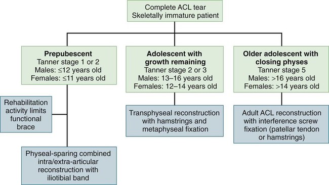
 Adolescent patient with significant growth remaining (Tanner stage 3 or greater) with a complete anterior cruciate ligament (ACL) tear and functional instability (Fig. 1)

 Adolescent patient with significant growth remaining with a partial ACL tear that has failed nonoperative treatment
 Adolescent patient with significant growth remaining with an ACL tear and other injuries such as meniscal tears or chondral injury
 Adolescent patient with significant growth remaining with congenital ACL insufficiency and symptomatic instability despite bracing
Examination/Imaging
 An examination under anesthesia is performed.
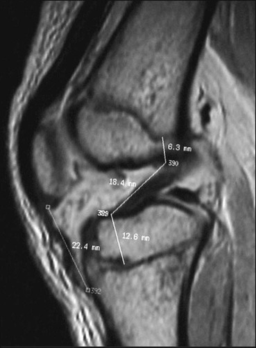
 Magnetic resonance imaging (MRI) (Fig. 2)

Surgical Anatomy
Portals/Exposures
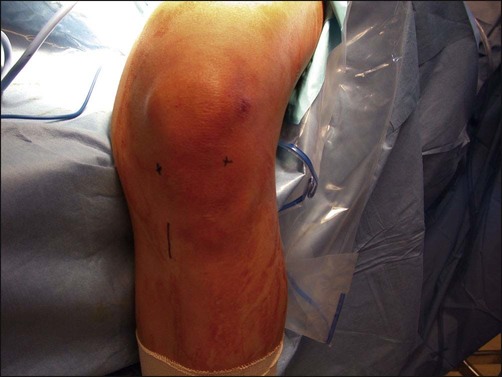
 Two standard arthroscopic portals are used (Fig. 3).

 Hamstring harvest/tibial tunnel
37: Transphyseal ACL Reconstruction in Skeletally Immature Patients Using Autogenous Hamstring Tendon


