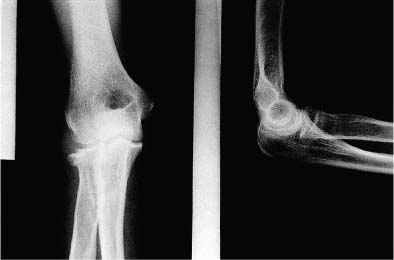
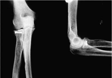
Case 36 A 29-year-old tennis player presents several hours following a fall onto his outstretched hand with his elbow extended. He complains of pain, loss of motion, and crepitation. Range of motion is limited to 60 to 95 degrees. He has 60 degrees of supination and 20 degrees of pronation, with an apparent mechanical block to pronation. Stability testing is difficult to assess because of the inability to extend his elbow. He is neurovascularly intact. Figure 36–1. Anteroposterior (AP) and lateral radiographs of the elbow. 1. Elbow dislocation 2. Medial ulnar collateral ligament disruption 3. Radial head fracture 4. Capitellum fracture Anteroposterior (AP) and lateral radiographs of the elbow demonstrate a displaced radial head fracture (Fig. 36–1). Displaced Radial Head Fracture. The patient’s history of pain, swelling, and crepitation, along with limited rotation of the elbow, leads the investigator to suspect a displaced radial head fracture. Radiographs confirm this suspicion. Displacement of the radial head fracture is important to assess radiographically. Displacement of greater than 2 mm is a relative indication for surgery, particularly if a mechanical block to rotation exists. Also, careful assessment of fracture comminution will help determine whether open reduction and internal fixation is feasible. Computed tomography (CT) scanning can be valuable in selected cases in helping to define the comminution and the displacement of the fracture. The authors prefer to treat all displaced, reconstructible radial head fractures that limit rotation with open reduction and internal fixation. Implants and hardware are available that will allow for excellent stability in many radial head fractures. Displaced two-part radial head fractures are often amenable to arthroscopic evaluation and treatment. Most investigators feel that if the displaced fragment blocks forearm rotation, operative intervention is indicated. Also, recently modified internal fixation instrumentation has allowed for improved treatment of these fractures, making either arthroscopic or open techniques more reliable. Arthroscopic evaluation of displaced radial head fractures can be accomplished with the patient in either the prone or supine position. Visualization through the proximal anteromedial portal allows for assessment of not only articular incongruity, but also fracture fragment stability through a range of forearm rotation. Any bony blocks to rotation are easily identified. Following a thorough evaluation, debridement of clot and fracture debris is easily accomplished arthroscopically. Next, a small Kirschner wire can be used as a joystick to manipulate and reduce some two-part radial head fractures. Following reduction, a cannulated guide wire can be drilled through the direct lateral (soft-spot) portal under fluoroscopic guidance. Following the confirmation of an adequate reduction, both radiographically and under direct visualization using the arthroscope, a small implant such as a Herbert-Whipple cannulated screw (Zimmer, Inc., Warsaw, IN) or cannulated AO screw, can be employed to secure fixation (Fig. 36–2). The Herbert-Whipple screw is self-compressing and provides good stability, which allows for early motion. Also, this implant can be completely buried beneath the articular rim of the radial head, thus avoiding impingement of the device in the proximal radioulnar joint. If arthroscopically assisted radial head fracture fixation is not possible (due to technical considerations or the inability to reduce the fracture fragment anatomically), open radial head fixation should be carried out. The surgical approach involves a lateral arthrotomy just anterior or posterior to the anconeus. Regardless of whether an arthroscopically assisted or open technique is used, anatomic realignment is very important. Also, when the radial neck is involved in the fracture pattern, a minifragment plate is sometimes required (Fig. 36–3). Placement of screws and wires across the fracture fragments must be done with care, so as to avoid implant interference with the proximal radioulnar joint. Figure 36–2. AP and lateral radiographs following arthroscopically assisted radial head fracture fixation using a small cannulated screw. • Despite radiographic evidence suggesting little or no comminution of the radial head, comminution is often identified intraoperatively. This may preclude effective radial head fracture fixation, making radial head excision necessary. If the radial head is excised in a patient with significant posterolateral ligamentous disruption and/or medial ulnar collateral ligament disruption, the instability pattern will worsen. For this reason, whenever concern exists over the competency of the collateral ligaments, the surgeon should have a variety of sizes of radial head spacers available for use if necessary. • The cannulated Herbert-Whipple screw system often eliminates the need for temporizing Kirschner-wire fixation, making radial head fracture fixation easier. PITFALLS • Failure to recognize significant ligamentous disruptions that commonly occur with radial head fractures, particularly when associated with posterior elbow dislocations, can lead to recurrent elbow instability. • Failure to respect the safe zone of the radial head during hardware placement can result in proximal radioulnar joint impingement. • Failure to adequately repair the posterolateral ligamentous complex at the time of radial head fracture fixation can result in posterolateral rotatory instability postoperatively.
History and Physical Examination
Differential Diagnosis
Radiologic Findings
Diagnosis
Surgical Management
Related posts:

Stay updated, free articles. Join our Telegram channel

Full access? Get Clinical Tree


