A. Progressive perilunate dislocation
B. Reverse progressive perilunate dislocation
Stage I. Tear of the SL/scaphoid fracture
Stage II. Capitolunate dislocation
Stage III. LT tear/triquetral bone fracture
Stage IV. Lunate dislocation, in turn classified into three types (type 1, 2 or 3) based on extent
Stage I. Tear of the LT
Stage II. Capitolunate dislocation
Stage III. Tear of the SL
Reagan et al. [7] suggested a second theory in which the wrist, following a distortion in hyperextension and radial deviation, is subjected to stress in the ulnar compartment, with resulting injury to the LT (reverse progressive perilunate instability). In this case the LT tear is stage I (Table 30.1-B)
However, the most common ligamentous injury in the wrist is an SL tear resulting in SL instability. If left untreated, this condition may give rise to several clinical forms [8] (Table 30.2).
Table 30.2
Different grades of an untreated SL dissociation
Predynamic SL dissociation | The SL ligament is only strained or partially torn |
Dynamic SL dissociation | Complete destruction of the entire SL ligament which appears still repairable; the secondary scaphoid stabilisers (scaphotrapezial-trapezoid and radioscaphocapitate ligaments) are intact or only slightly damaged. The cartilage is intact |
Static reducible SL dissociation | The ligament stumps are degenerated and not repairable; the secondary stabilisers are starting to collapse with resulting permanent (static instability), but reducible, instability. The cartilage is still intact |
Static irreducible (fixed) SL dissociation | Fibrosis develops in the space between the scaphoid and surrounding bones. In these cases the instability cannot be reduced. The cartilage is still intact |
Osteoarthritis secondary to SL dissociation (scapholunate advanced collapse, SLAC wrist) | The capitate migrates between the scaphoid and lunate as a result of the SL dissociation. The scaphoid, dislocated radially, creates an overload with the articular facet of the corresponding radius, with resulting damage to the cartilage |
30.2.3 Clinical and Diagnostic Examinations
30.2.3.1 Clinical Examination
Clinically, a proper diagnosis of SL ligament injury with possible dissociation is not easy to establish, especially if an associated lesion is present.
In the acute phase, the wrist presents with swelling and dorsal pain located 1 cm distal to Lister’s tubercle on the SL ligament. The grip is painful and weakened and joint motion is painful, especially in hyperextension. Palpation of the anatomical snuff box may induce pain, because of the radio-scaphoid overload due to the altered congruity between the scaphoid and radius resulting from instability. A clinical test that proves helpful in the diagnosis is the Watson test (scaphoid shift test). This is done by holding the wrist in neutral position and pressing on the scaphoid tubercle while moving the wrist in the radial and ulnar direction: the test is positive if it causes pain and a clunking sensation [1, 8].
30.2.3.2 Imaging
Radiography
A well-performed radiographic evaluation may provide valuable assistance in the diagnosis of SL dissociation. Standard radiography is unable to provide direct visualisation of the intrinsic ligaments, but dislocation of the carpal bones due to dissociation may provide a clear diagnosis.
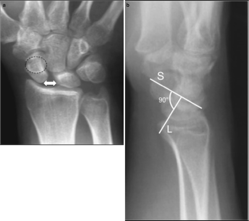
Several parameters exist that suggest scapholunate dissociation on wrist radiography. In our opinion, the most significant are (Fig. 30.1):


Fig. 30.1
X-ray signs of SL dissociation: scaphoid ring, SL distance and SL angle increase. The arrow means an increase of space between the scaphoide and lunate in the scapholunate dissociation. The circle is the scaphoid ring: appears in the presence of scapholunate dissociation, the scaphoid flexes abnormally and the scaphoid tuberosity appears ring-shaped on PA views
Increased SL space (Terry Thomas sign): seen on PA views when the space between the lunate and scaphoid is increased compared to the contralateral wrist. The space may increase on PA views obtained with clenched fist.
Scaphoid ring: in the presence of scapholunate dissociation, the scaphoid flexes abnormally and the scaphoid tuberosity appears ring-shaped on PA views.
Increased scapholunate angle: on the lateral view the angle formed by the lunate and scaphoid axes varies between 30° and 60°. When this exceeds 60°, a scapholunate dissociation should be suspected.
Magnetic Resonance Imaging (MRI)
Contrast-Enhanced Imaging (MRI Arthrography and CT Arthrography)
The intra-articular injection of contrast material considerably increases sensitivity and specificity as a result of the passage of contrast into the lesion itself. To avoid a ‘valve effect’ with failure of the contrast medium to pass through the lesion, the contrast should be injected into both the radiocarpal and midcarpal joints (two-compartment examination).
Diagnostic Wrist Arthroscopy
This is considered the gold standard examination for the diagnosis of intrinsic ligament injuries [1].
In addition to providing direct visualisation of the injury, it allows quantification and classification of the lesion. Geissler et al. [10] proposed an arthroscopic classification of SL ligament tears describing four grades of lesion (Table 30.2):
Grade 1
Partial tear of the SL ligament: the angles between the scaphoid and lunate are normal, the probe cannot penetrate the SL space.
Grade 2
Tear of the SL ligament: the angles between the scaphoid and lunate are modified, the tip of the probe passes into the SL space.
Grade 3
Tear of the SL ligament: there is malalignment between the scaphoid and lunate; the probe passes into and rotates in the SL space.
Grade 4
Massive tear of the SL ligament and malalignment between the scaphoid and lunate: the arthroscope passes through the SL space from the radiocarpal to the midcarpal joint and back.
30.2.4 Treatment Strategy
The surgical approach to SL ligament injuries depends on severity.
Whereas the more severe grades (static irreducible SL dissociation and SLAC lesion) require ‘salvage’ procedures, such as partial wrist arthrodesis or resection of the proximal carpal row, earlier grades (predynamic, dynamic and static reducible SL dissociation) can be treated with procedures aiming at repairing/rebuilding the SL ligament or restoring, as closely as possible, physiological carpal bone mechanics.
In the case of predynamic SL dissociation, up to Geissler’s grade 2/3, the SL can be stabilised by using K wires (pinning) in a closed procedure performed under fluoroscopic control or as an arthroscopy-assisted procedure, with excellent results [11]. In these cases, arthroscopy offers the advantage of allowing removal of scar tissue and fibrosis of the ligament tear in order to enhance vascularisation and healing. The K wires are removed after 8–10 weeks. When the membranous portion of the SL is involved, a debridement with shaver or radiofrequency waves should be performed to remove the ligament flap – a possible cause of painful impingement – and relieve the pain [12].
In dynamic SL dissociation, if the lesion is recent and therefore has good healing capability, direct repair of the ligament using various techniques is indicated [1–8]. It should be noted that injured intrinsic ligaments tend to degenerate very rapidly, and the optimal timing for repair is within 2 weeks, after which the likelihood of recovery decreases dramatically (the repair procedure should not be attempted after 4 weeks). After 2 weeks, capsulo-ligament reconstruction procedures can be carried out that aim either to avoid rotatory subluxation of the scaphoid [13] due to collapse of the secondary stabilisers or to strengthen the dorsal portion of the injured ligament [8]. Another proposed procedure is reconstruction of the dorsal portion of the SL ligament by using grafts fixed with bone-to-bone technique [14].
Wahegaonkar and Mathoulin [15] described an arthroscopic technique combining through suturing the dorsal portion of the scapholunate with capsulodesis with the dorsal interosseous ligament (DIL). The arthroscope is placed in the ulnar midcarpal (UMC) portal, and using a needle introduced through portal 3–4, a PDS 2/0 suture thread is passed through the DIL and lunate remnant of the torn SL and pulled out through the radial midcarpal (RMC) portal; the procedure is repeated for the scaphoid remnant of the SL. The two ends of the suture threads exiting from the RMC portal are tied into a knot and withdrawn within the midcarpal joint; finally, the proximal ends of the suture threads exiting from portal 3–4 are also tied, blocking the SL stumps with the DIL. According to the authors, this treatment should be reserved for cases with moderate diastasis, and the results appear encouraging. Del Pinal [16] proposed a similar technique, which uses a volar portal placed immediately ulnar to the flexor carpi radialis (FRC) tendon, to suture the volar portion of the SL ligament. Although these relatively recent techniques show encouraging preliminary results, they both require validation by studies carried out on larger series and with more adequate follow-up periods.
The techniques proposed for static reducible SL dissociation aim to treat rotatory subluxation of the scaphoid that has become stabilised in an attempt to restore the normal articular relations between the radius and scaphoid, altered by the SL dissociation. The most commonly used is the Brunelli technique [17] which has undergone numerous modifications over the years [8, 18]. In this technique, the scaphoid subluxation is reduced and stabilised in the correct position with the aid of a strip of the FRC tendon passed from palmar to dorsal through a hole in the distal pole of the scaphoid and secured dorsally first onto the scapholunate joint and then onto the radius. In static reducible SL dissociation, surgical arthroscopy has only limited efficacy: one option is to perform an arthroscopically assisted reduction and association of the scaphoid and lunate (RASL), described as an open procedure [19]. The technique involves stabilising the SL with a Herbert screw along the rotation axis with arthroscopic assistance. Arthroscopy offers the advantage of allowing debridement of fibrous tissue and thus greater chances of healing. However, the minimal error in centring the rotation axis or the prolonged permanence of the screw can cause serious damage at the articular level [20].
30.3 Instability of the Distal Radioulnar Joint
30.3.1 Aetiology
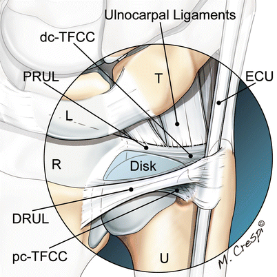
The distal radioulnar joint (DRUJ) is the distal joint of the articular system of the forearm, which allows pronation-supination. It is a joint with limited bony congruency, and it is stabilised by several anatomical structures [21, 22]: among them, the triangular fibrocartilage complex (TFCC) is considered the primary stabiliser. The complex structure of the TFCC comprises two ligaments that serve as true stabilisers for the DRUJ: the palmar radioulnar ligament (PRUL) and the dorsal radioulnar ligament (DRUL) (Fig. 30.2).


