Wrist Arthritis: Arthroscopic Synovectomy, Abrasion Chondroplasty, and Radial Styloidectomy of the Wrist

CHAPTER 29 Wrist Arthritis


Arthroscopic Synovectomy, Abrasion Chondroplasty, and Radial Styloidectomy of the Wrist


The wrist, with its many articular surfaces, is a crucial anatomic link between the hand and forearm. When afflicted by arthritis and conditions limiting the range of motion, the wrist greatly affects the daily lives of patients. The advent of arthroscopy has revolutionized the practice of orthopedic and hand surgeons. Newer techniques and significant application of wrist and small joint arthroscopy can be attributed to many pioneers.1,2


Wrist arthroscopy has provided a tool with which to examine and treat intra-articular abnormalities. Early results for arthroscopic synovectomy of the wrist showed that it reduced pain and swelling and improved joint function.36 The transitory or permanent effects depend mainly on the activities of the patient and underlying cause of arthritis. Abrasion chondroplasty of the wrist has not been described at length, but it is known that “repair cartilage” (i.e., fibrocartilage) replaces articular cartilage, as demonstrated in several canine models.7 Abrasion chondroplasty appears to have a therapeutic role in patients with proximal pole hamate arthrosis or radiocarpal arthrosis. Preliminary results of this procedure have been excellent.810 This chapter surveys the indications and techniques for arthroscopic synovectomy, abrasion chondroplasty, and radial styloidectomy.


ANATOMY AND PHYSIOLOGY


The anatomy of the wrist joint is probably the most complex of all the joints in the body. The wrist is a collection of many bones and joints, which allow the use of our hands in many different ways. The wrist must be extremely mobile to give hands a full range of motion. At the same time, the wrist must provide the strength for gripping heavy objects.


Fifteen bones form connections from the end of the forearm to the hand. The wrist itself contains eight carpal bones. These bones are grouped in two rows across the wrist. Beginning with the thumb side of the wrist, the proximal row of carpal bones is made up of the scaphoid, lunate, and triquetrum. The distal row is made up of the trapezium, trapezoid, capitate, hamate, and pisiform bones. One reason that the wrist is so complicated is because every small carpal bone forms a joint with the bone next to it. The wrist joint is composed of many small joints.


Articular cartilage is the material that covers the ends of the bones of any joint. Articular cartilage can be up to one fourth of an inch thick in the large, weight-bearing joints. It is thinner in joints such as the wrist that do not support much weight. Articular cartilage is white and shiny, and it has a rubbery consistency. It is slippery, which allows the joint surfaces to slide against one another without causing damage.


Articular cartilage absorbs shock and provides an extremely smooth surface to facilitate motion. Articular cartilage exists almost everywhere that two bony surfaces move against one another, or articulate. In the wrist, articular cartilage covers the sides of all the carpals and the ends of the bones that connect from the forearm to the fingers.


Matrix metalloproteinases and proinflammatory cytokines (e.g., interleukin-1) are important mediators of cartilage destruction in patients with primary osteoarthritis. Interleukin-1 increases the synthesis of matrix metalloproteinases and thereby plays an important role in osteoarthritis.


During the initial stages of osteoarthritis, the superficial layers of the articular cartilage fibrillate and crack. As degeneration progresses, deep layers become involved, resulting in erosions that produce bare subchondral bone. Denatured type II collagen is found in abundance in osteoarthritic articular cartilage, with decreased water content and decreased ratio of chondroitin sulfate to keratan sulfate constituents. In chronic injuries of the scapholunate ligament and in scaphoid nonunions, osteoarthritis starts in the radioscaphoid joint and progresses to the capitolunate joint. The radiolunate joint remains unaffected during the early stages.


Rheumatoid arthritis is a progressive inflammatory disease characterized by synovitis and joint destruction. Synovial cell proliferation results in pannus formation and fibrosis, which cause erosion of cartilage and bone. Cytokines, prostanoids, and proteolytic enzymes mediate this process. A cell-mediated immune response to an unidentified antigen appears essential in the pathogenesis of rheumatoid arthritis. Proinflammatory cytokines, such as interleukin-1 and tumor necrosis factor α, and T-cell initiation are the central mediators in rheumatoid arthritis.


In gouty arthritis, allantoin, the enzyme uricase that breaks down uric acid into a more soluble product, is deficient, resulting in tissue deposition of crystalline forms of uric acid. Although hyperuricemia is a risk factor for the development of gout, the exact relationship between hyperuricemia and acute gout is unclear. Acute gouty arthritis can occur in the presence of normal serum uric acid concentrations. Conversely, many patients with hyperuricemia may never develop gouty arthritis.


Secondary osteoarthritis resulting from previous trauma to the carpal bones or ligaments results in abnormal joint reaction forces with each movement of the wrist, causing misdirected forces that lead to some combination of loading forces. This process produces degeneration of the articular cartilage, resulting in radiocarpal arthritis, selective intercarpal arthritis, or pancarpal arthritis, depending on the initial injury and subsequent healing.


Scaphoid fractures can result in osteoarthritis by three mechanisms:


Kienböck disease results in lunatomalacia. The weakened lunate is subjected to a nutcracker effect between the prominent radius and the capitate head, causing progressive collapse. In its final stages, Kienböck disease leads to osteoarthritis in the radiolunate joint.


PATIENT EVALUATION


Diagnostic Imaging


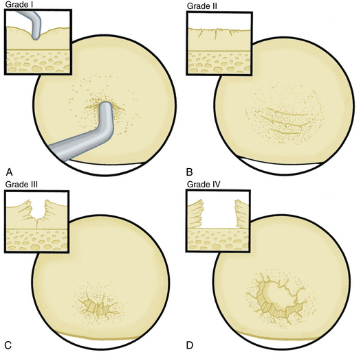
The radiographic evaluation system we use for wrist arthritis is the Outerbridge classification of cartilage defects (Fig. 29-1). We have found that with an appropriate clinical history, dedicated articular cartilage imaging has a sensitivity and specificity reaching the 95% confidence interval. This tool has been confirmed by arthroscopic findings and clinical correlations. However, Haims and colleagues think that magnetic resonance imaging (MRI) of the wrist (41 indirect MR arthrograms and 45 unenhanced [nonarthrographic] MR images) was not adequately sensitive or accurate for diagnosing cartilage defects in the distal radius, scaphoid, lunate, or triquetrum, as demonstrated by correlating MRI with arthroscopic findings.11 In cases of synovitis and ulnar-sided pathology, MRI results are a strong indicator of which areas need to be addressed with the arthroscope. Cartilage defects are often confirmed after diagnostic arthroscopy is completed.


Arthroscopic abrasion arthroplasty, subchondral drilling, and microfracture can be performed for focal chondral defects in patients with moderate degenerative wrist arthritis or when plain radiographs indicate vascular necrosis. MRI is a sensitive method for excluding the diagnosis of avascular necrosis and for evaluating the extent to which fibrocartilaginous repair tissue has formed postoperatively. These methods have been demonstrated for knee pathology, and we have established the same protocols for wrist defects.11.12


Jun 19, 2016 | Posted by in MUSCULOSKELETAL MEDICINE | Comments Off on Wrist Arthritis: Arthroscopic Synovectomy, Abrasion Chondroplasty, and Radial Styloidectomy of the Wrist

Full access? Get Clinical Tree

Get Clinical Tree app for offline access