More recently, hip shape has been defined using a quantitative measure of sphericity, termed the sphericity deviation score. This has been shown to closely correlate with the Stulberg outcome [13]. Quantitative measures of outcome, if reproducible, have particular advantages over qualitative measures in terms of greater efficiency in powering future studies of outcomes.
Radiographic Grade
The grade of disease is an attempt to predict long-term outcome, based on the radiographic appearance of the hip at a particular point in the cycle of disease. The grade of the disease is perhaps where greatest controversy exists. There are classifications that are used to help determine the prognosis: Catterall, Salter-Thomson, and Herring.
The Catterall classification is an assessment of the extent of the radiological involvement of the femoral head on the lateral radiograph [14]. Greater involvement of the head indicates a greater severity of disease, and a worse prognosis. Catterall suggested that involvement of >50 % of the head has poorer outcomes, compared to those with <50 % head involvement. However, the classification may only be applied at the point of maximal collapse, which is usually some months into fragmentation.
The Salter-Thompson classification recognises that approximately 30 % of hips will have a subchondral fracture early in disease [15]. If evident, a subchondral fracture reliably delineates the amount of collapse anticipated, which therefore maps onto Catterall’s descriptors, i.e. a subchondral fracture >50 % indicates a poor prognosis.
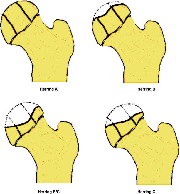
The Herring lateral pillar classifications assesses the integrity of the lateral portion of the epiphysis (Fig. 5.2). Herring et al. broadly described that hips with no lateral column collapse have good outcomes (‘Herring A Hips’), and those with greater than 50 % of lateral column collapse have poor outcomes (Herring ‘C’ Hips). Those with some collapse though less than 50 % (Herring ‘B’ Hips), may be amenable to intervention. However, like the Catterall classification, this may only be applied at the point of maximal collapse, which is some months into fragmentation [16].


Fig. 5.2
Herring classification
Early Predictors of Prognosis
In addition to the radiographic appearances of the hip there are other factors that must be considered when determining the eventual prognosis.
There is general consensus that younger children have better outcomes than older children. An arbitrary cut-off of 6 years old is often used, suggesting that children under 6 years have a good prognosis, and those over 6 years have a poor prognosis. This is clearly an oversimplification with many reports of young children with poor outcomes [19–22].
The sex of the child also appears to have a bearing on the prognosis. Boys generally have better outcomes than girls, which is thought to be a consequence of girls having greater skeletal development at any given chronologic age [14, 17].
The range of motion of the hip is also widely believed to have a bearing on prognosis, though it has seldom been investigated. A recent paper investigating outcomes amongst those under 6-years old, suggested range of motion to be more important than radiographic grade in predicting outcome [19].
Considerations When Forming the Treatment Plan
When considering the treatment of the patient one must therefore consider their age, sex, range of motion and the radiographic stage of disease. The radiographic grade is important; the optimal time to intervene may before significant deformity occurs, i.e. prior to the fragmentation stage because significant deformity has already occurred at this stage. It can therefore be argued that classifying a hip according to Catterall and Herring therefore occurs too late to guide treatment, although the presence of a subchondral fracture described by Salter-Thompson may have some useful bearing on the decision.
Critical Appraisal of the Evidence for the Management of Perthes’ Disease
We searched for studies that have reached level I or II evidence, i.e. prospectively collected cases with pre-defined inclusion and exclusion criteria and predefined outcomes, with or without an intervention.
There is no level I evidence in the management of Perthes’ disease, although there is an ongoing randomised controlled trial in Australia. This study is addressing whether the novel intervention of systematic bisphosphonate administration may prevent femoral head collapse [23].
There are two important level two studies addressing the question “What is the best treatment for Perthes’ disease?”
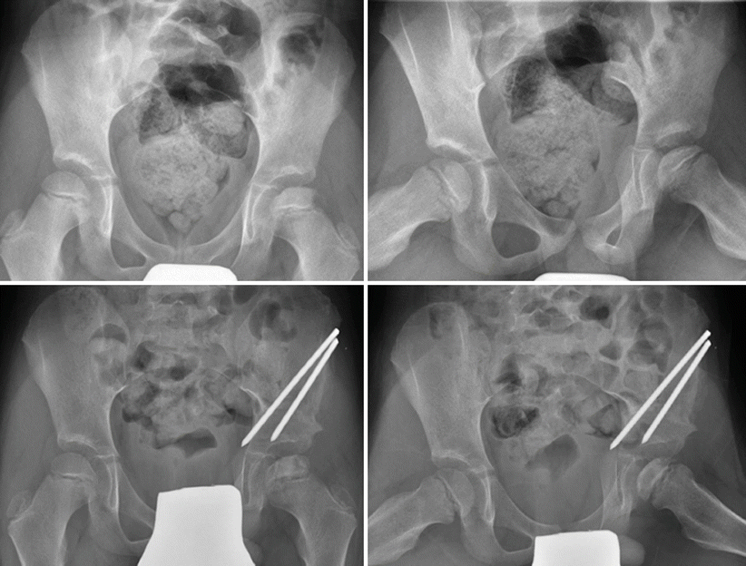
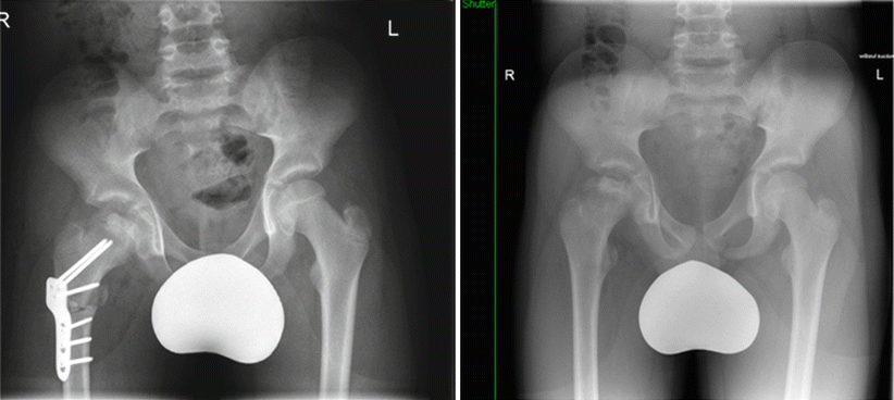
In 2004, Herring et al. reported findings from a prospective, multicentre study in North America and New Zealand. Thirty-nine paediatric surgeons enrolled 438 patients between 6 and 12 years of age across five treatment arms [17]. Hips that had reached the reossification stage at presentation were excluded. Stratification of disease severity was undertaken using the lateral pillar classification, and outcomes were defined according to Stulberg. 345 hips in 337 patients were followed to skeletal maturity. Each surgeon agreed to employ one of five treatment approaches for patients under their care (either no specific treatment, range of motion exercises, brace, Salter pelvic osteotomy or varus femoral osteotomy) (Figs. 5.3 and 5.4).


Fig. 5.3
Containment surgery using Salter’s pelvic osteotomy

Fig. 5.4
Containment surgery using femoral osteotomy
The majority (218, 63 %) of cases were Herring type B and 87 % were younger than eight at onset. The study found no superiority between the three non-surgical treatment options: range of motion exercises, Atlanta braces and no treatment. Further, no radiological difference was seen between femoral osteotomy and innominate osteotomy. Overall, patients receiving surgery had better outcomes than those treated non-operatively (p = 0.02), in particular those patients with Herring B and those older than eight at diagnosis. Herring C patients with severe disease (60, 17 %) generally had poor outcomes regardless of treatment. A new subgroup of the Herring classification was used to describe hips that were exactly 50 % collapse, or with a number of features deemed worse than a typical ‘Herring B’ hip – “the B/C border hip”. A summary of the recommendations from Herring is available in Table 5.1.
Table 5.1
Suggested treatment options dependent on severity of disease and age of onset
Age of onset | Lateral pillar A | Lateral pillar B | Lateral pillar B/C | Lateral pillar C |
|---|---|---|---|---|
Below 8 years | Good outcome | No superiority between treatment options | No superiority between treatment options | Poor outcome independent of treatment option |
Above 8 years | Good outcome | Osteotomy | Osteotomy | Poor outcome independent of treatment option |
This study is useful, however prone to selection bias as no attempt was made to randomise treatments. Although there were assurances that surgeons would employ a single treatment protocol, some patients crossed-over to other treatment regimens demonstrating that surgeons did not employ a single treatment supporting the possibility of selection bias. Eighty-percent of the hips operated on during this study received surgery prior to fragmentation, therefore prior to the maximum extent of lateral pillar collapse becoming apparent. Consequently it is not possible to advocate waiting for the lateral pillar collapse to become apparent based on this study. Perhaps the biggest criticism of this study is that there were no a-priori hypotheses relating to the grade of disease, which is particularly important to those hips that were newly classified to strengthen the statistical conclusions – the ‘B/C border hips’ [24]. The lateral pillar classification was chosen over Catterall and Salter-Thompson because it had a stronger correlation with outcome, and was therefore selected because of its statistical significance. However criticism can be made when using a dataset to derive a classification and then using this classification to make sense of the findings in the same dataset. This is therefore a hypothesis generating approach, rather than a hypothesis testing method. The overarching purpose of the study was to demonstrate the benefit of surgery, yet the results were far from conclusive.
Stay updated, free articles. Join our Telegram channel

Full access? Get Clinical Tree


