Fig. 49.1
A plain radiograph of a patient with multiple bony exostosis
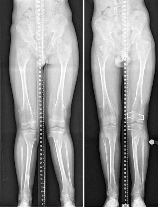
Fig. 49.2
Plain radiograph of a patient with MO and deformity of lower limbs. This patient with multiple bony exostosis developed left tibia knee valgus deformity and leg length discrepancy He was treated with guided growth using 8 plates
A field-change effect could cause both an increase in local osteochondroma formation and growth disturbance. Hence local osteochondroma ‘burden’ might be associated with deformity without invoking ‘cause and effect’. A putative ‘post-axial’ (ulnar and fibular) preference for a ‘field-change’ effect would result both in greater osteochondroma volume and the genetic effect would then result in more exostoses and a shorter ulna. However this theory becomes more difficult to sustain if some patients are observed to have a ‘pre-axial’ preference and others a post-axial one. This is especially so of different preferences were to be observed in opposite limbs of the same patient; in this case a neoplastic pathogenesis for local growth disturbance becomes more likely.
Solitary osteochondromas are not hereditary, but they appear identical to osteochondromas arising in the multiple form. Several studies have suggested that bi-allelic mutations can be found in solitary osteochondromas, however this is not ubiquitous and perhaps other non-EXT pathways are involved in the development of these benign tumours [2]. Nevertheless, their clinical behaviour and pathological features are indistinguishable from those in MO. The clonal nature of neoplasia means that the formation of a solitary osteochondroma will initially involve a genetic change in a single cell of the chondral mesenchyme of the peripheral physis. A field change is unlikely to be responsible as there is no germline defect such as occurs in MO. Hence growth disturbance caused by a solitary exostosis cannot easily invoke a field-change effect to explain its occurrence. Consequently, studies which identify growth disturbance in solitary exostoses can shed important light on the same effect in MO.
When a child with MO presents, the parents wish to know what the future is likely to hold. Without an understanding of pathogenesis there can be no certain advice about preventing the growth disturbance which in some children can be severe. If evidence favours solely a neoplastic pathogenesis then the surgeon will choose to remove exostoses in association with deformity as they develop at key sites (eg distal forearm, distal tibia and fibula). He/she may even decide NOT to remove some exostoses if that decision might result in more balanced growth disturbance affecting paired bones. On the other hand, if evidence favoured a field-change effect, then removal of exostoses could not be justified on grounds of amelioration of the disease process, only in order to reduce pain, improve range of movement or cosmesis. Finally it is possible that there is a mixed aetiology for growth disturbance; in which a field-change effect might cause growth restriction at preferred sites in the absence of osteochondromas yet additionally a local effect of a large osteochondroma could modify that effect. In that situation advice to parents about the result of surgery would be more guarded, and allow for both possibilities. If it is believed that the excision of exostoses might reduce growth-disturbance effects, then the question of timing (early or late) becomes important since early exostoses are confluent with the growth plate, but later ones may have already resulted in significant deformity.
Questions
Faced with a child with an established or developing growth disturbance at one of several sites (distal forearm, proximal femur, around the knee, distal tibia/fibula) and one or more local exostoses which are not of themselves painful or causing significant loss of function, the surgeon has the option to either to remove the local exostosis and observe the effect, or to remain vigilant and undertake deformity-correction surgery at a later date. The purpose of this review is to seek evidence to help the surgeon in this decision-making process.
Key questions to answer therefore are:
- 1.
Are local exostoses associated CAUSALLY with local growth disturbance?
- 2.
Does excision of exostoses result in amelioration of growth disturbance?
- 3.
Is early removal better than awaiting the onset of disturbance prior to excision?
- 4.
Even if excision does ameliorate the pace of deformity, are there balancing risks to consider?
Levels of Evidence
MO is a rare condition. Even the largest tertiary medical centres are unlikely to treat enough patients to allow for inter-subject randomisation. However intra-subject randomisation might be achievable, for example with one limb acting as a control. It is recognised, however, that most if not all studies are likely to provide Level IV evidence only.
Most research into MO is based on an investigation at an anatomical location. Hence we filtered our search to identify publications which had both a diagnosis under the headings below AND an anatomical location as shown in Table 49.1.
Table 49.1
Summary of keywords and search output
Diagnostic keyword | Anatomical keyword | Total number of abstracts | |
|---|---|---|---|
Cartilaginous exostoses | Forearm | 74 | 32 |
Diaphyseal aclasia | Ulna | 94 | 9 |
Diaphyseal aclasis | Radius | 32 | 1 |
Exostoses | Elbow | 101 | 6 |
Hereditary multiple exostoses | Wrist | 37 | 1 |
Hereditary multiple exostosis | Lower limb | 21 | 0 |
Multiple cartilaginous exostoses | Hip | 269 | 18 |
Multiple hereditary exostoses | Ankle | 211 | 18 |
Multiple osteochondroma | Knee | 233 | 6 |
Multiple osteochondromas | Tibia | 109 | 5 |
Multiple osteochondromatosis | Fibula | 82 | 15 |
Femur | 68 | 7 | |
Femoral | 94 | 5 | |
Malleolar | 8 | 3 | |
General | 11 |
This review has used Endnote® software (Thomson Scientific Inc) to identify publications found in Medline, PUBMED or EMBase at all time periods. Following deletion of duplications in each set and cross-set, this search generated searchable references and abstracts (Table 49.1). Each abstract was read to identify whether a growth-disturbance effect of MO might be identified within the publication. Single case-reports were included since it was anticipated that the quality of evidence would not exceed level IV. Full texts were sought and read (Table 49.1) in order to populate the data fields in Table 50.3, on which evidence the discussion section is written. In reading these full-texts, where reference was made to a publication of importance not already identified in the search, these were also sought and read and added to the database under the heading ‘General’ (Table 49.1).
There are no level I, II or III studies which address the question of growth disturbance in MO. Almost all evidence is found in case reports and case series. There are a very few retrospectively acquired longitudinal studies and two retrospective case-control study which do not reach the quality standard for classification at level III (Table 49.2).
Table 49.2
Study details with regard to questions on effect of exostoses and excision of exostoses on growth disturbance
Reference | LoE | Location | N | Exostosis associated with deformity | Exostosis and deformity linked by causality | Excision exostosis stopped deformity? | Timing of surgery and deformity? | Complications | Summary |
|---|---|---|---|---|---|---|---|---|---|
[3] | IV case series | Proximal fibula | 46 | n/a | n/a | n/a | n/a | n/a | No mention deformity |
[4] | IV case report | Distal femur | 1 | (Y) | (Y) | n/a | n/a | n/a | Solitary, Figs. 1 and 2 look like ante-curvatum but not mentioned in report |
[5] | V case report | Ulna | 1 | (Y) | n/a | n/a | n/a | n/a | Solitary exostosis proximal ulna. Dislocation by physical pressure |
[6] | V case report | Proximal femur | 1 | n/a | n/a | n/a | n/a | n/a | No deformity noted |
[7] | IV case series | Distal radius & ulna | 21(33), but only 10(11) had excision surgery | Y | n/a | N | Age 5–16 | None | 11 forearms in 10 patients had exostosis excision only at mean age 11. No description of extent of surgery. Average carpal slip, radial articular angle and ulnar variance all deteriorated at final follow-up, but no significant difference (in all groups mean 13 years FU). Unclear when was final follow-up. No control group |
[8] | IV case series | Radius and ulna | 36 had excisions | Y | n/a | n/a | n/a | One unhappy with scars | No procedure done ‘to prevent abnormal growth patterns of the involved bones’. Telephone questionnaire for function only |
[9] | IV case series | Distal tibia & fibula | 4(6) | n/a | n/a | n/a | n/a | n/a | Use of a screw for hemiepihyseodesis in ankle valgus 4 MO patients. No comment on exostosis site |
[10] | IV case series | Distal tib & fib | 6 | Y | n/a | n/a | n/a | n/a | Permanent epiphysiodesis only |
[11] | IV case series | Radius & ulna | 7(8) | Y | n/a | n/a | n/a | n/a | 8 forearms with ulnar lengthening & excision exostoses without separated procedures in Masada I & IIb(short ulna, ulnar exostoses) |
[12] | IV case series | Radius & ulna | 16(20) | Y | (Y) | n/a | n/a | n/a | Authors argue degree of forearm deformity associated with degree of metacarpal shortening. Only 1 Taniguchi type IIIA (radius short. This showed only a radial exostosis) |
[13] | IV case control | Radius & ulna | 35(65) | Y | N | n/a | n/a | n/a | X-sectional study. Relative forearm length compared with controls. Most forearms had both short radius and ulna cf controls. Exostosis presence evaluated (no tabulation) and found not to associate with relative length of the ulna or radius. Only 3 forearms had isolated radial exostosis. These had normal lengths compared with controls |
[14] | V case report | Whole body | 1 | n/a | n/a | n/a | n/a | n/a | Descriptive of multiple sites in one patient |
[15] | IV case series | Distal tibia/fibula | 23(19 solitary) | (Y) | (Y) | (Y) | Age 2–48 | n/a | Retrospective study. Age 8–48, Follow-up over 2.5–19 years. 19 had surgery. State that ‘deformity’ improved after excision, but no documentation of type of deformity. Only ‘pronation’ mentioned and fibular or tibial indentation or bony shortening. Tables do not quantify deformity, or correction, or association with exostosis position |
[16] | IV case Series | Radius & ulna | 4 | Y | n/a | n/a | n/a | n/a | 4 lengthenings with excision exostoses. No separated procedures |
[17] | V case report | Distal tibia | 1 | Y | n/a | n/a | n/a | n/a | Fibular indentation |
[18] | IV – case series | Shoulder | 145 | n/a | n/a | n/a | n/a | n/a | N = 172, clinical examination only. No correlation of exostosis number or location with deformity |
[19] | IV case series | Stature | 172 | n/a | n/a | n/a | n/a | n/a | N = 172 all ages with MO. Height is normal until the age of 10 when there is a diminution. Cross-sectional study is main weakness |
[20] | IV – case series | Whole body | 143 | n/a | n/a | n/a | n/a | n/a | 143 patients all ages, Genotype-phenotype with number of exostoses at different sites as main disease parameter.. No assessment of deformity. Clinical examination only |
[21] | IV case series | Radius & ulna | 106 | n/a | n/a | n/a | n/a | n/a | Adults. Clinical examination only. Exostosis number linked to range of movement and to presence of radial head dislocation |
[22] | IV case series | Radius & ulna | 168 | Y | (Y) | n/a | n/a | n/a | N = 172, clinical examination only. Number of distal femoral exostoses are independent factors for deformity (degree of valgus) on multivariate analysis |
[23] | IV case series | Knee | 3 | (Y) | (Y) | (Y) | N | N | Fibular bowing remodelled |
[24] | IV case series | Radius & ulna | 6 | Y | n/a | n/a | n/a | n/a | Study of ex fix in 6 children age 9–14. forearms all had ulnar exostoses. All had negative ulna variances of at least 10 mm. Only one had a distal radial exostosis |
[25] | V case report | Distal femur | 1 | Y | Y | n/a | n/a | n/a | 9/12 girl, genu valgum osteochondroma medial femoral condyle. Deformity only here (genu valgum) |
[26] | IV case series | Distal tib & fib | 19 | Y | n/a | n/a | n/a | n/a | Temporary hemiepiphyseodesis only. Never removed exostoses |
[27] | IV case series | Hip | 18(36) | Y | n/a | n/a | n/a | n/a | Observational study of deformity. No correlation of exostosis site/size with deformity |
[28] | V case report | Hip | 1 | n/a | n/a | n/a | n/a | n/a | Case report of CDH and MO |
[29] | IV case series | Distal tibia | 5 (3 solitary, 2 HME) | Y | n/a | n/a | n/a | n/a | Tibial exostoses. All indent fibular. Pre-op ‘ankle deformity’ and post-op ‘correction of angular deformity of fibula’ not quantified |
[30] | V case report | Hip | 1 | N | n/a | n/a | n/a | n/a | Case report of hip joint exostosis |
[31] | IV case series | Radius & ulna | 48 (76) | Y | Y | n/a | n/a | n/a | Retrospective x-ray review. Unselected so selection bias (e.g. age) possible. 102 MO patients, but only 48 (76 forearms) available for x-ray analysis. Forearm x-rays grouped into 5 groups BASED ON LOCATION. Group 1 (distal ulna only N = 33), Group 3 (distal radius only N = 9); these had different deformity-associated characteristics (ulnar variance, radial articular angle, radial bow all worse in ulna-only group, but not statistically so. Authors state they believe they behave differently. 13/14 radial head dislocations in ulna only group. Group 4 (diaphyseal exostoses) had least ulnar shortening but these were younger (median age 7 vs 13 for distal ulna alone), so may be due to age-related effects. Unclear what surgery done during period of radial head evaluation. FU 7 years, and based on this a qualitative appraisal of deformity in ulna-only group is described due to ulnar tethering of radius. Radial and ulnar shortening is measured and the DIFFERENCE between the two is least in radial exostoses (2 mm) cf 7 mm for both bones and 14 mm for ulna only, but no statistical analysis done |
[32] | V case report | Distal tibia | 1 | (Y) | n/a | n/a | n/a | n/a | Excision osteochondroma. Deformity is thin fibula only |
[33] | IV case series | Radius & ulna | 6 | Y | Y | n/a | n/a | n/a | Forearm lengthening in 4 MO and 2 distal unlar osteochondroma (implication is they are SOLITARY). Indication for surgery is radio-ulnar length discrepancy of > 2cm |
[34] | V case report | Prox femur | 1 | N | n/a | n/a | n/a | n/a | Age 24. Large femoral neck exostosis – solitary |
[35] | IV case series | Radius & ulna | 6 | Y | n/a | n/a | n/a | n/a | 6 children ulnar lengthening in Masada I (short ulna, ulnar exostoses, radial head located) |
[36] | IV case series | Radius and ulna | 13(14) | Y | Y | Y | Effective ages 4–12 (mean age 7) | Tumour recurrence in 8/14. No others volunteered | Natural history of MO following excision exostoses at distal ulna. Group 1 only distal ulna exostosis (6 forearms in 6 children, Mean age 7, FU 4 years). Group 2 touching distal ulna and distal radius exostoses (8 forearms in 7 children, mean age 8 years, FU 5 years). After surgery, group 1 showed significant improvement in % ulnar shortening (8.3–6.5 %) and % radial bow (10.2–7.3 %), but not radial articular angle or degree of carpal slip. Group 2 showed no significant improvement in any parameter and a deterioration in radial bow and radial articular angle. Tumour recurrence in 2/6 in group 1 and 6/8 in group 2, but recurrence not related to likelihood of continuing deformity. Possible that surgical adequacy had some effect |
[37] | IV case series | Hip | 2 | (Y) | n/a | n/a | n/a | n/a | 2 cases MO & hip subluxation. Surgical result not assessed for deformity improvement other than hip joint reduction |
[38] | IV case series | Distal radius & ulna | 10(12) | Y | n/a | n/a | n/a | n/a | Children with MO. Longitudinal radiographic study Combination of procedures including 4 excisions alone. Masada classification recorded, but small numbers and incomplete follow-up prevent analysis of long-term consequences |
[39] | V case report | Distal tibia | 1 | Y | n/a | n/a | n/a | n/a | Short fibula but varus ankle |
[40] | V case report | Distal humerus | 1 | N | n/a | n/a | n/a | n/a | Large exostosis, no deformity |
[41] | V case report | Distal tibia | 1 | N | n/a | n/a | n/a | n/a | Histological osteochondroma after raising a flap. No deformity |
[42] | V case report | Phalanx | 1 | Y | Y | n/a | n/a | n/a | 4th toe Proximal phalanx deformity in a SOLITARY exostosis |
[43] | IV case series | Distal tib/fib | 12 | Y | ? (see comment) | n/a | n/a | n/a | Lengthening fibular for valgus and talar shift. Table describes site of exostoses. 4 Fibula alone, 6 Tibia and Fibula. 2 Tibia alone. Authors state uncertain why an enormous tibial exostosis alone may cause fibular undergrowth |
[44] | V case report | Prox femur | 1 | Uncertain | Uncertain
Related posts: Evidence-Based Treatment of Flexible Flat Foot in Children
Evidence-Based Treatment for Congenital Dislocation of the Knee
Evidence-Based Treatment for Congenital Femoral Deficiency
Evidence-Based Treatment for Slipped Upper Femoral Epiphysis
Physeal Injury, Epiphysiodesis and Guided Growth
Evidence-Based Treatment for Feet Deformities in Children with Neuromuscular Conditions Evidence-Based Treatment of Flexible Flat Foot in Children
Evidence-Based Treatment for Congenital Dislocation of the Knee
Evidence-Based Treatment for Congenital Femoral Deficiency
Evidence-Based Treatment for Slipped Upper Femoral Epiphysis
Physeal Injury, Epiphysiodesis and Guided Growth
Evidence-Based Treatment for Feet Deformities in Children with Neuromuscular Conditions
Stay updated, free articles. Join our Telegram channel
Full access? Get Clinical Tree
 Get Clinical Tree app for offline access

|