Upper and Lower Extremity Prosthetics


Upper and Lower Extremity Prosthetics


Nicolas E. Walsh

Gordon Bosker

Daniel Santa Maria


Major limb amputation impacts multiple aspects of an individual’s life: body image, self-care activities, mobility, psychosocial health, vocational, and avocational opportunities. Successful rehabilitation allows the individual with an amputation to return to their highest level of activity and function. Tempering expectations with reality, balancing the use of prosthetic technology with the individual’s perceived needs, physical and cognitive capabilities, social support network, and financial resources are essential features of rehabilitative care.

Advances in the care and prosthetic restoration of the individual with an amputation have always come from multiple arenas: new surgical techniques, improvements in preoperative and postoperative management, advances in prosthetic technology, and better understanding of the psychosocial implications of limb loss. In the past decade, the greatest advances have taken place in the areas of prosthetic technologies, fabrication of prosthetic sockets, and improved components that more effectively replace the lost function of the extremity. Teams of health care providers in regional centers, who treat a large number of individuals with an amputation, are able to provide optimal prosthetic rehabilitation because of their combined experience (1). While such integrated teams are ideal, a less formal coordination of efforts between surgeons, physiatrists, prosthetists, and therapists in the community can provide effective care to most individuals with an amputation who lack access to specialized centers.

In this chapter, the causes of amputation, basic surgical issues, and the overall approach to the medical and physiatric care of the individual with an amputation are reviewed. A discussion on the prosthetic management of amputation at different levels in both the lower and upper limbs follows to aid the practitioner in organizing the myriad options available for restoring the function of the lost limb. Finally, common problems, medical complications, and special issues in pediatric amputation are discussed.


INCIDENCE AND ETIOLOGY


Acquired Amputation

The etiology of limb loss influences the clinical treatment, management, and functional expectations of the individual with an amputation. Data from the Agency for Healthcare Research and Quality (AHRQ) and the Veterans Health Administration (VHA) from the late 1980s to the late 1990s estimates that a total of 140,000 amputations are performed yearly in the United States (2,3). Acquired amputation accounts for 96% to 99% of all limb losses, with the remaining 1% to 4% being related to congenital causes.

The distributions of upper and lower extremity (LE) amputation by level are shown in Figure 74-1. In the LE, 75% to 93% of acquired amputations are the result of vascular disease (diabetic vascular disease, atherosclerosis, immunologic, and idiopathic). Diabetes alone contributes to two thirds of all LE amputations (2,3). Approximately 6% to 10% of acquired LE amputations result from traumatic injuries with the remainder due to benign or malignant tumors. While accounting for a smaller overall percentage of LE amputation, trauma is the most common cause for LE amputation in the second and third decades of life. Among those between the ages of 10 and 20 years, tumor is the most frequent cause of all amputations (4, 5, 6).

In the upper extremity (UE), trauma is the leading cause of limb loss, accounting for 80% of amputations with the vast majority of injuries limited to digital amputations. There are an estimated 10,000 to 15,000 upper limb amputations at the transradial level and above yearly in the United States (Table 74-1) (7). Most of these occur in individuals between the ages of 20 and 40 years. As in the LE, tumor is the most common cause of UE amputation in children.

From 1988 to 1996, the rates of trauma- and cancerassociated amputations declined by more than 40% (2,8). This decline likely reflects improved surgical reconstruction techniques for trauma, limb-sparing management of musculoskeletal tumors, and greater prevention through improved safety awareness. In contrast to the reduction in tumor- and trauma-related limb loss, dysvascular amputation rates have increased 10% to 19% over the past several decades. The increase in dysvascular-related amputation has occurred despite considerable evidence that comprehensive management of the diabetic foot at risk can substantially reduce or delay amputation. Factors that account for this adverse trend include the difficulty of implementing systematic comprehensive management strategies for the diabetic foot coupled with an increasing prevalence of diabetes, hypertension, and hypercholesterolemia placing more individuals at risk (2,9,10).


Congenital Amputation

The absence of part or all of an extremity at birth is more appropriately referred to as a congenital skeletal deficiency rather than a congenital amputation. The Birth Defects
Monitoring Program, a national program monitoring congenital malformations in the United States, reports the incidence of congenital upper and lower limb skeletal deficiency as 2.41/10,000 births (upper limb, 1.58/10,000 births; lower limb, 0.83/10,000 births) (11). Although a few genetically determined syndromes such as heart-hand malformation (Holt-Oram), renal dysfunction (Fanconi), thrombocytopenia (multiple named syndromes), absent radius and drugs (e.g., thalidomide) have been associated with skeletal deficiencies (12, 13, 14), no etiology can be determined for most congenital limb loss.


FIGURE 74-1. Percentage and level of amputations. (Data taken from the AHRQ and the VHA.)


TABLE 74.1 Numbers and Adjusted Rates of Limb Loss by Etiology and Level (7)


Level


Dysvascular No. (%) 1988-1996


Trauma-Related No. (%) 1988-1996


Cancer-Related No. (%) 1988-1996


Lower limb (total)


953,367 (97)


61,605 (31)


8,351 (76.1)


Toe


309,589 (31.5)


27,233 (13.9)


1,466 (13.4)


Foot


102,872 (10.5)


4,483 (2.3)


482 (4.4)


Ankle


7,478 (0.8)


823 (0.4)


164 (1.5)


Transtibial


271,550 (27.6)


14,244 (7.3)


1,501 (13.7)


Through-knee


4,237 (0.4)


921 (0.5)


133 (1.2)


Transfemoral


253,145 (25.8)


10,821 (5.5)


2,499 (22.8)


Hip disarticulation


3,554 (0.4)


418 (0.2)


726 (6.6)


Pelvic


469 (0.1)


52 (0.03)


1,369 (12.5)


Bilateral


0 (0)


1,504 (0.8)


0 (0)


Upper limb (total)


29,426 (3)


134,421 (68.6)


2,617 (23.9)


Thumb


2,344 (0.2)


24,325 (12.4)


352 (3.2)


Finger(s)


21,427 (2.2)


100,316 (51.2)


529 (4.8)


Hand


1,255 (0.1)


983 (0.5)


92 (0.8)


Wrist


514 (0.1)


415 (0.2)


21 (0.2)


Transradial


1,626 (0.2)


4,001 (2.0)


212 (1.9)


Through-elbow


385 (0.04)


346 (0.2)


123 (1.1)


Transhumeral


1,511 (0.2)


3,008 (1.5)


488 (4.4)


Shoulder


236 (0.02)


154 (0.1)


365 (3.3)


Bilateral


0 (0)


462 (0.2)


0 (0)


Forequarter


132 (0.01)


15 (0.01)


439 (4)


Multiple systems for classifying congenital limb deficiencies have been proposed but none has been universally accepted. A commonly used and preferred system is based on the International Society for Prosthetics and Orthotics recommendations that classify congenital limb loss as either a transverse or longitudinal skeletal deficiency. Transverse limb deficiency, also referred to as terminal deficiency, is defined as the loss of all skeletal components distal to a particular transverse axis (e.g., transverse forearm or transverse radial limb deficiency). Longitudinal limb deficiency, also referred to as intercalary limb loss, is defined as the loss (complete or partial) of one or more skeletal elements within the longitudinal axis of the limb, with preservation of some or all of the distal skeletal elements. Classification of congenital limb deficiencies is often confused further when congenital deficiencies are described in the same terms used for acquired amputations, such as “transradial” or “transtibial.” Similar terminologies are used either because the congenital deficiency appears similar to an acquired amputation or because a congenital skeletal deficiency has undergone a surgical conversion to accommodate appropriate prosthetic restoration. Surgical conversion has been estimated to be necessary in the management of 50% of LE congenital deficiencies and 8% of UE deficiencies (15).


Amputation Surgery

The underlying principle in choosing the amputation level is to preserve as much limb length as possible that will be consistent
with wound healing, an acceptable soft-tissue envelope, and functional prosthetic fitting. While in principle, decision making is straightforward, in practice many issues must be weighed including the severity of the underlying disease process, tissue viability, overall medical condition of the patient, the morbidity associated with limb salvage, expected functional level after amputation, and ultimately patient-physician preference. In patients with vascular disease, noninvasive vascular studies can assist in predicting wound healing; an absolute ankle Doppler blood pressure of 70 mm Hg or greater, an ankle-brachial index of 0.5 or greater, and transcutaneous oxygen pressure of greater than 20 to 30 mm Hg are all suggestive of a greater likelihood of healing at the transtibial level. Other nonvascular factors associated with compromised wound healing include poor nutritional status (albumin below 3.5 g/dL) or immunocompromised status (total lymphocyte count < 1,500). Despite these identified predictive factors, the final choice of amputation level in the vascular patient often cannot be made until the time of surgery, when the amount of blood flow in the relevant tissues can be observed (8,16, 17, 18). In most medical centers, the physiatrist plays a limited role in presurgical planning. In centers where multidisciplinary teams are involved, the physiatrist can offer useful insights into the likelihood of achieving various functional goals at differing levels of amputation. This information allows the health care team and the patient to weigh surgical options when uncertainty exists.

The decision between amputation and limb salvage following trauma is a complex one. Early or immediate amputation may be required in life-threatening multiple trauma situations when the physiologic demands associated with repeated surgery to salvage a limb would not be tolerated. The presence of preexisting medical conditions such as peripheral vascular disease (PVD) or neurologic injury that adversely affect function of the residual limb may also favor immediate amputation. However, when the injuries are not immediately life-threatening, the decision to amputate versus attempting limb salvage must be based on an assessment of the approach that will most effectively restore function and return the individual to his or her preinjury activities. Extensive soft-tissue loss, proximal arterial injuries, multiple arterial injuries, and sciatic or tibial nerve damage are poor prognostic indicators for successful limb salvage (17,19). In general, limb salvage of the LE requires more surgeries, involves longer hospital stays, delays weight bearing, and slows the return to preinjury activities compared to amputation (19). Because the functional demands of the UE are different from the LE, a bias towards limb salvage has developed in UE trauma surgery. The lack of weight-bearing forces, the ability to function with partial sensation, and the limited functionality of UE prostheses are reasons cited for greater effort directed toward limb salvage and reimplantation (20,21).

Amputation surgery must be approached as a reconstructive procedure. Bones are beveled to minimize the sharp edges that can cause tissue trauma and pain with weight bearing. The nerves are sharply transected and allowed to retract into proximal soft tissues so that they do not become adherent in scar or remain in a location subjected to high loading forces from a prosthesis. Appropriate myofascial closure of the muscle or myodesis provides for good control of the remaining bone in the residual limb, and appropriate placement of the skin incision line avoids bony prominences and adherence to underlying bone. Such attention to detail will result in a wellshaped residual limb that can be effectively fitted with a prosthesis. Ilizarov techniques or free fibular grafts (22, 23, 24) have been used to lengthen short residual limbs, though the indications for their use and their success are highly individualized. Skin grafts and myocutaneous free flaps (25,26) have been used successfully to preserve length in nonvascular individuals with an amputation, but in general the presence of skin grafts or insensate skin in the residual limb often results in recurring skin breakdown. When recurring skin breakdown occurs, stump revision, use of tissue expanders, and creative approaches to prosthetic design may be needed. The preferred levels of amputation are as follows.


Lower Extremity


  • Toe amputations


  • Ray resections


  • Transmetatarsal amputations


  • Syme amputation (i.e., ankle disarticulation)


  • Transtibial amputation (between the junction of the middle and distal thirds of the leg)


  • Knee disarticulation (KD)


  • Transfemoral amputation (8 cm or more proximal to the level of the knee joint)


  • Hip disarticulation (short transfemoral amputation at/ or proximal to the greater trochanter is functionally a hip disarticulation)


  • Hemipelvectomy


Upper Extremity


  • Digital amputation


  • Ray resection


  • Transmetacarpal resection


  • Wrist disarticulation (WD)


  • Transradial amputation


  • Elbow disarticulation


  • Transhumeral amputation (THA) (i.e., 6.5 cm or more proximal to the elbow joint)


  • Shoulder disarticulation


  • Forequarter amputation (interscapulothoracic disarticulation)


LE AMPUTATION


General Principles of LE Amputation Management

The interaction between the health care team and the patient to achieve the goal of prosthetic restoration and rehabilitation is referred to as prosthetic management (27). The process of prosthetic rehabilitation can be organized into a four-phase
process: preprosthetic management, postoperative care, prosthetic fitting and training, and long-term follow-up care. This staging permits the rehabilitation physician to assess the individual with an amputation and organize the rehabilitation program.


Preprosthetic Patient Evaluation and Management

Preprosthetic management begins when the decision to perform an amputation is made, when a patient is initially evaluated after a traumatic amputation, or when a child is born with a congenital skeletal deficiency. It ends with the fitting of a prosthesis. Optimal care is ensured when members of the prosthetic team can evaluate the patient before amputation, but often the events surrounding an amputation delay the rehabilitation assessment until the postoperative period.

The preprosthetic evaluation, whether performed preoperatively or postoperatively, should focus on identifying factors that will affect the ultimate functional status of the patient and optimize prosthetic fitting. Issues that need evaluation include assessing the premorbid functional status, identifying coexisting musculoskeletal, neurologic, and cardiopulmonary disease that will influence the rehabilitation potential, determining the available social support network, and understanding the patient’s goals and expectations. Education of the patient and family about the functional consequences of amputation and the steps involved in prosthetic rehabilitation will help allay some of the fears the patient may have about his or her future. Therapy programs for range of motion, conditioning exercises, correct positioning of the residual limb, ambulation with gait aids, relaxation techniques, and activities of daily living (ADLs) should be started as soon as medically appropriate. The patient is often better able to absorb and comply with a therapy program during the preoperative period than during the early postoperative period, when incisional pain, medication, or apprehension may interfere with the ability to participate.


Postoperative Care

The goals that direct the postoperative, preprosthetic management of the individual with an amputation are outlined in Table 74-2. During the immediate postoperative period, general medical care focuses on optimizing control of underlying disorders that can interfere with rehabilitation: diabetes, coronary artery disease, congestive heart failure, renal disease. Maintaining nutritional status is frequently neglected, nevertheless it plays a critical role in ensuring wound healing (8) and in facilitating the muscular strength adaptations needed for prosthetic mobility. The principles guiding residual limb care are based on ensuring primary wound healing, controlling pain, minimizing edema, and preventing contractures.


TABLE 74.2 Goals of Postoperative Management of the Individual with an Amputation


  • Successful healing of the amputation


  • Pain control


  • Maintaining range of motion in the remaining proximal joints of the amputated extremity


  • Strengthening of residual muscle groups needed for biomechanical compensation


  • Preparation of the residual limb for prosthetic fitting


  • Achieving independence in ADLs and mobility without a prosthetic limb


  • Education about the process of prosthetic limb fitting and expected functional outcome


  • Psychosocial support for the adaptations resulting from the amputation


Options for wound management include soft dressings, semi-rigid dressings (Unna casts), rigid dressings (plaster or fiberglass casts), and air splints. Each option has advantages and disadvantages. Soft dressings are typically used with an elastic bandage wrap (i.e., Ace bandage) or a compressive stockinette. Soft dressings have the advantage of being readily available, quickly applied, and allowing frequent wound inspection. However, they do not provide protection from external trauma and only have a limited ability to control edema. If poorly applied, elastic wraps can lead to tourniquet effect. Elastic bandages require considerable cooperation, skill, and attention on the part of the patient, family, and medical staff because the wraps need to be reapplied frequently and carefully to be successful. In practice their use is problematic enough and alternatives such as compressive stockinettes, elastic stump shrinkers, or roll-on gel liners are often a better choice of edema management. Despite a number of limitations, soft dressings remain the most commonly used wound care approach following amputation (28).

Rigid dressings have been reported to reduce wound-healing time and lead to more rapid and improved rehabilitation (29,
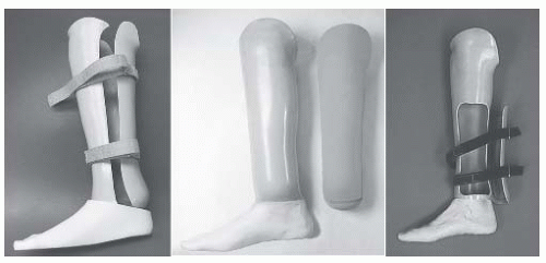
30). The primary concerns surrounding rigid dressings are the inability to inspect the wound and the potential increase in wound breakdown from incorrect application or early weight bearing in dysvascular individuals with an amputation. In spite of these limitations, postoperative rigid dressings may be the preferred method of wound care, especially for the transtibial individual with an amputation, but their clinical use and acceptance has been limited by the lack of expertise in their application. Rigid dressings can be fabricated in a removable form that resembles a transtibial prosthetic socket or as a nonremovable cast that extends to the mid-thigh level. The rigid dressing is most commonly made using standard orthopedic cast materials but commercially available prefabricated devices are also available. A nonremovable rigid dressing is typically applied during or shortly after surgery and replaced every 7 to 14 days. The mid-thigh length of the dressing prevents knee flexion contractures and is continued until adequate wound healing has occurred so that concerns over contracture development are minimized. Subsequent dressings are fabricated as removable rigid dressings (RRDs) that can be taken off whenever the wound needs to be inspected. Rigid dressings have been predominantly used in the individuals with a traumatic amputation because of lessened concern over wound healing or limb injury from the dressing. While rigid dressings can be used simply as a wound care strategy, they can also be used with a pylon attachment to which components
can be attached, creating a preparatory prosthesis that enables immediate or early weight bearing.

Because little objective data exist that clearly identify a superior wound dressing strategy, the choice of wound management appears to be driven largely by practice conventions, availability of skilled staff, and the personal experience of the surgeon. Greater attention is needed to facilitate rapid wound healing, especially in the individual with a dysvascular amputation in whom the effects of prolonged immobilization may substantively complicate rehabilitation effects. The preprosthetic phase of management, before preparatory prosthetic fitting, can typically last 6 to 10 weeks for the individual with a dysvascular LE amputation, a considerably shorter period of time for the individual with a traumatic LE amputation, and 3 to 6 weeks for the individual with a UE amputation (12).

Muscle imbalance and postoperative positioning to facilitate comfort leads to the development of knee flexion contracture in the transtibial residual limb and to hip flexion and abduction contractures in the individual with a transfemoral amputation. Contractures are preventable through a postoperative therapy program that emphasizes range of motion exercises and early mobilization. Strengthening of muscle groups that biomechanically substitute for the lost function of the limb is needed. Exercise programs are required to accomplish this task. In the individual with a LE amputation, the hip extensors (gluteus maximus and hamstrings), gluteus medius, hip flexors, and the contralateral ankle plantar flexors all contribute to restoring ambulation ability (31,32). In the individual with a UE amputation, proximal shoulder girdle muscle strengthening should be taught, emphasizing the trapezius, serratus anterior, pectoralis major, as well as any residual deltoid and biceps functions.

An individual’s psychological response to amputation may be compared to the grieving process that variably includes identifiable stages of denial, anger, depression, coping, and acceptance. Not every person ultimately adapts to limb loss. The individual’s ultimate response to the psychosocial impact of limb loss is determined by many factors, including the cause of amputation, personal life experience and inner strengths, the available social support system, the care provided by the prosthetic team, and the functional outcome that is achieved through rehabilitation.


Prosthetic Fitting and Training

An understanding of the functional needs of the individual with an amputation, his/her interest and motivation in pursuing prosthetic fitting, and an assessment of his/her ambulatory potential are required to set realistic goals for prosthetic fitting and training. Not all the individuals with an amputation are candidates for prostheses. Although the factors that predict success in prosthetic use are partially understood, a number of factors have been associated with a poor outcome in returning the individual with an amputation to functional ambulation at household or community levels. Negative prognostic factors include a delay in wound healing, the presence of joint contractures, dementia or cognitive disorders, medical comorbidities, and higher levels of limb amputation (transfemoral) (33, 34, 35). Age has inconsistently been identified as a predictor of prosthetic success, implying that except in advanced age (>80 to 85 years) other factors play a more important role in determining the rehabilitation potential of the individual with an amputation.

As a result of the uncertainty in identifying prosthetic candidates, considerable clinical judgment is required. Some general guidelines can be followed. An individual with an amputation should have reasonable cardiovascular reserve, adequate wound healing, and good soft-tissue coverage, range of motion, muscle strength, motor control, and learning ability to achieve useful prosthetic function. Individuals with an LE amputation who can walk with a walker or crutches without a prosthesis usually possess the necessary balance, strength, and cardiovascular reserve to walk with a prosthesis. Examples of poor candidates for functional prosthetic fitting would be an individual with a dysvascular LE amputation with an open or poorly healed incision, an individual with a transfemoral amputation with a 30-degree flexion contracture at the hip, or an individual with a transradial amputation with a flail elbow and shoulder. Generally, individuals with a bilateral, short, transfemoral amputation over the age of 45 years are considered unlikely candidates for full-length prosthetic fitting. Additional medical problems such as severe coronary artery disease, pulmonary disease, severe polyneuropathy, or multiple-joint arthritis may result in an individual with an amputation who could be fitted with a prosthesis but who may not be a functional prosthetic user. Patients in whom prognosis is poor, life expectancy is short, or with a disease that results in significant fluctuations in body weight are not good candidates. In borderline cases, it may be necessary to proceed with actual prosthetic fitting to determine the eventual prosthetic function. The use of a less costly, RRD with pylon and foot or a preparatory prosthesis is appropriate before a decision is made about fitting such a person with a more costly definitive prosthesis. The overall success rate in restoring functional ambulation in the individual with a lower limb amputation varies approximately from 36% to 70%. Amputation resulting from vascular disease is a manifestation of a severe systemic vasculopathy. The early mortality following major LE amputation is 15% to 20%, largely related to myocardial infarction. Overall, the individuals with dysvascular amputation have a 3- to 5-year 50% mortality, which underlies the importance of successful early rehabilitation to allow for an improved quality of life in their remaining years.

The timing of prosthetic fitting for the individual with an LE amputation remains controversial, reflecting the clinical uncertainty over early versus delayed weight bearing. Because the majority of LE amputations occur as a result of PVD, primary wound healing at the amputation site is of paramount importance. When the rigid dressing was introduced on a wide scale in the 1970s, it was used to implement immediate postoperative prosthesis (IPOP) (a rigid dressing with a pylon and foot) as a means to speed rehabilitation for individuals with LE amputation (36). Problems with wound healing and
residual limb trauma from poorly fabricated devices and a lack of experienced teams to manage this approach to early postoperative care led to abandoning their use in the individual with a dysvascular amputation. Despite these problems, in selected centers with adequate experience and a process to monitor closely the residual limb, an immediate or early postoperative prosthesis fabricated several weeks after surgery has been used safely in individuals with a dysvascular amputation (37). Immediate fitting in the younger patient with traumatic amputation has been more successful and is a reasonable method of treatment. Immediate and early postoperative prostheses are, in effect, an RRD with a pylon and foot attached. This device is used to achieve limited partial to full weight bearing, reduce edema, and accomplish initial gait training. Because the fit of these devices is always suboptimal compared to a custom-molded socket, they are not recommended for extended use.

When concern over wound healing dominates clinical care in the postoperative period, prosthetic fitting is delayed until the residual limb has healed adequately to allow unrestricted weight bearing. Providing a prosthesis is typically performed in two stages: a preparatory prosthetic limb phase is followed by the provision of a definitive prosthesis. The preparatory prosthesis is often of simple design, lower performance, and is more accommodating to changes in residual limb volume than is the definitive limb. It allows the individual with an amputation to gain skill and confidence in walking with prosthesis, facilitates residual limb maturation, and affords the rehabilitation team the opportunity to better define the ultimate functional level of the individual. When stump maturation has occurred, a definitive prosthesis is prescribed to meet the anticipated needs of the individual with an amputation.

Stump maturation is an imprecisely defined concept that occurs when the volume of the residual limb has stabilized, soft-tissue atrophy has occurred, and the residual limb has been molded into a cylindrical shape that optimizes prosthetic fitting. This can usually be determined when the individual with an amputation reports a plateau in the number of sock plies worn from day to day and by clinical exam that shows edema resolution. Residual limb maturation, typically, takes about 4 months (38) but may extend substantially longer depending on the activity level, amount of prosthetic limb use, and coexisting medical disease. After stump maturation occurs, a definitive prosthesis is prescribed to specifically meet the ADLs and vocational and avocational needs of the individual with an amputation. In the case of young children, the prosthesis prescription must also meet any needs related to the development of age-appropriate motor milestones. Although a two-stage approach (preparatory followed by definite limb) is commonly used, financial considerations are becoming increasingly important with many health insurance programs allowing for only a single limb. Under these situations, the prosthetic team may recommend as the initial prosthesis a limb that is projected to meet all the long-term needs of the individual with an amputation. Patients who are not candidates for functional prosthetic use may choose to have a cosmetic prosthesis that has an appearance similar to that of the opposite limb.


Gait Training

After completing the final prosthetic evaluation, the individual with a new amputation will require a period of gait training under the supervision of the physical therapist. The individual with an amputation is instructed on how to put on and take off the prosthesis, how to determine the appropriate number of limb socks to be worn, when and how to check the skin for evidence of irritation, and how to clean and care for the prosthesis. For the individual with a new amputation it is best if the initial gait training occurs while the prosthesis is still capable of being adjusted to permit alignment or length changes that may become apparent during gait training. Gait training often occurs on an outpatient basis and may last from weeks to months. The more proximal levels of amputation require lengthier gait training.

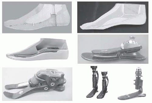
Gait training begins with weight shifting and balance activities while still in the parallel bars. Once weight shifting and balance activities have been mastered, a program of progressive ambulation begins in the parallel bars and progresses to the most independent level of ambulation possible with or without gait aids. Specific training should focus on transfers, knee stability, equal step lengths, and avoiding lateral trunk bending. Following mastery of ambulation on flat, level surfaces, techniques for managing uneven terrain, stairs, ramps, curbs, and falling and getting up off the ground are learned. Moving from a walker to less cumbersome gait aids can be achieved for most individuals with an LE amputation. For higher functioning individuals with an amputation, prosthetic training should include instruction and practice in driving, recreation, and vocational pursuits. Developing the optimal benefit from a prosthesis must take into account the specific mechanical attributes of the components used. For example, using a dynamic response (i.e., energy-storing) prosthetic foot requires loading the prosthetic toe during mid-stance and late stance to capture energy for push off assistance or to activate a prosthetic knee to initiate the swing phase.

Wearing tolerance for the prosthesis must be gradually increased. Initially, the individual with an amputation will wear the prosthesis only for 15 to 20 minutes, removing it to check the condition of the skin. As tolerance to weight bearing increases, the length of wearing time is gradually increased. Several weeks may be required before the individual with an amputation is able to wear the prosthesis full-time. The individual with an amputation may take the prosthesis home when safe and independent ambulation has been demonstrated and residual limb skin checks are assured. Common gait deviations and their causes are highlighted in Table 74-3.


LE Prosthetic Follow-up

During the initial 6 to 18 months, most individuals with an amputation will experience continued loss of residual limb volume, resulting in a prosthetic socket that will be too large. During this period, return visits should occur frequently enough to ensure that this loss of residual limb volume is being compensated for by the use of additional limb socks or by appropriate modifications of the prosthetic socket. It is usual for an individual with a new amputation to require replacement of the
prosthetic socket during this time because of the significant loss of soft-tissue volume. During follow-up visits, the condition of the residual limb, the prosthesis, the individual’s gait, and the level of function are reviewed (39). Appropriate medical treatment, prosthetic modifications, or additional therapies are prescribed as needed. When the residual limb volume has stabilized sufficiently and the patient is doing well with the prosthesis, yearly visits to the amputee clinic are appropriate. Once the residual limb has stabilized, the average life expectancy for an LE prosthesis before replacement should be 3 to 5 years.


TABLE 74.3 Abnormalities of Amputee’s Gait


Transtibial Amputee Gait


Gait Cycle


Observed Gait Abnormality


Possible Cause


Suggested Modifications


Initial contact to loading response


Abrupt heel contact, rapid knee flexion


Excessive heel levera


Realign prosthetic foot, change heel stiffness


Prolonged heel contact, knee remains fully extended


Inadequate heel leverb or heel worn out Improper socket flexion


Learned gait pattern, quadriceps weakness


Increase heel stiffness


Realign prosthesis


Gait training and strengthening


Jerky knee motion


Socket loose, poor alignment, inadequate suspension


Mid-stance


Medial or lateral socket thrust, lateral trunk shift over prosthesis


Foot too far outset or inset, socket loose


Realign prosthesis, replace socket or adjust socks


Pelvis drops or elevates


Prosthesis too short/too long


Adjust prosthetic length


Mid-stance to terminal stance


Early knee flexion or “drop off”


Inadequate toe leverc


Realign prosthesis, replace foot


Terminal stance


Heel off too early


Excessive toe leverd, too much socket extension


Realign prosthesis


Heel off excessively delayed


Inadequate toe leverc, too much socket flexion


Realign prosthesis


Swing phase


Prosthetic foot drags


Prosthesis too long, inadequate suspension


Shorten limb, modify suspension


Successive double support


Uneven step length


Hip flexion contracture, gait insecurity Uncomfortable socket


Physical therapy Adjust socket fit


Transfemoral Amputee Gait


Initial contact to loading response


Foot rotation at heel strike


Poor socket fit/rotation


Adjust socket fit, add belt for rotation control


Heel too firm


Reduce heel stiffness


Knee buckling


Excessive heel levera


Incorrect prosthetic knee alignment Weak hip extensors


Realign limb, reduce heel stiffness


Realign TKA relationship


Gait training and strengthening


Mid-stance


Lateral trunk bend or shift over prosthesis


Prosthetic limb abducted


Too much socket abduction, foot too far outset Prosthesis too long Medial groin pain


Realign prosthesis


Shorten prosthesis Adjust socket fit


Poor medial-lateral prosthetic control Poor socket fit Weak hip abductors Short residual limb


Prosthesis too short


Adjust socket fit Gait training and strengthening Accept, possibly add hip joint Adjust prosthetic length


Initial swing


Uneven heel rise


Knee friction too tight or loose Knee extension


Adjust knee friction or damping


Swing phase


Circumduction or prosthetic limb


Inadequate knee flexion, knee too stiff Prosthesis too long, inadequate suspension Poor gait pattern Improper knee rotational alignment


Adjust knee friction or damping Adjust prosthesis length Physical therapy Realign prosthesis


Whips


Excessive socket rotation


Adjust socket fit


Successive double support


Uneven step length


Hip flexion contracture Insufficient socket flexion


Physical therapy Realign prosthesis


a Causes of excessive heel lever—foot dorsiflexed too much, foot too far posterior, heel cushion too hard, shoe heel too hard.

b Causes of inadequate heel lever—foot plantarflexed too much, foot too far anterior, heel cushion too soft.

c Causes of inadequate toe lever—foot dorsiflexed too much, foot too far posterior, foot keel too soft/flexible.

d Causes of excessive toe lever—foot plantar flexed too much, foot too far anterior, foot keel too stiff.


TABLE 74.4 Medicare Guidelines for Functional Classification of Patients with Prosthesis


K Code Level


Functional Level


Activity Level


K0


Not a potential user for ambulation or transfer


Does not have the ability or potential to ambulate or transfer safely with or without assistance, and a prosthesis does not enhance their quality of life or mobility.


K1


A potential household ambulator including transfers


Has the ability or potential to use a prosthesis for transfer or ambulation on level surfaces at fixed cadence. Typical of the limited and unlimited household ambulator.


K2


A potential limited community ambulatory


Has the ability or potential for ambulation with the ability to traverse low level environmental barriers such as curbs, stairs or uneven surfaces. Typical of the limited community ambulator


K3


Community ambulator using variable cadence, including therapeutic exercise or vocation


Has the ability or potential for ambulation with variable cadence. Typical of the community ambulator that has the ability to traverse most environmental barriers and may have vocation, therapeutic, or exercise activity that demands prosthetic utilization beyond simple locomotion.


K4


High activity user which exceeds normal ambulation skills


Has the ability or potential for ambulation that exceeds basic ambulation skills, exhibiting high impact, stress, or energy levels. Typical of the prosthetic demands of the child, active adult, or athlete.


Source: DMERC medicare Advisory Bulletin, Columbia SC, 1994;12:95-145.


LE Prostheses

The LE prosthetic prescription must balance the individual’s need for stability, mobility, durability, and cosmesis with available resources and cost. Understanding the role and importance of prosthetic ambulation in achieving the mobility goals of the individual with an amputation is essential for correctly prescribing a prosthetic device. Prosthetic ambulation is usually the primary mode of mobility for the younger individual with an amputation as well as for other patients across a wider age range when the amputation is at the transtibial and more distal levels. For the elderly, with dysvascular amputation above the knee and a more proximal level of amputation, prosthetic ambulation is often limited to transfers, indoors, or short community distances. The prescription of the LE prosthesis is based on several principles: maximizing comfort, matching specific components to the mobility needs of the individual with an amputation, and providing acceptable cosmesis. Comfort is the most critical aspect and depends on achieving an appropriate distribution of forces between the residual limb and the socket. A poorly fitting or uncomfortable socket will limit the mobility and often lead to rejection of the prosthesis. Once comfort has been established, the appropriate choice of components facilitates achieving maximal independence and function during sitting, standing, transferring, walking, and running. Lastly, cosmetic concerns are considered. Cosmesis is influenced by personal preferences and psychosocial dynamics but is usually satisfactorily achieved using contoured foam and a nylon or rubber skin tone cover. Some individuals with an amputation prefer not to have their prosthesis covered because of the possible interference with prosthetic component function.

Medicare, a major funding source for prosthetic limbs in the United States, requires that the functional level of the individual with an amputation be taken into account when prescribing a prosthesis. The functional index is referred to as the Medicare “K” code and limits the components that can be used when fabricating the prosthesis. Although only required for Medicare, the “K” code classification is a simple but useful hierarchical framework for classifying the mobility potential of all individuals with an LE amputation (Table 74-4).


LE Prosthetic Components

The continual introduction of new component designs and the overlap of functional features of components from various manufacturers make it difficult to stay abreast of available prosthetic options. Collaboration between health care providers (physician, prosthetist, and therapist) is essential in developing an appropriate, individualized limb prescription. Seldom is there a single correct choice of components for a prosthesis, rather most individuals with an amputation can be successfully fit using components that span a reasonable range of mechanical and functional characteristics. Because objective data, linking prosthetic component characteristics to the demographics of individuals with an amputation, are limited, empiric approaches and experience play a major role in limb prescription. The prescription for an LE prosthesis should include the Medicare “K” code, diagnosis, type of prosthesis (with modifiers), socket type, liner, suspension method, foot, knee and hip systems (as required by amputation level), diagnostic or check socket, and supplies.


Prosthetic Feet

Prostheses for amputations at or proximal to the ankle require the use of a prosthetic foot. The selection of an appropriate prosthetic foot is complicated by the wide range of foot designs, marketing-driven claims of performance, and the limited availability of objective data comparing the relative biomechanical and functional advantages of different feet. In the clinical setting, the selection of a prosthetic foot is largely empirically based on the conceptual goal of matching the functional characteristics of the foot to the expected activity needs of the individual (40, 41, 42, 43, 44, 45). Within this approach, it is useful to group feet by their major functional feature(s) as belonging to rigid keel, flexible keel, single/multiaxial, or dynamic response (or energy-storing) categories. It is acceptable for the prescribing physician to define the functional features desired in the foot and to rely on the prosthetist who typically has a better working understanding of the commercially available feet to select the specific manufacturer and foot within the desired functional class. This multidisciplinary approach is increasingly important as foot designs become more sophisticated, more costly, and combine different functional characteristics into a single foot. Occasionally, another characteristic of a foot such as an adjustable heel height, cosmesis, or being waterproof is the primary determinate in its selection.

The solid ankle cushion heel (SACH) foot (Fig. 74-2) is the least expensive and most commonly prescribed prosthetic foot. It is durable and lightweight, which accounts in part for its usefulness. The SACH foot has no moving parts and consists of a wooden or composite keel with a compressible foam heel and toes that flex under load, allowing limited simulation of the effects of the heel and forefoot rocker mechanisms of the normal foot. A SACH foot is appropriate for individuals with an amputation who have a lower activity level (K1 to K2), with ambulation primarily limited to level surfaces. It can be used in a wide range of individuals with an amputation for the preparatory prosthesis and upgraded as the individual with an amputation progresses to a higher activity level. For a juvenile with an amputation, the SACH foot is often the most cost-effective foot due to the need for frequent foot changes because of rapid growth.


FIGURE 74-2. Prosthetic feet from the solid ankle cushion heel (SACH) and stationary attachment flexible endoskeletal (safe II flexible keel) foot (top). The impulse foot (dynamic response) and Luxon Max (dynamic response with multiaxis) (middle). The College Park TruStep (dynamic response, with some inversion, eversion, and transverse motion), the FlexFoot VSP (vertical shock pylon, dynamic response, multiaxis), and the Ceterus (VSP, dynamic response, multiaxis, and transverse motion) (bottom). The prosthetic manufactures have numerous feet available from the homebound to the paralympic patient. (Courtesy of Kingsley, Ohio Willow Wood, Otto Bock, CPI, OSSUR and Freedom Innovations. See prosthetic manufacture WEB Site Listings.)

The flexible keel foot (see Fig. 74-2) is designed to mimic the motion of the forefoot rocker mechanism by replacing the rigid keel of the SACH foot with a flexible keel. The keel bends with controlled stiffness as the foot moves from mid-stance through preswing. Several versions of flexible keel feet are commercially available, each with different construction but sharing similar function. The stationary-ankle-flexible endoskeletal (SAFE) II foot is a commonly used flexible keel foot. The flexible keel foot allows some inversion and eversion, and gives a smoother rollover than a SACH foot, making it appropriate for general mobility needs in the individual with an amputation with a low to moderate activity level. However, the more active individual with an amputation may perceive the flexible keel foot as being too soft, especially for fast walking or running activities.

Articulating prosthetic feet include both single axis and multiaxis designs. The single axis foot allows controlled movement in the sagittal plane (plantar-flexion and dorsiflexion), adjusted by using different durometer bumpers. The primary advantage of the single axis foot is its ability to reduce kneebending movements during limb loading, thus improving knee stability. Disadvantages include a greater weight than many other feet and more maintenance to ensure correct function. This foot is primarily used in the individual with a proximal amputation that requires better knee stabilization, such as the elderly individual with a transfemoral amputation or the individual with a transfemoral amputation and a short residual limb.

Multiaxial foot designs allow for varying degrees of controlled movement in the sagittal, coronal, and transverse planes (plantar/dorsiflexion, inversion/eversion, some degree of transverse rotation). Multiaxis feet can use mechanical joints to supply motion such as the Greissinger foot or the College Park foot (see Fig. 74-2), but increasingly rely on the inherent flexibility of rubber and polymer materials to provide multiaxial motion. Using material flexibility improves durability and reduces both weight and maintenance compared to mechanical jointed feet. Multiaxis “ankle” motion can be integrated into the foot (e.g., Endolite foot, Luxon) or added to other feet through the use of separate multiaxial ankle components (e.g., impulse ankle—Ohio Willow Wood, Mt. Sterling, Ohio). Multiaxis capabilities are appropriate for the individual with an amputation who needs improved ankle motion to accommodate to uneven terrain and for the active individual with an amputation who requires greater ankle movement to adjust to different speeds or for cutting and pivoting quickly.


Dynamic response (i.e., energy-storing) prosthetic feet incorporate elastic (spring-like) elements that store energy in the foot during limb loading and mid-stance as the elastic material compresses or flexes. Energy is returned at the time of push-off as the spring components of the foot returns to its normal shape or configuration. Examples include the Flex-foot, the Springlite feet, Seattle foot, and the impulse feet (see Fig. 74-2). The dynamic energy characteristics of these feet make them particularly suitable for individuals with an amputation involved in activities requiring running and jumping. Many individuals with an amputation believe that they are more functional with a dynamic response foot. Dynamic elastic response (DER) feet were expected to make ambulation more efficient by reducing the oxygen consumption of individuals with an amputation but the results of objective studies have been mixed (44,46). The metabolic benefits of DER designs are limited and primarily seen at faster walking speeds.


Prosthesis by Level of Amputation


Partial Foot Amputation

Toe amputations, ray resections, and transmetatarsal amputations require minimal prosthetic/orthotic intervention. At the more distal foot amputation levels and for the less active individual with a transmetatarsal amputation, accommodative shoes with custom insoles, arch supports, and toe fillers are usually adequate. More active individuals with a transmetatarsal amputation may benefit from orthotic modifications that better substitute for the lost anterior foot lever arm. Options include the addition of carbon fiber or spring steel sole shanks, rocker soles, or short ankle foot orthosis. Partial foot amputations at the tarsal-metatarsal and transtarsal levels (e.g., Lisfranc, Chopart) are relatively uncommon and have historically been associated with equinovarus contracture of the hind foot, increasing the likelihood of skin breakdown over the plantar surface of the foot. However, improved surgical techniques that include Achilles tendon lengthening/resection and anterior tibialis and peroneus tendon transfers have reduced equinovarus deformities and result in a functional and useful amputation level (47,48). Prosthetic/orthotic devices for the individual with a proximal partial foot amputation need to supply medial-lateral stabilization of the hind foot and substitute for the lost forefoot lever. Options include: (a) an extra-depth shoe with toe filler, steel shank, and rocker bottom modifications; (b) custom posterior leaf-spring ankle-foot orthosis with toe filler; or (c) a custom prosthetic foot with a self-suspending rear-opening split socket (47,48). A major advantage of all partial foot amputations is the ability to be fully end-bearing, allowing ambulation without any devices.


Syme Amputation

Similar to the hind foot amputation, the Syme (tibiotarsal disarticulation) amputation is capable of full weight end bearing. The heel flap is anchored to the distal end of the tibia and fibula, and following healing, allows short distance ambulation without a prosthesis. The substantial leg length discrepancy makes long distance ambulation impractical. Over time, posterior migration of the distal heel pad occurs in some individuals with a Syme amputation leading to problems with skin breakdown and difficulty in prosthetic fitting (49, 50, 51). The relatively bulbous distal end of the residual limb has the advantage of enabling the use of self-suspending prosthetic designs; however, it also contributes to the major disadvantage of the Syme amputation—poor cosmesis due to the bulkiness of the prosthesis around the ankle joint. There are several different types of prostheses available for individuals with a Syme amputation.


FIGURE 74-3. From left to right: The posterior opening Symes for bulbous distal end; PTB Symes with Pelite liner; Canadian-type Syme prosthesis as modified by the Veterans Administration Prosthetic Center. (Courtesy of PSL Fabrication, Fulton, MO.)

May 25, 2016 | Posted by in PHYSICAL MEDICINE & REHABILITATION | Comments Off on Upper and Lower Extremity Prosthetics

Full access? Get Clinical Tree

Get Clinical Tree app for offline access