Triple Arthrodesis


Triple Arthrodesis


John A. Ruch

Lopa Dalmia

Patrick B. Hall


The triple arthrodesis performed today is a variation of the procedure described by Ryerson in 1923 (1). Modifications have evolved out of the need to meet new challenges as the triple arthrodesis has been applied to a greater variety of disorders (2,3,4,5,6,7,8,9,10,11 and 12). The basic aim of a triple arthrodesis is to improve foot function by providing stability, correction of deformity, and elimination of pain. Providing the patient with a stable, pain free platform for ambulation through triple arthrodesis offers gratifying and predictable results for a variety of foot deformities (13,14,15,16,17,18,19,20,21,22 and 23).


PREOPERATIVE CONSIDERATIONS

Certain considerations should be made before triple arthrodesis is performed. These include patient expectations, the desired goal of the fusion and its functional effect, timing of the surgical intervention, biomechanical and positional considerations of the subtalar joint (STJ) and midtarsal joint, the position and alignment of the ankle and leg, bone quality, soft tissue quality, the patient’s age, and the anticipated recovery time.

Candidates for triple arthrodesis usually possess conditions that have proven resistant to conservative therapy, or they have a condition that cannot be expected to respond to conservative measures and one in which the surgeon can expect an adequate result with fusion. The elimination of STJ and midtarsal joint motion may restrict the ability of the patient to adapt to uneven surfaces and terrain; however, in candidates for triple arthrodesis, this motion is often either painful or absent preoperatively. Additionally, the existing deformity often prevents the motion from serving any benefit for the patient, due to either painful arthritis or uncontrollable instability.

Evaluation of ankle joint range of motion is a critical part of the preoperative assessment. This may reveal either an arthritic limitation or a soft tissue equinus contracture, altering the surgical plan. Careful examination needs to be performed in a patient with a severely collapsed pes valgus deformity because significant amounts of dorsiflexion may occur at the midtarsal joint level. In patients with rigid pes valgus conditions, it is difficult to position the foot adequately to assess the true amount of ankle dorsiflexion until the joints have been resected at the time of surgery. In contrast, ankle joint dorsiflexion in patients with a pes cavus deformity may first appear inadequate because of the increase in the osseous height from the maximally supinated position of the midtarsal joint and STJ. Upon restoring a more plantigrade osseous alignment after fusion, one may note a suitable increase in the dorsiflexory motion at the ankle.

Perhaps the most critical aspect of triple arthrodesis is the ultimate position of the foot after fusion. Poor or inappropriate positioning of the foot may be one of the primary reasons for residual pain and the creation of adjacent arthritis postoperatively. The heel should be aligned to rest in a neutral to slightly everted position. The greatest success in triple arthrodesis has been achieved with the midtarsal joint positioned in slight valgus when fused, that is, with the medial column slightly plantarflexed relative to the lateral column. This position increases the stability of the medial column and first ray, permitting enhanced first metatarsophalangeal joint motion. The valgus positioning may also be more easily accommodated with an orthotic device postoperatively. If the medial column is dorsiflexed relative to the lateral column, the patient is left with a fixed forefoot varus deformity for which no suitable compensation exists.

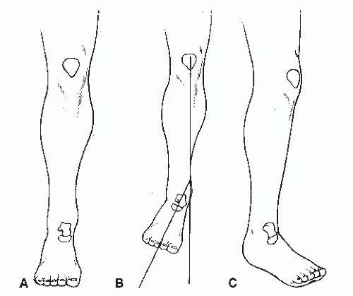
It is important to plan the alignment of the forefoot to the rearfoot and the rearfoot to the leg (Fig. 58.1). This is especially critical in determining the final position of fusion. The foot normally exhibits 10 to 15 degrees of abduction from the line of progression in gait. In arthrodesis of the rearfoot, the surgeon must know the position of the knee during gait as well as during the surgical procedure. If the knee functions when medially rotated at 15 degrees, then it would be desirable to abduct the foot on the leg 30 degrees, thus resulting in a 15-degree abduction from the line of progression. It is not advisable to abduct a foot if the patient already possesses 15 to 30 degrees of lateral position of the knee in gait. In the latter instance, the foot may be aligned directly with the knee.

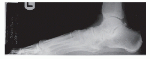
These preoperative assessments are aided by a series of weight-bearing radiographs (Fig. 58.2) including dorsoplantar, medial oblique, lateral, and calcaneal axial views. Weightbearing films allow a more representative view of osseous alignment. The degree of deformity should be evaluated in each of the cardinal planes prior to proceeding with surgical reconstruction.


TECHNIQUE


MEDIAL INCISION/DISSECTION

Landmarks for the medial approach to triple arthrodesis include the medial gutter of the ankle joint proximally and
the inferior aspect of the navicular cuneiform joint (Fig. 58.3). This oblique orientation provides full exposure of the talonavicular joint and allows for screw fixation of the STJ and the talonavicular joint. A dorsal to plantar fixation of the STJ utilizes insertion of the large cancellous screw at the dorsal medial aspect of the talar neck. Fixation of the talonavicular joint with a large cancellous screw is directed from the distal inferior aspect of the navicular up into the head and neck of the talus.


TABLE 58.1 Conditions That May Benefit from Triple Arthrodesis


Idiopathic collapsing pes planovalgus deformity


Peroneal spastic flatfoot


Tarsal coalition


Congenital vertical talus


Chronic pain


Rheumatoid arthritis


Degenerative arthritis


Posttraumatic arthritis


Charcot arthropathy


Tibial posterior tendon dysfunction


Idiopathic cavus and cavovarus deformities


Residual or uncorrected clubfoot


Poliomyelitis


Spina bifida


Friedreich ataxia


Charcot-Marie-Tooth disease


Muscular dystrophy


Cerebral palsy


Myelodysplasia


Arthrogryposis


Joint instability


Figure 58.1 Relationship of the knee position to the foot. A: Rectus knee and foot. B: Rectus knee with the foot abducted 30 degrees. C: Internal knee position with the foot adducted 25 degrees.


Figure 58.2 Preoperative radiograph.


TABLE 58.2 Indications for Triple Arthrodesis


Valgus foot deformities


Collapsing pes planovalgus deformity


Tibial posterior tendon dysfunction


Tarsal coalition


Arthritic conditions


Rheumatoid arthritis


Degenerative arthritis


Posttraumatic arthritis


Chronic pain


Varus foot deformities


Cavus and cavovarus


Talipes equinovarus


Miscellaneous conditions


Joint instability


Neuromuscular disease


Hereditary familial sensorimotor neuropathies


Paralytic deformities


Cerebral palsy


Charcot arthropathy


Other diseases affecting the spinal cord and brain


Medial skin incision for exposure of the talonavicular joint and insertion of the TN screw and the talocalcaneal screw extends from the medial gutter of the ankle to inferior aspect of the navicular cuneiform joint (Fig. 58.4A). The greater saphenous vein will usually be encountered during dissection through the subcutaneous layers. Inferior tributaries may be transected and ligated and the main portion of the vein reflected superiorly (Fig. 58.4B). The primary incision for exposure of the talonavicular joint is made through the deep fascia and capsule along
the dorsal medial aspect of the joint. The incision extends from the medial gutter of the ankle joint to the navicular cuneiform joint (Fig. 58.4C).


Figure 58.3 Landmarks for the medial approach to triple arthrodesis include the medial gutter of the ankle joint proximally and the inferior aspect of the navicular cuneiform joint.


Figure 58.4 A: Medial skin incision. B: Dissection through the subcutaneous layers. C: Primary incision for exposure of the talonavicular joint.

The capsular incision for the talonavicular joint is a T incision (Fig. 58.5A and B). The dorsal medial longitudinal incision allows for reflection of capsular tissues for the dorsal aspect of the talonavicular joint. The vertical medical incision allows for deliverance of the head of the talus without reflecting capsular tissues of the medial aspect of navicular. A secondary incision is made vertically along the proximal medial edge of the navicular but does not usually transect the tibialis posterior tendon (Fig. 58.5C and D). This modification in the talonavicular incision leaves capsule and periosteal tissues intact over the medial aspect of the navicular. The capsule
is reflected from the dorsal surface of the talonavicular joint and will routinely release the dorsal talonavicular ligament (Fig. 58.5E). This modification in the arthrotomy of the talonavicular joint provides full exposure and minimizes soft tissue or periosteal reflection.


Figure 58.5 A,B: Capsular incision for the talonavicular joint.


Figure 58.5 (Continued) C,D: Secondary incision is made vertically along the proximal medial edge of the navicular but does not usually transect the tibialis posterior tendon. E: Reflection of the capsule from the dorsal surface of the talonavicular joint.

Jul 26, 2016 | Posted by in MUSCULOSKELETAL MEDICINE | Comments Off on Triple Arthrodesis

Full access? Get Clinical Tree

Get Clinical Tree app for offline access