Trigger Digits
CASE PRESENTATION
An 18-month-old child presents for evaluation of bilateral “thumb deformities” (Figure 12-1A). The patient’s grandmother noticed “deformities” after examining the child’s hand after a fall down stairs. Both thumbs have fixed interphalangeal (IP) joint flexion contractures; they are not spontaneously or manually reducible but are not painful or tender. Physical examination demonstrates a palpable nodule at the volar aspect of the metacarpophalangeal (MCP) joints and mild hyperextension laxity of the MCP. No treatment has yet been instituted.
CLINICAL QUESTIONS
What is a “congenital trigger thumb”?
Is it congenital? Hereditary? Acquired?
What is the natural history of pediatric trigger thumbs?
Is there a role for splinting, stretching, or occupational therapy?
What are the surgical indications for trigger thumb release?
How is pediatric trigger thumb release performed?
What are the anticipated results and potential complications?
Are trigger fingers similar to trigger thumbs?
What are the indications for surgical release of trigger fingers?
How is a trigger finger release performed? Does A1 pulley release suffice?
What are the anticipated results and potential complications?
THE FUNDAMENTALS
First master the fundamentals.
—Larry Bird
Etiology and Epidemiology
The estimated prevalence of pediatric trigger thumbs is thought to be 3:1,000 live births at the age of 1 year.1
While the exact etiology is unknown, several studies evaluating thousands of newborns for the presence of trigger thumbs have clearly demonstrated that this condition is not present at birth.2, 3, 4 and 5 Therefore, the term “congenital trigger thumb” is a misnomer and its use discouraged.
Triggering or locking is due to a size mismatch between the flexor pollicis longus (FPL) tendon and the tendon sheath, typically at the level of the first annular (A1) pulley. It has been speculated that the constant flexed position of the thumb IP joint during the pre- and post-natal periods results in collagenous degeneration, fibrosis, and synovial proliferation, leading to thickening of the FPL tendon and subsequent constriction.6, 7 and 8 Prior histopathologic studies have demonstrated no degenerative or inflammatory changes in the tendon or sheath; therefore, trigger thumbs are not likely due to an inflammatory, infectious, or degenerative process. To date, the true cause of pediatric trigger thumbs remains unknown.
Fingers are not thumbs, and triggering of the lesser digits should be considered an entirely different clinical entity, with its own set of clinical considerations and pitfalls. Trigger fingers are approximately 10 times less common than trigger thumbs. While the etiology is even less well understood, they are likely due to anatomic abnormalities of the flexor mechanism.8, 9, 10, 11, 12, 13, 14, 15, 16 and 17 Most commonly, a proximal decussation of the flexor digitorum superficialis (FDS) slips or aberrantly distal lumbrical muscle origin leads to triggering as the digits move from flexion to extension. Nodular tendon thickening and intratendinous calcification or granulation tissue have also been described. Finally, deposition diseases such as the mucopolysaccharidoses are known to cause secondary triggering.
Children are not small adults. While the temptation can be great, adult rules should not be applied to the pediatric game. Inflammatory or overuse etiologies should not be implicated, nor should treatment modalities such as corticosteroid injections be utilized.
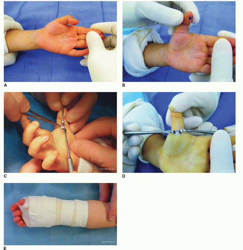 FIGURE 12-1 Surgical technique of trigger thumb release. A: Clinical photograph of a pediatric trigger thumb. B: Prior to surgical release, marks are made on either side of the palpable Notta nodule in the MCP flexion crease, marking the extent of the transverse skin incision. C: After spreading and retraction of the soft tissues and neurovascular bundles, the tendon sheath is exposed. D: The A1 pulley is incised. Note the IP joint hyperextension following pulley release. E: A bulky soft dressing is applied after wound closure. |
Clinical Evaluation
Patients with trigger thumbs typically present during the first 18 months to 3 years of life with either triggering or a fixed flexion contracture of the IP joint. Often the abnormality is noted incidentally by family, teachers, or other medical care providers. There are times when it is recognized with some sort of trauma; then, it can be mistaken by parents, primary and emergency room physicians, and caregivers as an acute dislocation. They can be further mislead by a flexed posture on the lateral radiograph with unossified epiphysis of the distal phalanx; the sense of “joint reduction” with manipulation of the nodule distal to the first annular (A1) pulley; and frustration of almost immediate recurrent “dislocation” in their postreduction splint. When locked, the thumbs are not painful, nor do patients inhibit spontaneous use of the thumb and hand. When spontaneously or manually reducible, abrupt extension of the thumb IP joint may be
associated with transient pain, which may alert the family to the condition. One-third of patients will demonstrate bilateral thumb involvement, though this may be metachronous with variable severity.18 Careful inspection and palpation will identify the Notta nodule within the FPL tendon, which will demonstrate excursion with passive IP joint motion. Often in long-standing cases, the MCP will demonstrate compensatory hyperextension laxity or instability, as the child attempts to open the first web space during cylindrical grasp. Nodules and triggering are not seen in the index through small fingers in the typical trigger thumb.
associated with transient pain, which may alert the family to the condition. One-third of patients will demonstrate bilateral thumb involvement, though this may be metachronous with variable severity.18 Careful inspection and palpation will identify the Notta nodule within the FPL tendon, which will demonstrate excursion with passive IP joint motion. Often in long-standing cases, the MCP will demonstrate compensatory hyperextension laxity or instability, as the child attempts to open the first web space during cylindrical grasp. Nodules and triggering are not seen in the index through small fingers in the typical trigger thumb.
In trigger fingers, there too will be characteristic triggering or less commonly fixed flexion contractures, typically at the proximal interphalangeal (PIP) joint and associated with what feels like a Notta nodule (Figure 12-2). Both single and multiple digital involvements occur commonly, and bilateral hand involvement is not uncommon. Care should be made to identify associated medical conditions; in cases of associated mucopolysaccharidoses, evaluation for carpal tunnel syndrome and/or median neuropathy should be performed.19
Natural History
Say you were standing with one foot in the oven and one foot in an ice bucket. According to the percentage people, you should be perfectly comfortable.
—Bobby Bragan
Definitive statements about the natural history of pediatric trigger thumbs


Related posts:

Stay updated, free articles. Join our Telegram channel
Full access? Get Clinical Tree
