Total Wrist Arthroplasty
Ian A. Trail
John K. Stanley
Indications
The development and usage of total arthroplasty for the wrist has slowly followed that of the other major joints. Even today, however, it has not found widespread acceptance in that most surgeons prefer to recommend arthrodesis to their patients. Certainly, arthrodesis is simpler to perform and does not have the same number of potential long-term complications when compared with arthroplasty. With the advent of newer prosthesis, attitudes are gradually changing, however, particularly for patients with bilateral inflammatory arthritis of the wrist. The latter is undoubtedly the pre-eminent indication for total wrist replacement. It has always been our policy to replace the dominant wrist and fuse the nondominant wrist in these patients.
Few comparative studies appear in the literature. The first was reported by Vicar and Burton (1), who reviewed 48 patients, 33 with a wrist fusion and 37 with silicone wrist arthroplasty. Of the arthrodesis group, 97% showed good to excellent results, whereas the arthroplasty group had a similar outcome (78%). All the arthrodesis patients had reduced pain, a stronger wrist, but reduced dexterity. The complication rate was 18%. Most of the arthroplasty group had improved dexterity and adequate strength. Movement averaged 61 degrees of flexion–extension. Here, the complication rate was 25%, which included four revisions. Most problematic radiologically, 14% of the arthroplasty group had evidence of bone resorption around the stems of the implant, with 11% showing evidence of settling. As a consequence, these authors recommended arthroplasty only for patients who did not use ambulatory aids or crutches, and had good bone stock, no deformity, and intact wrist extension.
In 2003, Murphy et al. (2) compared 24 arthrodesis and 27 arthroplasties retrospectively with a follow-up ranging from 26 to 48 months. Using the Disability of the Arm Shoulder and Hand (DASH) score, Patient Rated Wrist Evaluation (PRWE), and an in-house questionnaire, they found a trend toward greater ease of certain activities, specifically personal hygiene and fastening buttons, although no statistically significant difference reported in the arthroplasty group. In addition and while the follow-up was short, particularly for the arthroplasty group, there was no difference in complications, with 56% of the arthrodesis and 52% of the arthroplasty groups having some form of complication.
Finally, research undertaken at Wrightington into the survivalship and the mode of failure of the Biaxial wrist replacement (Depuy) identified a number of long-term problems, although many of these patients still preferred arthroplasty to arthrodesis. A group of 27 patients who underwent replacement on one side and arthrodesis on the other mostly preferred arthroplasty, despite a number demonstrating what were felt objectively to be poor results.
Contraindications
Although the principal indication for total wrist arthroplasty is inflammatory arthritis, wrist replacements have also been used in osteoarthritis, following trauma, and for a number of other chronic wrist conditions, including Kienböck disease. It should be noted, however, that published long-term outcome data on all these indications are currently not available.
Potential absolute contraindications include infection, instability, deformity, muscle weakness, and inadequate bone stock. An active ongoing infection would be a clear contraindication to any
joint replacement or, indeed, metal work being inserted in the wrist. It may be possible, however, to undertake a joint replacement if the infection has been eradicated, although even then the risk of subsequent infection must be higher. Instability is a relative contraindication in that if a wrist is unstable before surgery then the risk is that it may be unstable subsequently. Much will depend on the skill of the surgeon. Similarly, any deformity should be corrected at surgery by a combination of bone resection, soft tissue release, and subsequent repair. Muscle weakness or tendon rupture would obviously impair wrist movement, in which case a repair or tendon transfer may be required.
joint replacement or, indeed, metal work being inserted in the wrist. It may be possible, however, to undertake a joint replacement if the infection has been eradicated, although even then the risk of subsequent infection must be higher. Instability is a relative contraindication in that if a wrist is unstable before surgery then the risk is that it may be unstable subsequently. Much will depend on the skill of the surgeon. Similarly, any deformity should be corrected at surgery by a combination of bone resection, soft tissue release, and subsequent repair. Muscle weakness or tendon rupture would obviously impair wrist movement, in which case a repair or tendon transfer may be required.
Finally, if insufficient bone stock exists to support a prosthesis, then this should not be attempted. The principal area of concern is on the radius, which can be eroded, particularly around the sigmoid notch in cases of rheumatoid arthritis. Although a more proximal radial bone cut can be undertaken, this may result in the residual bone being too narrow to support even the smallest component. Small defects can be filled with bone graft, but little is known about the long-term outcome.
As experience develops, the list of contraindications will diminish and more will be possible. The safer option in difficult cases, particularly for the inexperienced surgeon, is still arthrodesis, however.
Surgical Technique
Review of literature indicates that most, if not all, wrist replacements have been inserted by a dorsal approach. Initially, the skin incisions were planned with the aim of not crossing the skin creases in a perpendicular fashion. However, contracture has rarely if ever been described and, as a consequence, most surgeons now perform a longitudinal incision. This has the added advantage of allowing both radial and ulnar flaps to be easily developed, thus reducing the risk of skin edge necrosis. It is of equal importance to lift the skin edges and underlying soft tissue down to and including deep fascia (i.e., superficial to the extensor retinaculum as one flap). This should safeguard the blood supply to the skin.
During this process, ligate any large veins that cross the incision.
Identify and dissect free the extensor retinaculum, usually starting on the ulnar aspect over the ulnar head, raising it across to the first compartment. In the authors’ experience, when elevating the extensor retinaculum it is better to expose all the tendons on the dorsum of the wrist, including those of the first compartment. It is also important to take great care when elevating the extensor retinaculum overlying the extensor pollius longus tendon. Remember that this tendon crosses the wrist diagonally distal to Lister’s tubercle.
Then, mobilize all the tendons, allowing them to be retracted in a radial or ulnar fashion. Lister’s tubercle is removed.
Identify the posterior interosseous nerve and vessels lying in the base of the fourth compartment and resect or ligate them, as appropriate.
Then open the dorsal capsule of the wrist to expose the distal radius and carpal bones. The method of exposing the carpus can vary and is often surgeon preference. A reversed T-incision allows better midline exposure as well as ease of repair (Fig. 39-1). Again, however, this depends on the implant used and the degree of exposure required.
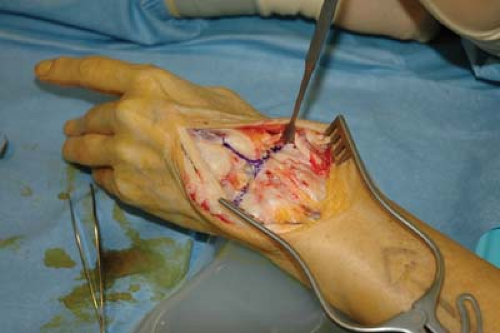
Figure 39-1 Reverse T-incision of the dorsal capsule.
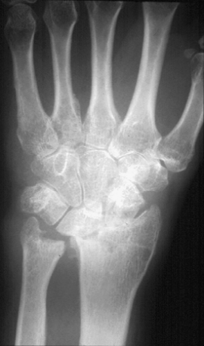
Figure 39-2 Lunate erosion of the radial articular surface.
Remove the proximal row of the carpus as well as the distal radial articular surface. The degree of bone resection depends on the implant to be inserted. It is of importance, however, not to penetrate the anterior capsule, thus safeguarding the structures on the volar aspect of the wrist; the only exception being when an anterior release is required. Rheumatoid or inflammatory arthritis is the most common indication for joint replacement. In light of this, it is not unusual to find significant joint erosion, which makes identification of the radiocarpal and mid-carpal joints more difficult. More specifically, the lunate can erode into the lunate fossa (Fig. 39-2). For this reason, preoperative planning is crucial, such that the removal of the distal radial articular surface does not involve the removal of so much bone that an implant cannot be inserted. In addition, many of these patients have problems at the distal radioulnar joint (DRUJ), which frequently are dealt with by a resection arthroplasty (Darrach’s procedure). In certain cases, particularly in a more active patient, this can lead to instability of the distal ulnar joint with resultant weakness. Alternatively, leave the ulnar in situ or replace the ulnar head. The former can lead to problems with exposure, as well as persisting pain and discomfort. As far as the latter is concerned, currently little published experience exists.
With regard to the actual insertion of a particular implant, see the various manufacturers’ surgical techniques. It is imperative that surgeons are completely familiar with both the implant and its instrumentation before surgery.
Closure is relatively straight forward, although it is sometimes difficult to close the capsule completely. In such cases, then it is possible to suture part of the extensor retinaculum beneath the tendons supplementing the capsule repair. It is inadvisable, however, to suture all the extensor retinaculum under the tendons; a strip should be sutured superficially to prevent bowstringing. A sound repair of the capsule of the DRUJ is more important to prevent instability of the ulna stump. Usually, this closure is double-breasted, thus closing any dead space.
Postoperatively, most patients who have had total wrist arthroplasty experience severe pain, at least initially. To counteract this, it is important that the patients are prescribed adequate analgesia, including regional nerve blocks. We have found the latter to be particularly useful. In addition, the hand and wrist can become extremely swollen for several days after the procedure;
in light of this, elevation with early movement of the fingers is important. It is also crucial to monitor the neurovascular status of the hand because a few patients develop an acute carpal tunnel syndrome after total wrist replacement, some of whom require urgent carpal tunnel release. At this time, however, it is not the authors’ practice to release the carpal tunnel routinely. However, once any swelling has subsided, the pain is under control, and the wound is seen to be healing satisfactorily, consider wrist mobilization. This early mobilization is somewhat controversial in that a number of authors prefer to wait several weeks before advocating movement. The rationale for this being that, in their opinion, early mobilization can lead to instability. In the authors’ opinion, however, if the implant was felt to be stable at surgery, then it should be possible to begin passive and subsequently active movement of the wrist somewhat earlier. At this stage, however, no evidence indicates that either technique gives better results.
Pearls and Pitfalls
The role of wrist arthroplasty is currently still in its infancy and, as such, significant improvement of both implant design and surgical technique will continue. The current implant design, particularly that of the Universal II and SBI, have reached a watershed for uncemented fixation. The latter seems reproducible and reliable with regard to prolonged fixation, especially for the carpal component. In addition, new instruments have been designed to aid bony preparation specifically to improve alignment of the implant. Any surgeon undertaking this procedure should be fully conversant with all the instruments available. The use of radiologic control has also proved invaluable, particularly for initial alignment, but also to assess the position of the components before fixation. The authors would strongly recommend its usage during this procedure.
Exposure of the radiocarpal joint is greatly facilitated by removal of the ulna head (Darrach’s procedure). This is usually necessary in cases of inflammatory arthritis because it is usually involved in the disease process. In cases of osteoarthritis, however, merit is found in retaining the ulna head to prevent postoperative ulna stump instability.
Distally, the main difficulty often occurs in removing the distal pole of the scaphoid. This lies deep in the incision on the volar side and close to some terminal branches of the radial artery. As a consequence, take great care when working in this area.
With regard to sizing the prosthesis, it is obviously important that no overhang exists, particularly distally, because sharp edges can result in further bone erosion and soft tissue damage. Johnson et al. (3) reported a case of erosion of the pisiform bone on a distal metal component.
With regard to screw fixation, traditional teaching is to cross the carpometacarpal (CMC) joint only at the index and middle fingers and not at the ring and little fingers. Although under most circumstances, the authors would adhere to that, remember that the aim is to obtain sound fixation and, as such, if it is absolutely necessary, CMC joints on the ulna side can be transgressed.
The key to a successful wrist replacement is management of the soft tissues (i.e., by way of appropriate capsular releases, repair, and tendon transfer, if appropriate). Capsular release is a release of both radial and ulnar capsular as well as volar attachments to allow free movement of the prosthesis. This should obviously follow the bony resection. At that stage, if the implant feels tight then either remove more bone or insert a smaller implant; the new, modular implants facilitate this. At the end of the procedure, the implant should be stable and balanced. Avoid any over tightening of the posterior capsule on closure. Also undertake any tendon transfers that restore wrist movement.
Finally, postoperative management, the tradition after total wrist replacement has been to immobilize the wrist for a period of 6 weeks or sometimes longer to achieve stability. This has changed in so much that, as with hip, shoulder, and knee replacements, many surgeons believe that if a prosthesis is stable at the end of surgery there is no reason why the patient cannot begin early mobilization. In practice, this often begins at 3 to 4 days after surgery when swelling has receded. In addition, it is also wise to provide the patient with a splint for use at night or between therapy sessions. It is the authors’ view (as yet unproved) that early mobilization will result in better long-term range of motion.
Postoperative Management
The objective of any therapy program is to obtain a stable, pain-free joint with a functional arc of movement. In addition, for long-term survival of the implant, teach the patient joint protection principles to minimize external forces being transmitted through the prosthesis. In the initial
postoperative period, the wrist is elevated to reduce bruising and edema, which can last for several days. It is also important to give adequate analgesia because a wrist replacement can be a particularly painful procedure. As to when mobilization is begun, it is a decision taken in consultation with the surgeon, therapist, and most importantly the patient. Certainly, early mobilization should not be started if problems with wound healing exist, or if the joint is particularly painful and swelling persists. At our institution, when the wound has healed and the sutures have been removed, if it is felt collectively that the joint is stable, then mobilization—specifically passive and active assist flexion–extension, as well as radioulna deviation—is initiated. Pronosupination is also begun. A splint is worn between exercise periods and at night. It is also important generally to continue active movements of the fingers and thumb, as well as the rest of the upper limb. It should also be noted that currently there is a move to shorten the period of rigid immobilization further to a matter of 3 to 5 days.
postoperative period, the wrist is elevated to reduce bruising and edema, which can last for several days. It is also important to give adequate analgesia because a wrist replacement can be a particularly painful procedure. As to when mobilization is begun, it is a decision taken in consultation with the surgeon, therapist, and most importantly the patient. Certainly, early mobilization should not be started if problems with wound healing exist, or if the joint is particularly painful and swelling persists. At our institution, when the wound has healed and the sutures have been removed, if it is felt collectively that the joint is stable, then mobilization—specifically passive and active assist flexion–extension, as well as radioulna deviation—is initiated. Pronosupination is also begun. A splint is worn between exercise periods and at night. It is also important generally to continue active movements of the fingers and thumb, as well as the rest of the upper limb. It should also be noted that currently there is a move to shorten the period of rigid immobilization further to a matter of 3 to 5 days.
At weeks 3 to 4, increase the number of exercise sessions and repetitions and encourage the patient to use the hand and wrist for functional activities without a splint—for instance, dressing, using a knife and fork, hygiene. At this time, however, power and weight-bearing activities are avoided.
Between weeks 4 and 6, instigate strengthening exercises, particularly for the wrist extensors and fingers, which will increase functional activities out of the splint. It is also important that joint protection principles are taught and used. At this stage, however, wrist extensors cannot normally maintain the wrist in neutral alignment while holding an object. This should be avoided or at least only undertaken with a splint in situ.
In weeks 6 to 12, continuous active mobilization is undertaken, with diminished use of splintage, which, however, is continued at night for 3 months. Throughout this period, it is important to emphasize to the patient that a total wrist replacement is a non–weight-bearing joint and that heavy activity (household or occupational), the use of a walking aid, or repetitive staccato movements (hammering) will increase the likelihood of loosening of the implant and ultimate failure.
If at the time of surgery it is felt that the implant may be unstable, then the period of immobilization should be prolonged to 4 weeks before mobilization is started. With ongoing concern, then this period could be lengthened further.
Potential problems within this period are obviously early dislocation of the implant. In our experience, any dislocation often presents in the first 6 week period. In addition, owing to swelling or edema the patient can present with acute carpal tunnel syndrome. If this is the case and symptoms do not subside within a 24-hour period, then carpal tunnel release is advocated. Finally, stiffness of the surrounding fingers and thumb can also be seen, particularly because most of these patients also have rheumatoid arthritis. Again, it is important that patients regain as much movement as possible as soon as possible.
Complications
Complications of wrist arthroplasty undoubtedly have many similarities to the complications of other major joint replacements; however, they do appear, at least in the early reported series, to occur with significant frequency. The major complications remain infection, dislocation, and loosening, including fracture. The management of these complications is similar to other joints and will be discussed later. A number of complications are specific to the wrist joint. Aside from surgical injury to structures (e.g., major arteries, nerves, tendons), problems with skin closure and subsequent necrosis have been described. Effectively, these can be prevented by using the appropriate surgical techniques, specifically a dorsal mid-line incision and elevation of flaps down to and including the deep fascia. With this technique, the authors have encountered no significant skin problems, even with revision surgery. This approach can also be used even with additional healed surgical scars on the dorsum and over the distal ulnar, provided they are long standing. At this time, no rationale exists for using oblique or curved incisions because a skin contracture at this site rarely occurs. For wrists that may require multiple procedures a dorsal mid-line approach should be taken for all.
A not uncommon local complication after wrist arthroplasty is median nerve compression or carpal tunnel syndrome. Certainly, this should be expected in the early postoperative period, although it revolves quickly for most patients. If this is not the case, then undertake decompression, including release of the carpal ligament, as an urgent procedure. In addition if the patient has had preoperative symptoms suggestive of carpal tunnel syndrome, then a prophylactic carpal tunnel release at the time of surgery would seem appropriate. A hematoma or edema around causes most median nerve compression. Compression from direct impingement of the implant on the nerve has
been described (4). This is obviously a significant design fault for any implant. Within the same article, damage to the long flexor tendons was also seen. In the authors’ experience with current implant design this is very rare.
been described (4). This is obviously a significant design fault for any implant. Within the same article, damage to the long flexor tendons was also seen. In the authors’ experience with current implant design this is very rare.
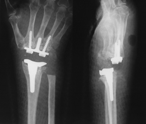 Figure 39-3 Dislocated total wrist arthroplasty. |
Damage to the long extensor tendons can occur, either at the time of surgery or as a result of chronic attrition. In the authors’ experience, this is again rare. Other authors, however, have reported this problem and, again, this may be as a result of poor implant design; more specifically, there should be no sharp edges to the implant, which could lead to soft tissue abrasion, particularly tendons. What is important, however, is the reconstruction of the dorsal wrist capsule at the end of surgery. In the authors’ practice, great care is taken to reconstruct the dorsal capsule of the wrist, always placing a layer of soft tissue between the implant and extensor tendons. At the same time, a strip of the extensor retinaculum is repaired to prevent bowstringing—a thorough synovectomy having been previously undertaken, with removal of the ulnar head, if appropriate.
During insertion of the implant, fractures of the radius and metacarpal bones, usually the third can occur. Generally, however, these are not displaced and do not require internal fixation. In the authors’ practice, however, this has resulted in a prolonged period of immobilization, often 6 weeks or more, with the potential for a poorer clinical outcome. Dawson (5), in 1989, reported four cases of radial fracture as a result of a fall 18 or more months after a total wrist arthroplasty. All were treated nonoperatively; however, three went on to nonunion or healed with deformity. In two cases, revision arthroplasty was undertaken with satisfactory results.
Dislocation can occur as with all major joint replacements. Effectively, however, this occurs more often in the immediate postoperative period and is easily recognized both clinically radiologically (Fig. 39-3). Treatment, in the authors’ experience, has involved a closed reduction under general anesthesia. This, combined with a longer period of immobilization of between 6 and 8 weeks, has usually resulted in no long-term problems. Indeed, a number of patients often go on to have good or excellent results. For the rare, persistent case, if necessary, either revision surgery (i.e., realignment of the implant, if appropriate) or the insertion of a thicker component with soft tissue reconstruction and tendon rebalancing, is usually successful. Temporary stabilization can also be obtained by the use of an external fixator (6). Finally, Cooney et al. (7) performed either an excision arthroplasty or revision to Silastic interposition arthroplasty in particularly difficult cases. The authors have also seen some success with the former.
Of the other major complications, mainly in the earlier postoperative period, infection can be catastrophic. Treatment for this is excision arthroplasty, one- or two-stage revision, or alternatively arthrodesis.
 Figure 39-4 Typical loosening of the biaxial carpal component. |
With regard to revision surgery in general, a number of published articles have dealt with this. In 1992, Ferlic et al. (8


Related posts:

Stay updated, free articles. Join our Telegram channel
Full access? Get Clinical Tree
