Total Hip Arthroplasty in the Young Active Patient With Arthritis

CHAPTER 35 Total Hip Arthroplasty in the Young Active Patient With Arthritis


Introduction


The treatment of disabling hip pain as a result of degenerative joint disease in the young patient has many surgical options, the least palatable of which has traditionally been total hip arthroplasty (THA). The innate desire of the orthopedic surgeon to preserve the native hip joint in this population has led to the successful development of a variety of procedures, such as hip arthroscopy, surgical dislocation, and hip resurfacing. At the same time, the evolving technology and long-term success of THA have engendered increased confidence in the durability of this procedure and led to expanded indications.


Although indicating a young patient for THA is always disheartening, the good news is that patients can usually expect excellent pain relief and a high level of performance in a reproducible and predictable manner. Long-term results are tempered by wear-induced failures, which are a function of the increased number of loading cycles as a result of increased activity levels (Table 35-1) and increased life expectancies (Table 35-2). This data shows that there is a higher risk of failure in a younger patient during the subsequent 15 years after THA.


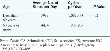
Table 35–2 15-Year Survival of Patients with Total Hip Arthroplasties


Age at Surgery Percentage of Patients Still Alive Percentage of Arthroplasties Still in Place
45 years 96% 77%
65 years 72% 92%

From Berry DJ, Harmsen WS, Cabanela ME, Morrey BF. Twenty-five-year survivorship of two thousand consecutive primary Charnley total hip replacements: factors affecting survivorship of acetabular and femoral components. J Bone Joint Surg Am. 2002;84-A:171–177.


Sir John Charnley articulated this problem well: “The challenge comes when patients between 45 and 50 years of age are to be considered for the operation, because then every advance in technical detail must be used if there is to be a reasonable chance of 20 or more years of trouble free activity.”


This chapter will discuss the inherent challenges involved when performing a THA in a young patient.


History and physical examination


The patient history determines the source of the patient’s symptoms, with the understanding that a wide spectrum of musculoskeletal and nonmusculoskeletal diseases can present with a chief complaint of hip pain. Detailed questioning is performed to clarify the character, location, duration, and severity of the symptoms; the resultant functional disability; and the realistic expectations of the patient. Concurrent spine pathology, ipsilateral knee disease, and a history of inflammatory arthritis should be investigated. A history of childhood or adolescent hip disease, previous hip surgery, hip trauma, risk factors for avascular necrosis, and current medical problems and medications should also be determined.


The physical examination includes the patient’s general condition and body habitus. In general, large, muscular males are more likely to have difficulty with exposure than are obese patients. The posture and gait pattern are observed. Hip abductor function is assessed with the use of the Trendelenburg test and side-lying abduction strength testing. Functional and real limb-length discrepancy should be distinguished and quantified. While assessing the range of motion, it is important to steady the pelvis to appreciate the endpoint and the forced motion of the pelvis through the hip. The impingement test is a very useful and sensitive test for any intra-articular pathology, and the McCarthy test can indicate labral pathology. It is also important is to evaluate the previous surgical interventions as well as the current neurovascular status of the limb.


Preoperative templating


Because these patients are usually healthy, the most important issue is usually preoperative planning. Careful consideration must be given to proximal femoral and acetabular deformities to ensure the availability of modular implants. Preoperative radiographs are important when planning the type and the size of the prosthesis and when making decisions about the position and orientation of the components with the aim of restoring the biomechanics of the hip in terms of the center of rotation, the offset, and the limb length. In addition, the distance from the top of the lesser trochanter to the center of the femoral head is also measured, and every attempt is made to match this length with that of the normal side during surgery. It is crucial to consider the effect of magnification during these measurements.


The next step is to template the implants. In cases of unilateral hip disease, the goal is to match the opposite leg in terms of limb length and offset. Thus, the templating should be performed on the normal side, provided that the socket would reproduce the normal hip center. A template is made of the acetabulum for the approximate component size and position and to find out its effect on the center of rotation. The acetabular template is placed just lateral to the lateral edge of the teardrop at an angle of 40 degrees to 45 degrees. Ideally, the cup should be completely covered by bone, and it should span the distance between the teardrop and the superolateral margin of the acetabulum. In most cases, this will restore the anatomic center of rotation. Templating of the femoral canal is performed to determine the type and size of the component to be selected, its position relative to the lesser trochanter, the level of the neck osteotomy, and the selection of the head length to reproduce both leg length and femoral offset. The component size that best accomplishes this is chosen. All of the preoperative sizes for which templates have been made should be noted and correlated with the operative findings.


Anesthesia and surgical technique


We prefer hypotensive regional anesthesia in the form of a single spinal shot. Regional anesthesia has shown advantages with respect to blood loss, deep vein thrombosis, postoperative pain management, and rehabilitation. We usually supplement the anesthesia with 1000 IU of intravenous heparin during femoral preparation to minimize the risk of deep vein thrombosis.


Patient positioning and its assessment before patient preparation and draping are crucial. The relative position of the knees and feet with symmetric flexion of the hips and knees can provide an idea about the starting leg-length relationship. Usually the superior side when the patient is in the lateral position would appear to be slightly shorter because of the adduction. Moreover, correct patient positioning is extremely important if external acetabular guides are used.


The choice of approach and the implant used are subjective and often geographic. Most surgeons in North America prefer cementless fixation, whereas cemented fixation is still popular in Europe. We use a posterolateral approach with the patient in a lateral position. We prefer a tapered wedge, a proximally hydroxyapatite (HA)-coated non-cemented femoral stem, and an HA-coated non-cemented acetabular cup. The choice of bearing surface is often discussed at length with the patient; in our practice, it is most often ceramic on highly cross-linked polyethylene or metal on metal. Ceramic-on-ceramic articulations have fallen out of favor in our practice as a result of their tendency to squeak.


We prepare the acetabulum first; some surgeons who use computer navigation prefer to prepare the femur first to have an idea of the femoral anteversion and to then adjust the combined anteversion on the acetabular side. In many cases, 1 mm to 2 mm of acetabular over-reaming is necessary to compensate for the hard bone associated with younger, healthier patients. This technique is helpful for avoiding acetabular insertional rim fractures and the incomplete seating of the component.


Uncemented femoral preparation is also critical for the young patient, because it can be complicated by fracture, subsidence, or leg-length discrepancy. We use a burr to open up the posterolateral neck and to remove the overhanging trochanteric bone. This not only avoids varus position and calcar fracture, but it also prevents tip contact with the posterior femoral cortex. “Touch, feel, and pitch” change are learned skills of broaching that come with experience. Once again, we recommend overbroaching to ensure the proper fit and seating of the tapered wedge component. The use of selective distal broaching may be necessary if the diaphyseal fit is too tight.


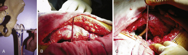
For the assessment of limb-length discrepancy, we prefer the method described by Ranawat and colleagues. This technique involves the use of the posterior approach. After the initial dissection and release of the short external rotators, the inferior capsule is incised at the 6-o’clock position to expose the posteroinferior lip of the acetabulum. A image-inch Steinmann pin is inserted into the posterior infracotyloid groove (Figure 35-1, A through C); this represents the bony groove inferior to the posteroinferior lip of the acetabulum. The pin is placed initially at an angle of approximately 60 degrees until it touches the ischium, and it is then made vertical and allowed to slide along the bone into the infracotyloid groove. The pin is kept vertical and viewed end-on from above, and then a mark is made on the greater trochanter before hip dislocation (see Figure 35-1, B). The hip is then dislocated. The center of the femoral head is marked with electrocautery, and the distance between the center and the lesser trochanter is noted and compared with the calculation made during the preoperative planning (Figure 35-2, A). The neck resection is now completed in accordance with preoperative templating.


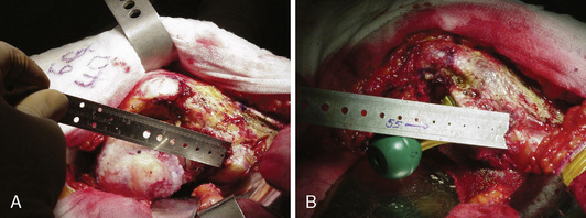
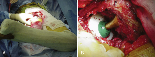
After bone preparation and the placement of the trial implants, the offset and the distance from the center of the head to the lesser trochanter are assessed (see Figure 35-2, B). After reduction, the component position is assessed with the use of the combined anteversion test (Figure 35-3, A and B). This test measures the angle of internal rotation required for the femoral head to be coplanar with the face of the acetabulum with 10 degrees of flexion and 10 degrees of adduction. Normally, the angle is between 35 degrees and 45 degrees of internal rotation for women and between 25 degrees and 35 degrees for men.


< div class='tao-gold-member'>

Stay updated, free articles. Join our Telegram channel

Jul 24, 2016 | Posted by in MUSCULOSKELETAL MEDICINE | Comments Off on Total Hip Arthroplasty in the Young Active Patient With Arthritis

Full access? Get Clinical Tree

Get Clinical Tree app for offline access