Supine Approach to Hip Arthroscopy

CHAPTER 9 Supine Approach to Hip Arthroscopy


Introduction


Two standard positions have been used for hip arthroscopy: lateral and supine. The supine approach is often favored for its familiar positioning with easy reference to the anatomic landmarks to which most surgeons are accustomed. The maneuverability and functionality of the arthroscopic equipment are also most similar to that used for other arthroscopic procedures with patients in the supine position. The anatomic orientation required for this position appears to be easier to attain from the surgeon’s standpoint than that required for the lateral position. With the patient in the supine position, the operative extremity can be mobilized (i.e., dynamic testing in hip flexion–extension and rotation) during the procedure, and it also allows for capsular relaxation and the dynamic visualization of the joint and the peripheral compartment. With the supine approach, traction may be applied with the use of a standard hip fracture table or other device, whereas the lateral position requires a more specialized setup. In addition to the position being more familiar to classic total joint surgeons, the main benefit of the lateral position is that it uses gravity more effectively to allow the soft tissue to fall away from the greater trochanter as the primary landmark for instrumentation. This is most significant for the larger, moderately obese patient. However, some feel that the lateral approach is more time consuming and that using fluoroscopic imaging with a C-arm may be insufficient to get around an obese patient under the operating room table. In the end, both of these positions are effective, with surgeon preference and patient comfort being the determining factors as far as which one is chosen. Our preferred method is the supine position for the reasons outlined previously. This chapter describes our general surgical technique for patients in the supine position.


General considerations


The hip joint is not a true space. To perform hip arthroscopy of the central compartment, which is the area within the acetabulum, traction must be applied. Usually 8 mm to 12 mm of joint space opening is required to perform hip arthroscopy safely. If there is too little traction, then iatrogenic injury to the articular cartilage may occur while trying to travel through the joint. If there is too much traction, then there is increased potential risk of injury to the neural structures around the joint and the perineum as well as risk to the knee, foot, and ankle. Traction may be applied with the use of a standard fracture table or with the use of other commercially available attachments that have been devised for regular operating room tables, although some authors have reported using weight over the end of a standard operating room table as a means of applying traction. Usually 25 lb to 50 lb of force is needed to apply adequate traction for hip arthroscopy.


To facilitate traction, we prefer the use of general anesthesia with paralysis. This allows for adequate traction without undue force to reduce the risk of injury to the nerves and the perineum. Arthroscopy of the hip may be performed with spinal anesthesia if there is adequate muscle relaxation. As an adjunct to general anesthesia, we have also used a lumbar plexus block to reduce the amount of narcotic intraoperatively, thereby reducing the amount of nausea postoperatively and the amount of pain during the immediate postoperative period (usually 12 to 18 hours). We prefer hypotensive anesthesia to reduce bleeding within the joint, thereby allowing for better visualization.


Hip arthroscopy is facilitated by the use of specialized instrumentation. Although extra-long arthroscopes and cannulas may be used, there is a risk of damage to these instruments. A standard-length hip arthroscope with a cannula that has a modified bridge is most commonly used, and this has been used almost exclusively by the senior author for the past 15 years. Only in the particularly obese patient is the extra-long arthroscopic lens needed. Cannulated hip arthroscopy systems that make use of Nitinol wires and blunt trochars have significantly reduced complications and aided in the development of arthroscopy portals. Many specialized extra-long instruments have been developed to perform therapeutic hip arthroscopy, including longer arthroscopic motorized shavers and burrs. Some shavers are curved to help facilitate access to areas around the femoral head. Other instruments include modified hand instrumentation, such as arthroscopic knives, rongeurs, meniscal biters (i.e., forward and backward, straight and upbiting), curettes, and even microfracture awls. Radiofrequency devices that are narrow and that bend at the tip are quite useful for tissue ablation and tissue shrinkage. In addition, instrumentation for labral repair (e.g., suture anchors that fit through narrow cannulas) is also available.


The senior surgeon generally uses 5.0-mm cannulas for the three central compartment portals, although, in tight hips with limited distraction, a 4.5-mm cannula may be used. However, when using gravity inflow for fluid, the fluid dynamics with the 4.5-mm cannula are suboptimal, and the cannula is changed, when possible. Alternatively, the introduction of irrigation fluid with a pump may overcome the limited dynamics of the smaller cannula. Frequently, 5.5-mm or 5.6-mm cannulas are necessary for the introduction of many instruments, including labral repair instruments, curettes, microfracture awls, and some labral biters. Alternatively, a slotted cannula (i.e., half-pipe) can allow for the maintenance of the portal while allowing larger instruments to be introduced (e.g., curved shaver, microfracture awl).


Saline or lactated Ringer’s solution with epinephrine is usually used during hip arthroscopy as irrigation fluid. Irrigation fluid is introduced with the use of a gravity inflow system to reduce the risk of clinically significant fluid extravasation, because there have been reported cases of intra-abdominal fluid extravasation that have resulted in cardiac arrest. Visualization is enhanced with the use of hypotensive anesthesia and 5.0-mm or larger cannulas. However, on occasion, a pump is needed to perform arthroscopy (in the senior author’s experience, this happens with less than 1% of cases).


To ensure the adequate visualization of the entire joint, the senior author prefers using three portals for central compartment arthroscopy on all patients and using both the 30-degree and 70-degree lenses in all three central compartment portals. To help with the maneuverability of both the arthroscope and the instrumentation, capsulotomies made with an arthroscopic knife or a radiofrequency probe do allow for significant freedom for access and visualization.


Surgical technique


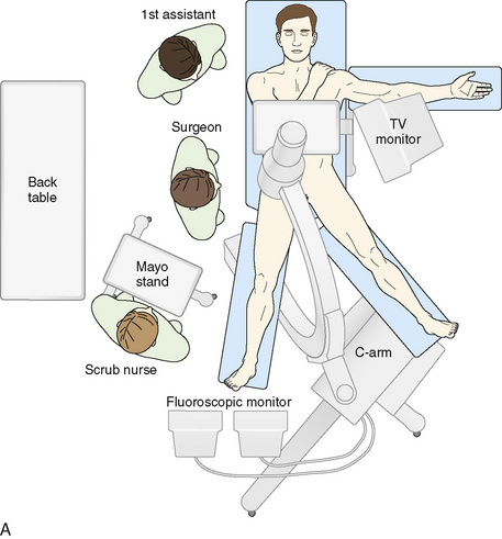
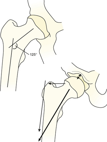
For the supine position technique, the patient is given a general anesthetic that provides muscle relaxation. Paralysis is recommended, because this allows less force to be used to distract the hip, with the purpose of reducing the risk of pudendal nerve injury from pressure between the pelvis and the perineal post of the traction table. The patient is placed supine on the fracture table with both feet secured to traction boots or mobile spars, depending on the specific table used. A well-padded perineal post is placed, and the patient is brought into position so that the post is firmly situated against the perineum and lateralized toward the operative hip, with care taken to protect the genitalia. The operative leg is positioned in 10 degrees of abduction, neutral flexion–extension, and neutral rotation. The nonoperative extremity is positioned in 45 degrees to 60 degrees of abduction, neutral flexion–extension, and neutral rotation to serve as countertraction for lateralization. Gentle traction is applied to the abducted nonoperative leg, which lateralizes the patient’s pelvis and results in the perineal post resting on the inner upper thigh of the operative extremity (Figure 9-1). This allows for the pressure of the perineal post to be diverted away from the perineum itself to minimize the risk of neuropraxia to the pudendal nerve as traction is applied to the operative leg. In addition—and quite important—this also helps to generate the appropriate vector of force for a uniform distraction both laterally and distally (Figure 9-2). Traction to the operative leg straight distally would be met with unnecessary resistance to overcome the inferior transverse acetabular ligament. Lateralization of the hip with the use of the post helps to pull the femoral head laterally and distally from the socket without having to overcome the ligament as a barrier to distal translation.
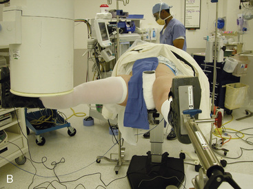


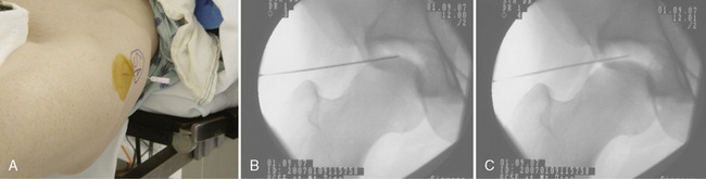
After the patient has been positioned appropriately and slight traction has been applied to the nonoperative extremity, traction on the operative limb can be applied. This should be incremental, and it can be monitored with serial images from an image intensifier (i.e., C-arm). A tensiometer can also be used; however, the senior author has not found this to be useful. Jim Glick has shown that the risk of nerve injury as assessed by somatosensory evoked potentials is associated with the duration of traction rather than the amount of traction (Glick, personal communication). Thus, the absolute amount of traction is apparently not important. Some surgeons use the tensiometer to evaluate any changes in tension. We use the fluoroscopic image intensifier routinely; we bring the base of the machine in from the foot of the table in between the patient’s abducted legs, and we center the column over the operative hip (see Figure 9-1). Incremental traction is then applied until approximately 8 mm to 10 mm of femoroacetabular joint distraction is generated. The area of the proposed anterolateral portal is identified and marked; the area around this proposed portal site is prepared with Betadine solution; and a spinal needle is then used to enter the hip joint to verify the correct path of the anterolateral portal, to ease joint access, and to perform an air arthrogram to release negative intra-articular pressure. In a cadaver study, Dienst and colleagues demonstrated that positioning the hip in 20 degrees of flexion and performing an air arthrogram (i.e., disrupting the vacuum seal and distending the hip joint) reduced the amount of traction required to distract the joint for safe entry, thereby further reducing the risk of neuropraxia. The needle is placed from the intended anterolateral portal with the guidance of the fluoroscopic image intensifier (Figure 9-3). The needle needs to enter the central compartment to effectively reduce the intra-articular pressure. We have shown that placing the needle onto the femoral neck does not release the negative intra-articular pressure within the joint. Care is taken to keep the spinal needle close to the femoral head (to reduce the risk of injury to the labrum) and to keep the longer part of the tip away from the femoral head. When the suction seal is broken, the joint will open more widely. The needle position relative to the femoral head is evaluated before and after the seal is broken. If the needle moves proximally when the negative pressure is released, the labrum may have been violated by the needle. If the needle moves with the femoral head, the labrum has likely not been injured.


< div class='tao-gold-member'>

Stay updated, free articles. Join our Telegram channel

Jul 24, 2016 | Posted by in MUSCULOSKELETAL MEDICINE | Comments Off on Supine Approach to Hip Arthroscopy

Full access? Get Clinical Tree

Get Clinical Tree app for offline access