Arthroscopic Management of Pediatric Hip Disease

CHAPTER 25 Arthroscopic Management of Pediatric Hip Disease


Introduction


Injuries of the hip and pelvis in the pediatric population are increasing and receiving more attention. The majority of injuries are soft-tissue or apophyseal injuries that heal with nonoperative treatment. However, with the advent of hip arthroscopy and the development of more advanced imaging of the hip that involves magnetic resonance arthrography, internal derangements of the hip (e.g., labral tears, loose bodies, chondral injuries) are being diagnosed and treated with increased frequency. Hip arthroscopy has obvious advantages as compared with arthrotomy and surgical dislocation for the pediatric population. It is significantly less invasive than arthrotomy, and it allows for a quicker recovery and a sooner return to activities. Most of the experience in hip arthroscopy has been with hip disorders in adults; the role of hip arthroscopy for children and adolescents has been less well characterized.


Historically, arthroscopy of the hip in the pediatric population was used as a diagnostic and therapeutic tool for slipped capital femoral epiphysis, septic arthritis, and various arthropathies. The most common indication has been in cases of Legg-Calvé-Perthes disease, both for the diagnosis of severity and treatment, including the removal of loose bodies. In a review of 24 hip arthroscopies performed in 21 patients between the ages of 11 and 21 years old, Schindler and colleagues concluded that hip arthroscopy was effective for synovial biopsy and loose-body removal; however, as a diagnostic procedure, the arthroscopy failed to correlate with the presumptive cause of symptoms in 11 hips (46%). In many of the general series reported in the current literature, the role of hip arthroscopy for pediatric and adolescent athletes is rarely addressed. As our understanding of the mechanisms and anatomic abnormalities of hip pathology increases, the role of hip arthroscopy for the pediatric population will continue to expand.


Brief history and physical examination


Pediatric and adolescent patients are different from adults in that they frequently do not complain about hip pain. It is common for hip pathology to be discovered only after the examination of a young patient who complains of knee pain: up to a quarter of patients with stable slipped capital femoral epiphysis (SCFE) report knee pain. Another frequent presentation is in the limping child who has not experienced any known injury. As with any orthopedic patient, a detailed history is mandatory. On average, less than half of patients can recall a traumatic event that led to the injury. Running, jumping, and kicking activities are frequently involved in labral pathology, and patients often report an acute twisting injury. A gradual history of pain is frequently more common. Subjective complaints of catching, locking, and giving way point to mechanical symptoms that can benefit from surgical intervention. These symptoms should be carefully distinguished from popping or snapping sensations, which may indicate extra-articular pathology (e.g., psoas tendon bursitis, coxa saltans externa or interna).


The physical examination of the hip begins with the examination of the range of motion. A decreased range of motion as compared with the contralateral unaffected hip is a useful sign of hip pathology. Loss of flexion and internal rotation are usually the earliest signs in these patients. Hip contractures should be examined, and leg lengths and pelvic obliquity should be noted. An impingement test with flexion, adduction, and internal rotation appears to be predictive of intra-articular pathology. An examination of the low back, including straight-leg raising, should be conducted to rule out any contribution of the spine.


Surgical technique


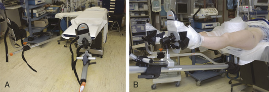
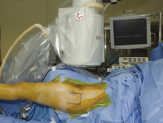
Hip arthroscopy can be performed with the patient in the supine or lateral decubitus position. Both positions allow for adequate visualization and positioning, and the one that is used is based on surgeon preference. We perform hip arthroscopy with the patient in the supine position, and it is generally performed as an outpatient procedure for the pediatric population. General anesthesia is used with muscle relaxation, although supplementation with regional anesthesia may be used, depending on the anesthesia available and patient preference. A standard fracture table or a specialized hip-scope table is used with a traction device and a large padded perineal post (Figure 25-1). The affected leg is prepped and draped along with a C-arm (Figure 25-2). The nonoperated side is placed in approximately 30 degrees of abduction, and firm traction is applied to that side so that the patient remains firmly against the perineal post. The operated leg is placed in neutral abduction to maintain a constant relationship between the topographic landmarks and the joint. A slight 10 degrees of flexion is used to relax the capsule; however, excessive flexion and traction can place tension on the sciatic nerve. Maximal internal rotation is used to make the femoral neck parallel with the floor. Traction is applied to achieve joint distraction between 5 mm and 10 mm, and this is confirmed by fluoroscopy; this will typically demonstrate the “vacuum sign” when viewed with the use of fluoroscopy. In general, approximately 50 lb to 75 lb of force is needed to distract a joint. The goals are to use the minimal force necessary to achieve distraction and to keep traction times as brief as possible (in general, less than 2 hours is considered optimal).


Portals


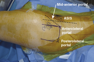
Two to three operating portals are typically used. The anterolateral and posterolateral portals are placed at the superior margin of the greater trochanter at the anterior and posterior borders (Figure 25-3). The mid-anterior portal is made by first drawing a sagittal line from the anterosuperior iliac spine and another line at the anterolateral portal; a sagittal line is then drawn equidistant to these two lines. The entry point for the mid-anterior portal is approximately 45 degrees from the anterolateral portal on the middle sagittal line. With careful attention paid to these landmarks, the portals can be safely made away from neurovascular structures. The lateral femoral cutaneous nerve typically lies just medial to the anterosuperior iliac spine; the femoral nerve and artery are even more medial.


< div class='tao-gold-member'>

Stay updated, free articles. Join our Telegram channel

Jul 24, 2016 | Posted by in MUSCULOSKELETAL MEDICINE | Comments Off on Arthroscopic Management of Pediatric Hip Disease

Full access? Get Clinical Tree

Get Clinical Tree app for offline access