CHAPTER 59 Total Elbow Arthroplasty for Distal Humerus Nonunion and Dysfunctional Instability
INTRODUCTION
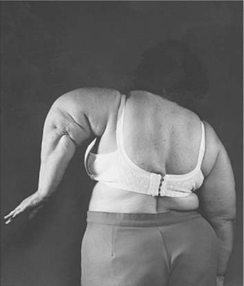
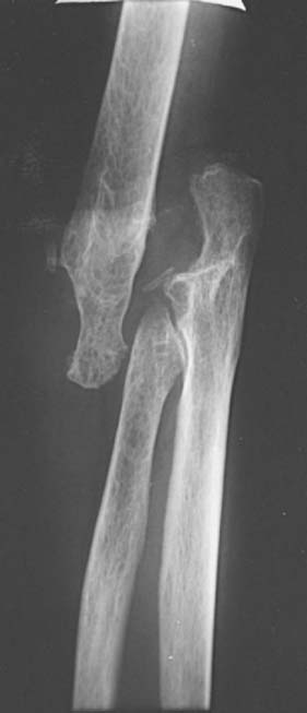
Dysfunctional elbow instability may be defined as a condition in which the elbow joint has lost its fulcrum properties and is no longer able to provide enough support for the hand to be functionally controlled in space (Fig. 59-1).10 In the most extreme circumstances, destruction of the elbow leads to a flail extremity. In less severe cases, stability may be maintained with the arm adducted against the body, but not in other positions (Fig. 59-2). This condition may result from situations such as distal humerus nonunion, severe rheumatoid destruction of the humerus, extensive post-traumatic bone loss, and bone resection for the treatment of deep infection or tumors. Linked elbow arthroplasty provides a dramatic improvement in the function and quality of life of these patients, but the improvements need to be balanced against the risk of complications and mechanical failure.

FIGURE 59-2 Patients with dysfunctional instability are unable to control the position of the arm in the space.
ELBOW ARTHROPLASTY FOR DISTAL HUMERUS NONUNION
As noted in Chapter 23, nonunion is one of the most challenging complications of distal humerus fractures. Internal fixation is the treatment of choice whenever possible. Modern series have reported a high union rate when internal fixation is used, but the reoperation rate has remained high and function is not always re-established.6,11 Total elbow arthroplasty is an excellent surgical alternative for the salvage of distal humerus nonunions when fixation is considered to be impractical or expected to be associated with a high rate of failure.
RATIONALE, INDICATIONS AND CONTRAINDICATIONS
Joint replacement is a well-accepted treatment modality for fractures in other locations, such as the femoral neck or the proximal humerus. The good track record of some elbow implants for patients with rheumatoid arthritis and other conditions prompted the use of elbow replacement for distal humerus nonunion.5 Elbow arthroplasty is indicated only in a selective group of elderly patients who present with either preexistent symptomatic pathology (for example, a rheumatoid elbow) or low nonunions with substantial osteopenia and severe damage to the articular surface. It is contraindicated in the presence of infection, as well as in nonunions amenable to stable internal fixation and in patients with anticipated high physical demands. An associated nonunion of an olecranon osteotomy complicates the surgical technique but should not be considered a contraindication for the procedure.
SURGICAL TECHNIQUE
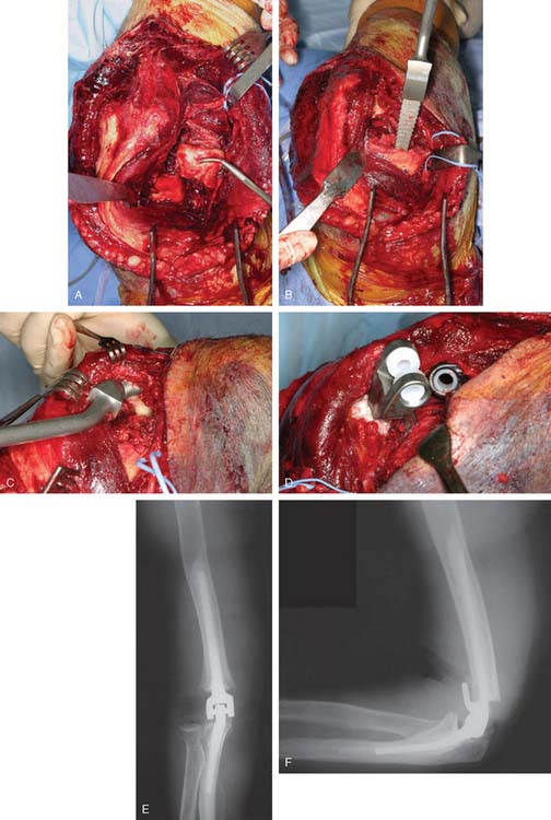
The elbow is exposed through a posterior midline skin incision and the ulnar nerve is identified and treated according to the location of the nerve and preoperative symptoms as mentioned in Chapter 23 on internal fixation for distal humerus nonunions. The extensor mechanism is left undisturbed and the procedure is performed working on both sides of the triceps unless an associated olecranon nonunion, or triceps detachment provides the opportunity for exposure (Fig. 59-3) (see also Chapter 7, Surgical Exposures). Retained hardware is removed, and the nonunited distal humerus is resected subperiosteally and saved for bone grafting behind the flange of the humeral com-ponent. Tissue samples are sent for pathology and microbiology.
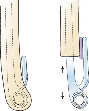
The working space created by resection of the distal humerus is ample enough to instrument the canals andimplant the components. The surgical technique for implantation of a linked elbow arthroplasty is detailed in Chapter 52. A capsular release should be associated routinely. Use of a humeral component with an intermediate length stem provides secure fixation. In the presence of severe humeral bone loss, an implant with a longer flange may be cemented proud to make up for part of the lost humeral length (Fig. 59-4). On the contrary, a regular humeral component may be cemented in a deeper position to elevate the joint line and correct flexion contractures. Exposure of the ulnar canal may be improved by partial detachment of the triceps from the olecranon on the medial side. The implants may be fully seated before interlocking, because the articular windows at the medial and lateral side of the elbow after removal of the united distal humerus bone allow interlocking. At the end of the procedure, the common extensor and common flexor muscle groups are sutured to the lateral and medial triceps fascia to seal the joint space and maintain the strength of the forearm muscles.
Stay updated, free articles. Join our Telegram channel

Full access? Get Clinical Tree


