Total Ankle Arthroplasty in the Varus Ankle
James K. DeOrio
INTRODUCTION
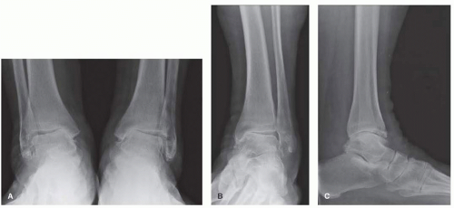
The most common deformity presentation with arthritis in the ankle is the varus ankle. Given the large number of ankle sprains sustained worldwide on a daily basis, this is not surprising. Once the lateral ligaments are torn, the ankle moves in a less congruous way. This change in mechanics shears away the remaining cartilage until eburnated bone appears along with pain. It is for this reason that we strongly advocate early repair of an unstable ankle.
Trauma is another frequent cause of a varus deformity because an inverted position of the ankle is so much more common in an injury. This not only tears the lateral ligamentous tissues but also results in trauma to the medial side of the ankle. Further deterioration of the medial cartilage over a period of time also results in a varus deformity.
INDICATIONS
Indications for treatment of the arthritic varus ankle are pain and/or a significant alignment problem which leads to increased deformity and pain unrelieved with conservative measures.
CONTRAINDICATIONS
Contraindications for treatment of these deformities have been previously cited in this book and include poor skin integrity, peripheral vascular disease, generally patients younger than 35 years, type 1 diabetes, neuromuscular paralysis, and avascular necrosis of the talus.
PREOPERATIVE PREPARATION, PLANNING, AND CONCEPTS
The preoperative planning should include standing anteroposterior (AP), lateral, oblique, and Saltzman axial calcaneal x-rays of both ankles. In addition, standing lower leg x-rays should be evaluated when there is a deformity above the ankle, vascularity tests should be performed when there are no or faintly palpable pulses (consider a vascular consultation as well), a computed tomography scan should be obtained when looking for adjacent joint arthritis, and a magnetic resonance imaging scan should be reviewed when assessing the talus or distal tibia for vascularity.
STEPWISE TECHNIQUE
The first thing to consider when a patient comes in with a varus ankle and pain from arthritis is whether or not there is enough cartilage to salvage the ankle and delay the need for an ankle replacement or arthrodesis. If there is moderate instability and narrowing of the medial tibial-talar plafond joint space, but significant erosion into the subchondral bone has not occurred medially, then salvage procedures can be attempted. Mann et al.1 propose an opening wedge tibial osteotomy from the medial side. A medial longitudinal incision is placed above the articular cartilage. A guide pin is aimed toward the ankle mimicking the angle of the osteotomy. Next, three transverse K-wires are placed at the bottom of the proposed osteotomy site. This allows an opening wedge osteotomy to be carried out. The opening is backfilled with cancellous bone, and a plate is applied to the medial side of the tibia. The study was a retrospective review of 19 patients, 18 of whom received a lateral ligament reconstruction in the form of a modified Chrisman-Snook: 4 patients failed (2 went on to arthrodeses and 2 required ankle replacements) and the other 14 were satisfied or very satisfied. Although significant mention was made of cleaning out the lateral gutter, no patient received a fibular osteotomy to make room for the talus laterally. Perhaps this was a reason why the tibiotalar articular angle was not significantly improved following the procedure, resulting in 10° varus alignment despite the good clinical results.
If the patient has too much ankle erosion to make an osteotomy realistic in providing the patient pain relief, then the next decision to be made is arthrodesis versus replacement. The decision to proceed with one or the other is dependent on a lot of factors, including the age of the patient and the source of the varus angulation. For example, I would tend to lean toward ankle arthrodesis in a young active patient less than 35 years old with a posttraumatic injury and an ankle replacement in a 45-year-old patient with inflammatory arthropathy affecting
the ankle and subtalar and talonavicular (TN) joints. Since this manual is technically oriented toward ankle replacement, let us pursue in detail the various adjunctive procedures done in conjunction with total ankle replacement in the varus ankle, the most common presentation of the arthritic ankle.2
the ankle and subtalar and talonavicular (TN) joints. Since this manual is technically oriented toward ankle replacement, let us pursue in detail the various adjunctive procedures done in conjunction with total ankle replacement in the varus ankle, the most common presentation of the arthritic ankle.2
The emphasis in this chapter will be to produce a wellaligned ankle with the replacement in a balanced position. The reason for this is to (1) make it easier for the patient to walk and (2) remove abnormal stresses that occur across a malaligned ankle causing the polyethylene to wear out or the bone-metal interface to fail.3 That is what makes total ankle arthroplasty (TAA) such a difficult operation: getting the correct balance.
SURGICAL APPROACH
There is no special approach for the varus ankle. It is the standard anterior incision between the extensor hallucis longus tendon and the anterior tibial tendon. The tissue is swept off the anterior tibia allowing good exposure of the malleoli. A deep retractor is now placed to avoid excessive or repetitive tension on the skin. Furthermore, to avoid excessive skin tension from retraction, I often extend the exposure to the TN joint.
DELTOID PEEL AND LATERAL LIGAMENT RECONSTRUCTION
For mild to moderate tightness medially, determined preoperatively with fluoroscopy as well as intraoperative judgment, a deltoid peel is (along with a gastroc release [described subsequently]) usually all that is needed on the medial side of the ankle to allow it to be balanced (Figs. 9.1 and 9.2).4 This was the most common adjunctive procedure in a review of 67 ankle replacements—21 times—and is routinely done for the varus ankle. On first opening of the ankle, traction is applied to the foot and calcaneus. If ankle opening is more than 1 cm, then less bone can be taken from the tibia to avoid too much laxity. Under no circumstances, however, should a compensating cut be made. In other words, all cuts in the coronal plane are at 90° to the tibia. Furthermore, it must be remembered that the polyethylene component (poly) from the different manufacturers is limited in thickness, and it is possible to resect too much bone and not have a thick enough poly to restore the normal tension in the ankle, albeit this is more common in a revision total ankle.5 The periosteum and deltoid fibers are sharply cut off the tibia all the way back to the posterior tibial tendon as well as inferiorly to the tip of the medial malleolus. No fibers are cut off the talus for fear of endangering the blood supply of the talus. One must be careful not to cut the posterior tibial tendon. No fibers are left attached to the medial malleolus. The deltoid fibers, superficial and deep, are not cut transversely once they are peeled off the medial malleolus, but rather they are elevated off the medial malleolus all the way around to the intra-articular portion of the medial malleolus.
Others think that in routine TAAs, release of the superficial and deep posterior deltoid ligament may improve the range of motion, whereas release of the tibiocalcaneal portion of the deltoid may help correct varus talar tilt.6 However, if you are going to get true freedom medially, in my opinion all the fibers need to be released. Thus, this is an “all or none” phenomenon. The lateral gutter is cleaned out, and when all the cuts have been made, the ankle is trialed with the poly spacers. If no more than 1 mm difference in opening is obtained on stressing the ankle in varus and valgus with the components in position, this is acceptable and no lateral ligament procedure needs to be performed. If there is more than 1 mm difference and you have reached the maximum height of the poly, then a lateral ligament reconstruction is performed. I prefer a Brostrom ligament reconstruction.7 Others do a modified Chrisman-Snook reconstruction.8 I now remove the trial poly and leave the poly out when doing this lateral ligament surgery to provide maximum tightness
laterally. This allows me to collapse the ankle laterally in valgus while doing the repair. Then, when the real poly is reinserted, the repaired fibers are stretched to the maximum. The incision for the ligament reconstruction is a midline AP fibular incision about 3 cm long. The capsule is cut and peeled off the anterior 3 mm of the fibula and inferior fibula. The peroneal tendons are inspected through this incision and repaired, debrided or transferred depending on their condition (see below); the lateral gutter is cleaned out with removal of talar osteophytes and loose bodies.9 Then two suture anchors are placed in the fibula and the anterior and inferior capsules are pulled securely to the fibula. Now the ankle is retrialed with a poly thickness normally about 2 to 4 mm less than the thickest poly trialed earlier. If this is acceptable, the real poly is now inserted. And in ankles such as the Salto-Talaris, the poly may be inserted in the tibial component and the final tibial component inserted. Closure is as per usual with a well-padded cast applied at surgery.
laterally. This allows me to collapse the ankle laterally in valgus while doing the repair. Then, when the real poly is reinserted, the repaired fibers are stretched to the maximum. The incision for the ligament reconstruction is a midline AP fibular incision about 3 cm long. The capsule is cut and peeled off the anterior 3 mm of the fibula and inferior fibula. The peroneal tendons are inspected through this incision and repaired, debrided or transferred depending on their condition (see below); the lateral gutter is cleaned out with removal of talar osteophytes and loose bodies.9 Then two suture anchors are placed in the fibula and the anterior and inferior capsules are pulled securely to the fibula. Now the ankle is retrialed with a poly thickness normally about 2 to 4 mm less than the thickest poly trialed earlier. If this is acceptable, the real poly is now inserted. And in ankles such as the Salto-Talaris, the poly may be inserted in the tibial component and the final tibial component inserted. Closure is as per usual with a well-padded cast applied at surgery.
Stay updated, free articles. Join our Telegram channel

Full access? Get Clinical Tree


