Conversion to Fusion
Christopher E. Gross
Selene G. Parekh
INTRODUCTION
Ankle arthrodesis has long been considered the surgical gold standard for ankle arthritis that has failed to respond to conservative management. A tibiotalar arthrodesis has been favored partly because of its consistent and reproducible results in returning functionality and decreasing pain and also partly because of high rates of complications associated with first-generation total ankle replacement (TAR) systems.1 An ankle fusion has good functional outcomes with low complication rates. However, the arthrodesis is not without its long-term consequences. Other hindfoot and tarsal joints must compensate for the loss of tibiotalar motion and may subsequently lead to adjacent joint arthritis.2,3
The third-generation ankle arthroplasty systems have taken significant strides in improving the biomechanical and material designs. Such improvements include minimizing bone resection, retaining ligamentous support, and anatomic balancing. TAR indications are expanding as the technology matures.
COMPLICATIONS AND RISK FACTORS
Invariably, as ankle arthroplasty becomes more widely used and the time since implantation increases, surgeons face serious dilemmas over managing its complications and failures. Complications from a TAR can range from the innocuous to devastating. Glazebrook et al.4 conducted a systematic review of 2,386 TARs. They identified three high-grade, three intermediate-grade, and three low-grade complications. Short- and intermediate-term failure ranged from 1.3% to 32.3%, with an average of 12.4% at 64 months. Deep infections, aseptic loosening, and implant failure were considered high grade since they resulted in failure more than 50% of the time. Implant subsidence, technical error, and postoperative periprosthetic fracture were considered intermediate grade as they caused failure less than 50% of the time. Intraoperative periprosthetic fractures and wound healing problems were considered low grade as they resulted in no failures. Numerous other complications include malalignment, component sizing mismatch, polyethylene dislocation, ligamentous instability, malleolar impingement, idiopathic pain, and an inadequate soft tissue envelope.
The host also plays an important role in the complication rate of TAR. Patient factors such as diabetes, renal failure, rheumatoid arthritis (RA), obesity, tumors, immune-modulating medications, and hemophilia can predispose patients in developing a deep infection.5,6,7,8 and 9 It is worthwhile to note that most of the literature regarding host factors come from total hip and knee replacement studies. A patient’s lifestyle and habits, such as smoking, may contribute to poor outcomes. Obesity has not been seen as an independent factor in TAR failure. Barg et al.10 looked at the results of 123 patients with a minimum body mass index (BMI) of 30 kg/m2, who underwent a TAR. There were no statistical differences in functional outcomes, complications, or failures at 68 months between patients who are considered obese and those who are not.
Two other factors that need to be considered when suspecting an implant failure are implant design and surgeon experience. There have been dozens of total ankle implants with various permutations of designs that rely on cement or those that require biologic ingrowth. The implant-bone interface may be a source for potential aseptic loosening. The type of coating material (trabecular metal, grit-blasted, hydroxyapatite)11 on the implant may impact how well the device osteointegrates. The amount of bone resected for implantation and the surface area by which the implant interfaces with bone may also affect the outcomes. Currently, there is no literature to suggest that an implant’s coating affects the outcomes. A surgeon’s experience plays an important role in a TAR complication rate.12,13 Henricson et al.13 looked at 531 prostheses implanted between 1993 and 2005. The overall survival rate was significantly higher after the first 30 cases had been performed (0.78 vs. 0.86). Clement et al.14 reviewed a single surgeon’s experience with third-generation implants and discovered that certain complications such as intraoperative fractures and component malpositioning decreased with time, while others (wound complications) did not. Other surgical technical factors that can influence outcome include anatomic and soft tissue balancing; coronal, axial, and sagittal plane alignment; respecting the soft tissue; choosing the correct implant size; and addressing other presurgical varus or valgus deformities.
PREOPERATIVE CONSIDERATIONS
PHYSICAL EXAMINATION
Pain is the most common presenting symptom for a patient dealing with a failed TAR.11 A thorough history and physical examination in patients with pain following a total ankle arthroplasty are essential in guiding a clinician’s diagnostic path.
Pain is often described as start-up or associated with increased physical activity. The quality and location of the patient’s pain must be identified. First, one must identify whether the pain is originating from the subtalar joint. On examination, one must evaluate the motion at the subtalar joint and compare it to the contralateral extremity. Using provocative maneuvers, one might find restricted and painful range of motion if the subtalar joint is arthritic. A fluoroscopic-guided injection of corticosteroid and anesthetic into the subtalar joint may prove to be both diagnostic and therapeutic. Other important physical examination concerns are alignment and gait. The static and dynamic assessment of varus and valgus malalignment must be evaluated by observing the patient from the front and back.
A thorough vascular examination should include the palpation of the dorsal pedis and posterior tibial pulses with observation for any signs of venostasis or vasculopathy. Poor distal blood flow may serve as a predictor for future wound complications should the patient warrant a surgery. An observation of prior surgical incisions and wounds should also be noted. This can become important when considering approaches for surgical options.
One must also be vigilant for signs and symptoms of a deep space infection. One should ascertain whether the patient had a prior wound healing issue, prior draining sinuses, recent infection, acute increase in ankle pain, or other signs of systemic illness. A standardized approach for ruling out infection includes obtaining a complete blood count and erythrocyte sedimentation rate. If one of the laboratory values is elevated, then a joint aspiration under sterile conditions should be performed. The synovial fluid should be sent for cell count, cultures, and crystal analysis.
RADIOGRAPHY
All patients should obtain weight-bearing views of the foot and ankle along with a hindfoot alignment or long axial view.
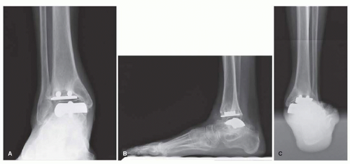 Figure 15.1. (A) AP, (B) lateral, and (C) long axial radiographs of failed total ankle arthroplasty. Notice gross component loosening, peri-implant radiolucency and cysts, and instability. |
These views will help to visualize adjacent joint degenerative changes or malalignment, which needs to be taken into consideration for preoperative planning. These radiographs may show periprosthetic osteolysis, implant failure, component subsidence, and lucent lines that are related to loosening (Fig. 15.1). Gross component loosening is readily apparent, but in the cases of subtle instability or loosening, one should look carefully at previous radiographs. Particular attention should be paid to the remaining talar bone stock as this may determine future hardware or bone graft needed. In patients with normal radiographs, one must consider gutter impingement and soft tissue strain.
Oftentimes, radiographs can underestimate the amount of bone stock available for reconstruction. Computed tomography scans are routinely used to evaluate the subtalar joint, bone stock, alignment, and presence of cyst and its size. All of this information becomes important in planning for the surgical approaches, techniques, and procedures required during the reconstruction.
PREOPERATIVE PLANNING
If the patient is deemed appropriate for revision surgery, there are three surgical options: revision TAR, arthrodesis of the tibiotalar or tibiotalocalcaneal (TTC) joints, or amputation. Ideally, a surgeon tries to revise to another TAR in the hopes of preserving ankle range of motion and preventing adjacent joint arthritis.11 When a surgeon is faced with severe prosthesis failure, subsidence, loosening, or a septic joint, a total ankle revision is not advisable. One must consider a patient-customized device, an arthrodesis with or without structural auto- or allograft, or an amputation. The standard of treating severe osteolysis, gross implant subsidence, soft tissue compromise, infection, or loss of bone stock is primary or staged arthrodesis.15,16 and 17
A surgeon has several considerations when an arthrodesis is finally chosen: mechanism of failure, bone stock available, implant to be removed, skin quality, and approach. Technical considerations include how to remove the implant, should the subtalar joint be preserved, and should the fusion be in situ or account for a deformity. In order for any successful fusion, the surgeon must create broad, congruent cancellous surfaces using curettes or acetabular reamers. Osteotomes are useful in feathering and penetrating into subchondral bone. Fundamentally, the two main issues that a surgeon faces when confronted with converting an arthroplasty to fusion are how to fill in the large bony void and how to best fixate the fusion mass.
SUBTALAR FUSION
One must first decide whether the subtalar joint should be included into the fusion construct. Ideally, one should limit the fusion to the ankle in the hopes of preserving hindfoot motion, limiting complications, and maximizing functional outcomes.18,19,20,21 and 22 A surgeon must always consent for and plan to fuse the subtalar joint if an intraoperative assessment of the talus after implant removal reveals poor or nonviable bone stock. At times, preoperative radiographs underestimate the amount of bone loss and viability of the talus.18 A TTC fusion is recommended in the setting of subtalar arthritis and pain, talar bone loss, and subsidence of the implant into the subtalar joint.
BONE GRAFTING
Bone is lost during many of the stages in surgically treating ankle arthritis. Intraoperatively, bone is removed from the tibia and talus to implant the ankle replacement. Osteolysis and implant subsidence contribute to large amounts of bone loss postsurgically. Bone stock is further diminished with each failed revision procedure, implant removal, and preparing the joint surfaces for arthrodesis. If a chronic infection is present, a two-staged procedure is utilized to help maximize the chances of eradicating the infection. Removing the implant and debriding nonviable bone in an infection setting may result in large bone defects.
Berkowitz et al.23 recommend different bone grafting strategies for talar bone stock more and less than 2 cm. Once the implant and nonviable bone are removed and the joint surfaces are prepared for fusion, the residual talar bone stock should be assessed. If a small defect is present (less than 2 cm) and there is no significant limb length shortening, a simple tibiotalar arthrodesis can be performed. This fusion may be supplemented with local autograft or allograft from the local bone or iliac crest. It is essential to take note of any malleolar impingement that occurs when compression between the joints is applied. Impingement may prevent an adequate correction of malalignment and be a source of significant postoperative pain.
The talus is essential in achieving fusion after a failed TAR. Complications such as nonunion may cause significant pain and functional limitations which may require another revision.24 Internal fixation fails if the talus is avascular, collapses, or is absent. If the talar bone stock is inadequate, then structural bone grafting is necessary. Structural bone graft allows for a stable fusion construct, restores limb length, and keeps the muscles or tendons about the ankle to work in a biomechanically advantageous position.25,26 Common structural bone graft choices include tricortical iliac crest autograft,18 fibular autograft,27 distal tibia allograft,18 iliac crest allograft, and femoral head allograft.28 A distal tibia allograft can be precisely cut to fill the defect left by the tibial and talar components. However, much time, skill, and patience are required in order to achieve a perfect fit with the allograft.
Our preference is to use a large bulk allograft such as a femoral head allograft. This readily available allograft can accommodate massive bone defects. An acetabular reamer can be used to prepare the host bone for femoral head allograft placement. Our technique will be discussed later in the chapter.
Biologic agents, such as bone morphogenic protein (BMP), have been used29 to supplement and enhance arthrodesis. Bone marrow aspirate concentrate (BMAC) and a bone stimulator may also enhance fusion rates. These adjuncts’ usage is purely anecdotal with minimal literature support.
HARDWARE CONSIDERATIONS
There are numerous options of internal fixation to consider when attempting to fuse the ankle or TTC joint. For an isolated ankle arthrodesis with minimal deformity and bone loss, an in situ fusion can be attempted with anterior, lateral, or posterior plating (Fig. 15.2) or screws. It is important to consider whether or not a structural allograft is going to be used since they require a more rigid construct in order for their successful incorporation by creeping substitution.30,31 Some surgeons utilize parallel screws inserted in opposite directions and autologous cancellous bone graft,32 while others use one or two lowcontact dynamic compression (LCDC) plates supplemented with screws.33 Oftentimes, these fusion constructs are supplemented with linear or circular external fixation.17,29,34 External fixation may be used to help compress across the fusion site17 or to help equalize limb length discrepancy with distraction osteogenesis.29 Unfortunately, these patients may be placed in a frame anywhere from 6 to 38 weeks and may have frequent complications associated with external hardware such as pin site pain and infections.
Related posts:
 Walking Mechanics Following Surgical Interventions for Ankle Arthritis
Patient Selection, Surgical Indications, and Preoperative Planning
Management of Anterior Translation of the Talus During a Total Ankle Replacement
Complications After Total Ankle Replacement
Polyethylene
Conversion of Painful Ankle Arthrodesis to Total Ankle Replacement
Walking Mechanics Following Surgical Interventions for Ankle Arthritis
Patient Selection, Surgical Indications, and Preoperative Planning
Management of Anterior Translation of the Talus During a Total Ankle Replacement
Complications After Total Ankle Replacement
Polyethylene
Conversion of Painful Ankle Arthrodesis to Total Ankle Replacement
Stay updated, free articles. Join our Telegram channel

Full access? Get Clinical Tree


