Chapter 89 Tibial Spine Fractures
An avulsion fracture of the tibial intercondylar eminence usually occurs in individuals between the ages of 8 and 14 with no predilection for gender. This is still a relatively rare injury, accounting for about 2% of knee injuries and occurring in 3 per 100,000 children per year.29,48 Classically, pediatric tibial spine fractures have resulted from bicycling accidents, although they have also been seen with pedestrian–motor vehicle accidents (MVAs) or sports injuries.36,41,42 Although far less common, tibial spine fractures can occur in adults and frequently involve lesions of the meniscus, capsule, or collateral ligaments because they are associated with higher-energy mechanisms.*
Fractures of the tibial spine are avulsion fractures of the anterior cruciate ligament (ACL) insertion; in addition to disrupting ACL continuity, these fractures may, depending on their size, involve the articular surface of the tibia.40,58 Noyes has shown that as the subchondral bone fails, elongation or stretch of the ACL occurs.40 This has led many authors to equate this injury to midsubstance ACL rupture in adults.†
Historically, treatment has evolved from closed treatment of all fractures to operative treatment of certain types. Garcia and Neer12 reported 42 fractures of the tibial spine in patients ranging in age from 7 to 60 years with successful closed management in half their patients. Meyers and McKeever36 recommended arthrotomy and open reduction for all displaced fractures, followed by cast immobilization with the knee in 20 degrees of flexion. Gronkvist and associates14 reported late instability in 16 of 32 children with tibial spine fractures, and recommended surgery for all displaced tibial spine fractures, particularly in children older than 10 years of age, because of increased demand on the ACL–tibial spine complex. In a comparison of displaced tibial spine fractures, McLennan32 reported on 10 patients treated with closed reduction or with arthroscopic reduction with or without internal fixation. After a second-look arthroscopy at 6 years, those treated with closed reduction had greater knee laxity than those treated arthroscopically.
Modern treatment is based on fracture type. Fractures that are able to be reduced can be treated closed. Hinged and displaced fractures that do not reduce require open or arthroscopic reduction with internal fixation. A variety of treatment options have been reported with the goal of treatment to obtain a stable, pain-free knee. The prognosis for closed treatment of nondisplaced and reduced tibial spine fractures and for operative treatment of displaced fractures is good. Most series report healing with an excellent functional outcome despite some residual knee laxity.* Potential complications include nonunion, malunion, arthrofibrosis, residual knee laxity, and growth disturbance.†
Mechanism of Injury
The most common mechanism of tibial eminence fracture in children has been a fall from a bicycle. However, with increased participation in youth sports at earlier ages and higher competitive levels, fractures resulting from sporting activities are being seen with increased frequency. The differential injury patterns of an ACL tear versus a tibial eminence fracture in the skeletally immature knee may be due to loading conditions, biomechanical properties, and anatomic differences.22,40,55,58 The most common mechanism of tibial eminence fracture is forced valgus and external rotation of the tibia, although tibial spine avulsion fractures can also occur from hyperflexion, hyperextension, or tibial internal rotation. Slower loading rates, relative weakness of the incompletely ossified intercondylar eminence compared with the ligament midsubstance, greater elasticity of the ACL, and a wider intercondylar notch are believed to preferentially result in tibial spine avulsion fracture.22,40,55,58
In a biomechanical cadaver study, a fracture of the anterior tibial eminence was simulated by an oblique osteotomy beneath the eminence and traction on the ACL. In each specimen, the displaced fragment could be reduced into its bed by extension of the knee, likely affected by the lateral femoral condyle.49 In experimental models, midsubstance ACL injuries tend to occur under rapid loading rates, whereas tibial eminence avulsion fractures tend to occur under slower loading rates.40
Additionally, intercondylar notch morphology may influence injury patterns. In a retrospective study of 25 skeletally immature patients with tibial spine fractures and midsubstance ACL injuries, Kocher and colleagues found narrower intercondylar notches in those patients sustaining midsubstance ACL injuries.22
Imaging
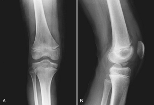
Standard roentgenograms and anteroposterior, lateral, and notch radiographic views are usually diagnostic. The fracture is best seen on lateral and notch views (Fig. 89-1). Radiographs should be carefully scrutinized, as the avulsed fragment may be mostly nonossified cartilage with only a small, thin ossified portion visible on the lateral view. If necessary, computed tomography (CT) scanning allows refined definition of the fracture anatomy.
Associated Injuries
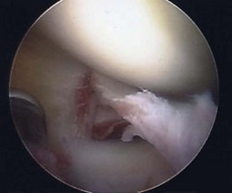
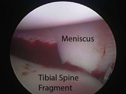
Associated intra-articular injuries are relatively uncommon. Intercondylar eminence fractures may include or may be associated with any combination of bone, chondral, meniscal, and ligamentous injuries.10 However, in a series of 80 skeletally immature patients who underwent surgical fixation of tibial eminence fractures, Kocher and coworkers found no associated chondral injuries and associated meniscal tear in only 3.8% (3/80) of patients (Fig. 89-2).23 Associated collateral ligament injury or proximal ACL avulsion in conjunction with a tibial spine fracture has also been reported.15,45
Classification
The classification system of Meyers and McKeever is based on the degree of displacement and is widely used to classify fractures and to guide treatment (Fig. 89-3).35,36 Zaricznyj later modified this classification to include a fourth type—comminuted fractures of the tibial spine59:
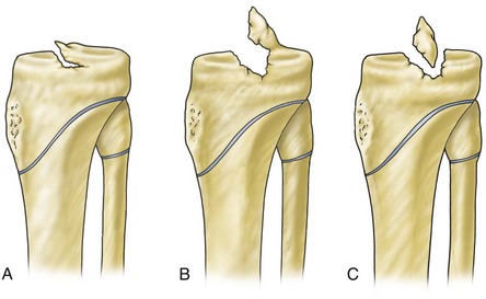
Interobserver reliability with type 1 and type 2/3 fractures is good; however, differentiation between type 2 and 3 fractures may be difficult.22
Surgical and Applied Anatomy
Between the condyles, the intercondylar eminence or the spine is the insertion point for portions of the menisci and the anterior and posterior cruciate ligaments. The tibial eminence is triangular and refers to the portion of the proximal tibia that includes two ridges of bone and cartilage. In the immature skeleton, the proximal surface of the eminence is covered entirely with cartilage. The ACL attaches distally to the anteromedial portion of the tibial intercondylar eminence (Fig. 89-4). The posterior cruciate ligament (PCL) inserts on the posterior aspect of the proximal tibia, distal to the joint line. Both menisci insert into the tibia in the region between the lateral and medial eminences, but no direct connection exists between the ACL and the menisci. In 12 patients with displaced tibial spine fractures that were unable to be reduced closed, Lowe and colleagues reported that the anterior horn of the lateral meniscus and the ACL were attached simultaneously and were pulling in different directions.27
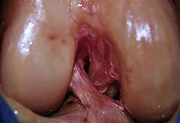
Figure 89-4 Anterior cruciate ligament (ACL) insertion onto the anteromedial portion of the tibial eminence.
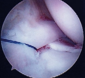
Meniscal or intermeniscal ligament entrapment under the displaced tibial eminence fragment can be common and may be a rationale for considering arthroscopic or open reduction in displaced tibial spine fractures (Fig. 89-5).6,7,10,23 Meniscal entrapment can prevent anatomic reduction of the tibial spine fragment, which may result in increased anterior laxity or a block to extension and knee pain after the fracture has healed.14,17,32,41,42 Mah and coworkers found medial meniscal entrapment preventing reduction in 8 of 10 children with type 3 fractures undergoing arthroscopic treatment.31 In a consecutive series of 80 patients who underwent surgical fixation of tibial eminence fractures that were not able to be reduced closed, Kocher and associates reported entrapment of the anterior horn medial meniscus (n = 36), the intermeniscal ligament (n = 6), or the anterior horn lateral meniscus (n = 1) in 26% of type 2 fractures and 65% of type 3 fractures.23 The entrapped meniscus typically can be extracted with an arthroscopic probe and retracted with a retaining suture (Fig. 89-6).
Current Treatment Options
Current treatment options include cast immobilization,24,37 closed reduction with immobilization,41,57 open reduction with immobilization,37 open reduction with internal fixation,38,57 arthroscopic reduction with immobilization,33 and arthroscopic reduction with a variety of fixation methods, including suture fixation,* wire3 and screw fixation,5,24,33,44 anchor fixation,54 and bioabsorbable nail fixation.46 Many options regarding fixation of the fracture are available, and all have been used with good success, most commonly, suture and screw fixation. Study findings are equivocal in terms of strength of fixation, although suture fixation may be favored because it offers the advantages of eliminating the risks of comminution of the fracture fragment and posterior neurovascular injury and the need for hardware removal.2,25,46,47
The goal of treatment of a tibial spine avulsion is anatomic reduction; however, controversy continues regarding whether the tibial spine should be over-reduced. Theoretically, over-reduction may lead to excessive tightening of the ACL and limited knee motion.28 On the other hand, it is likely that permanent intersubstance stretching of the ACL occurs before the fracture40; therefore, over-reduction could be considered. Although further clinical or in vitro research is required, long-term evaluation of well-reduced tibial eminence fractures shows subtle increases in anteroposterior knee laxity without functional deficit.*
Closed reduction can be successful for some type 2 fractures but frequently is not successful for type 3 fractures. In their series, Kocher and associates reported closed reduction in approximately 50% of type 2 fractures (26/49) and unsuccessful closed reduction in all 57 type 3 fractures.23 Arthroscopic or open reduction with internal fixation of type 2 and 3 tibial eminence fractures, which do not reduce, has been advocated because of the potential for clinical instability and loss of extension associated with closed reduction and immobilization, the ability to evaluate and treat injuries, and the opportunity for early mobilization.6,7,21,31 For displaced type 2 and 3 fractures, Wiley and Baxter found a correlation between fracture displacement and measured knee laxity despite good patient function.55
Related posts:


Stay updated, free articles. Join our Telegram channel

Full access? Get Clinical Tree


