Thoracolumbar Spine
EPIDEMIOLOGY
Neurologic injury complicates 15% to 20% of fractures at the thoracolumbar level.
Sixty-five percent of thoracolumbar fractures occur as a result of motor vehicle trauma or fall from a height, with the remainder caused by athletic participation and assault.
Most isolated thoracic and lumbar spine fractures are related to osteoporosis and involve minimal or no trauma.
Osteoporosis accounts for approximately 750,000 vertebral fractures annually in the United States and far outnumbers the 15,000 traumarelated thoracic and lumbar spine fractures.
Thoracolumbar trauma occurs most frequently in male patients between 30 and 39 years of age.
Ninety percent of vertebral fractures occur in the thoracolumbar spine.
Neurologic injury occurs in approximately 20% of thoracic and lumbar fractures.
Sixty percent of thoracolumbar fractures occur between T11 and L2 vertebral levels.
ANATOMY
See Chapter 8 for a general definition of terms.
The thoracolumbar spine consists of 12 thoracic vertebrae and 5 lumbar vertebrae.
The thoracic level is kyphotic, and the lumbar region is lordotic. The thoracolumbar region, as a transition zone, is especially prone to injury.
The thoracic spine is much stiffer than the lumbar spine in flexionextension and lateral bending, reflecting the restraining effect of the rib cage as well as the thinner intervertebral discs of the thoracic spine.
Rotation is greater in the thoracic spine, achieving a maximum at T8-T9. The reason is the orientation of the lumbar facets, which limit the rotation arc to approximately 10 degrees for the lumbar spine versus 75 degrees for the thoracic spine.
The conus medullaris ends at the L1-L2 level. The cauda equina, which comprises the motor and sensory roots of the lumbosacral myelomeres (Fig. 10.1), lies caudal to the conus.
The corticospinal tracts demonstrate polarity, with cervical fibers distributed centrally and sacral fibers peripherally.
The ratio of the spinal canal dimensions to the spinal cord dimensions is smallest in the T2-T10 region, which makes this area prone to neurologic injury after trauma.
Neurologic deficits secondary to skeletal injury from the 1st through the 10th thoracic levels are frequently complete deficits, primarily related to spinal cord injury with varying levels of root injury. The proportion of root injury increases with more caudal injuries, with skeletal injuries caudal to L1 causing exclusively root (lower motor neuron) injury.
The region between T2 and T10 is a circulatory watershed area, deriving its proximal blood supply from antegrade vessels in the upper thoracic spine and distally from retrograde flow from the purported artery of Adamkiewicz, which can be variably located between T9 and L2.
Most thoracic and lumbar injuries occur within the region between T10 and L2, commonly referred to as the thoracolumbar junction. This increased susceptibility can be explained by a variety of factors. The thoracolumbar junction is a transition zone between the relatively stiff thoracic spine and the more mobile lumbar spine.
MECHANISM OF INJURY
These generally represent high-energy injuries, typically from motor vehicle accident or falls from a height.
They may represent a combination of flexion, extension, compression, distraction, torsion, and shear.
CLINICAL EVALUATION
1. Patient assessment: This involves airway, breathing, circulation, disability, and exposure (ABCDE). Please see also Chapter 9.
2. Initiate resuscitation: Address life-threatening injuries. Maintain spine immobilization. Watch for neurogenic shock (hypotension and bradycardia).
3. Evaluate the level of consciousness and neurologic impairment: Glasgow Coma Scale
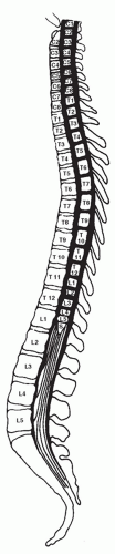 FIGURE 10.1 The relationship between myelomeres (spinal cord segments) and the vertebral bodies. (From Benson DR, Keenen TL. Evaluation and treatment of trauma to the vertebral column. Instr Course Lect 1990;39:577.) |
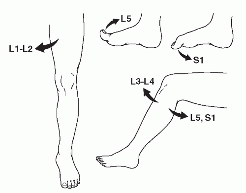 FIGURE 10.2 A screening examination of the lower extremities assesses the motor function of the lumbar and first sacral nerve roots: hip adductors, L1-L2; knee extension, L3-L4; knee flexion, L5-S1; great toe extension, L5; and great toe flexion, S1. (From Benson DR, Keenen TL. Evaluation and treatment of trauma to the vertebral column. Instr Course Lect 1990;39:583.) |
4. Assess head, neck, chest, abdominal, pelvic, and extremity injury.
5. Ascertain the history: Assess the mechanism of injury, witnessed head trauma, movement of extremities/level of consciousness immediately following trauma, etc.
6. Physical examination
Back pain and tenderness
Lacerations, abrasions, and contusions on back
Abdominal and/or chest ecchymosis from seat belt injury (also suggestive of liver, spleen, or other abdominal injury)
7. Neurologic examination
Cranial nerves
Complete motor and sensory examination (Figs. 10.2 and 10.3)
Upper and lower extremity reflexes
Rectal examination: perianal sensation, rectal tone (Fig. 10.4)
Bulbocavernosus reflex (Fig. 10.5)
8. In the alert and cooperative patient, the thoracic and lumbar spine can be “cleared” with the absence of pain or tenderness or distraction mechanism of injury and a normal neurologic examination. Otherwise, imaging is required.
RADIOGRAPHIC EVALUATION
Anteroposterior (AP) and lateral views of the thoracic and lumbar spine are obtained.
Abnormal widening of the interpedicular distance signifies lateral displacement of vertebral body fragments, typical of burst fractures.
Vertebral body height loss can be measured by comparing the height of the injured level with adjacent uninjured vertebrae.
Quantification of sagittal plane alignment can be performed using the Cobb method.
Chest and abdominal radiographs obtained during the initial trauma survey are not adequate for assessing vertebral column injury.
Computed tomography (CT) and/or magnetic resonance imaging (MRI) of the injured area may be obtained to characterize the fracture further, to assess for canal compromise, and to evaluate the degree of neural compression.
CT scans provide finer detail of the bony involvement in thoracolumbar injuries, and MRI can be used to evaluate for soft tissue injury to the cord, intervertebral discs, or for posterior ligamentous disruption.
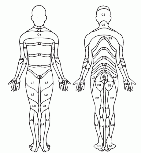 FIGURE 10.3 A pain and temperature dermatome chart. These sensory modalities are mediated by the lateral spinothalamic tract. Note that C4 includes the upper chest just superior to T2. The rest of the cervical and T1 roots are located in the upper extremities. There is overlap in the territories subserved by each sensory root and variation among individuals. (From Benson DR, Keenen TL. Evaluation and treatment of trauma to the vertebral column. Instr Course Lect 1990;39:584.) |
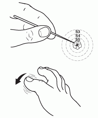 FIGURE 10.4 Sacral sparing may include the triad of perianal sensation, rectal tone, and great toe flexion. (From Benson DR, Keenen TL. Evaluation and treatment of trauma to the vertebral column. Instr Course Lect 1990;39:580.) |
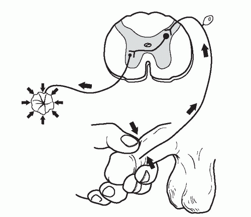 FIGURE 10.5 The bulbocavernosus reflex arc is mediated by the conus medullaris and the lower three sacral roots. Stimulation of the glans penis, glans clitoris, or gentle traction on a Foley catheter to stimulate the bladder will evoke contraction of the rectal sphincter. (From Benson DR, Keenen TL. Evaluation and treatment of trauma to the vertebral column. Instr Course Lect 1990;39:578.) |
CLASSIFICATION
Orthopaedic Trauma Association Classification of Thoracic and Lumbar Spine Injuries
See Fracture and Dislocation Classification Compendium at http://www.ota.org/compendium/compendium.html.
McAfee et al.
Classification is based on the failure mode of the middle osteoligamentous complex (posterior longitudinal ligament, posterior half of vertebral body, and posterior annulus fibrosus).
Axial compression
Axial distraction
Translation within the transverse plane
This led to the following six injury patterns in this classification:
1. Wedge-compression fracture
2. Stable burst fracture
3. Unstable burst fracture
4. Chance fracture
5. Flexion-distraction injury
6. Translational injuries
McCormack et al.
This is a “load-sharing classification.”
A point value is assigned to the degree of vertebral body comminution, fracture fragment apposition, and kyphosis. Based on their primary outcome of hardware failure, McCormack et al. concluded that injuries with scores >6 points would be better treated with the addition of anterior column reconstruction to posterior stabilization. A recent study demonstrated very high interobserver and intraobserver reliability of this classification system.
Thoracolumbar Injury Classification System
This is an injury scoring system to categorize lumbar burst fractures by morphology, posterior ligamentous complex disruption, and neurologic involvement rather than by anatomy or injury mechanism (Vaccaro et al., 2005a).
Thoracolumbar Injury Classification System (TICS) is designed to grade and predict acute spine stability, risk of future deformity, and progressive neurologic compromise.
It facilitates treatment recommendations for surgical or nonoperative management. This system has demonstrated good inter- and intraobserver reliability (Harrop et al., 2006).
The most important determinants of stability are neurologic deficit as in incomplete spinal cord injury and involvement of all three columns of the spine.
Denis
Minor Spinal Injuries
Articular process fractures (1%)
Transverse process fractures (14%)
Spinous process fractures (2%)
Pars interarticularis fractures (1%)
Related posts:


Stay updated, free articles. Join our Telegram channel

Full access? Get Clinical Tree


