Thoracic Spine
Matthew Morrey
Mark B. Dekutoski
Steve Cassivi
Ziya L. Gokaslan
ANTERIOR CERVICAL THORACIC APPROACHES
The four approaches used for the upper thoracic region include first the high thoracotomy at the fourth rib which provides excellent access to the lateral aspect of the anterolateral cervical thoracic junction especially the sympathetic chain. This is, however, a difficult approach for placement of anterior fixation. Second is the modified anterior approach which resects the medial clavicle. This exposure is fraught with significant shoulder morbidity albeit downplayed for patients who are anticipated not to survive their pathology. These exposures have fallen out of favor with the increasing familiarity with the manubrial split accompanied by a lateral traverse sternal split between the third and fourth rib. The full manubrial split cervicothoracic approach allows anterior extension as low as the diaphragmatic attachments. The modified anterior and manubrial split are limited by the aortic arch which traverses approximately T3 vertebra. Fortunately, the aortal caval window approach can be used to reach the anterior aspect of T3-T5, allowing for direct anterior decompression, and by passing the plate under the aortic arch direct anterior plate fixation is possible.
Indications
Anterior approaches of the cervical thoracic junction are often necessary to address tumor pathology, deformity and anterior column support. These approaches are often avoided due to limited familiarity of the surgeon with the approach, which is paramount to successful surgical intervention and avoidance of complications.
Contraindications
Contraindications or limitations to the cervicothoracic approaches are primarily related to the nature of the pathology of spinal structures such as tumor pathology and with vascular involvement postradiation changes which can substantially increase the risk of intraoperative scarring compromising exposure and esophageal tissue integrity.
Anterior cervical thoracic approaches for tumor involvement may require ligation of vascular and potential for a dysfunctional limb.
Preoperative Planning
Collaboration between the access surgeon and the spinal surgeon is useful. Adequate visualization of the neurologic structures for direct decompression most commonly defines the caudal extent of the exposure. When anterior fixation is only required to T1 or T2 an extensile anterior lateral neck approach may be adequate. However, for decompression of the T1 vertebra, a manubrial split is required to gain adequate anterior access and fixation. The authors’ preference is to conduct a right anterior neck approach to avoid the lymphatic duct and the dominant left carotid artery. However, in the final analysis, the specific pathology most commonly determines the approach selection.
High Thoracotomy
Position
Typically the patient is placed prone with an axillary roll and with the arm elevated and flexed anteriorly to allow for full prepping of the arm and axilla.
Technique
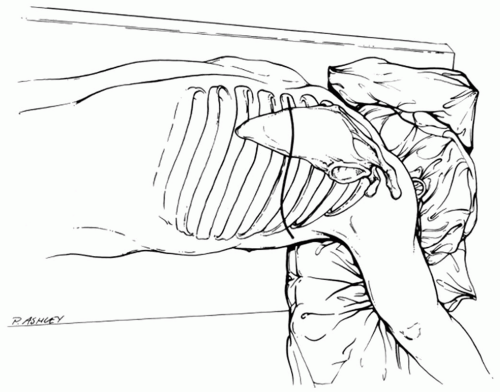
Incision: with the inferior border of the scapula defined, the incision courses over the mid-body of the scapula (Fig. 13-1).
The inferior muscular attachments of the scapula are mobilized after transection and/or mobilization of the trapezius dorsally. The latissimus dorsi muscle is retracted laterally through this window (Fig. 13-2).
The rhomboids are released and the fourth rib is identified by palpation.
An incision over the fourth rib is conducted while protecting the intercostal neurovascular structures anteriorly and cephalad (Fig. 13-3).
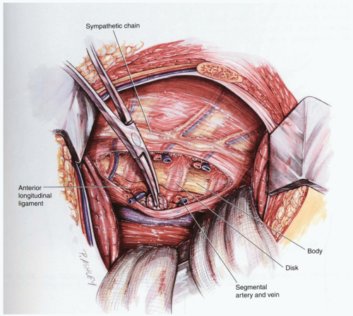
A dorsal osteotomy of the rib allows further mobilization and retraction of the scapula. The scapula retractor, along with chest wall retractors, allows for direct visualization from C7 to the diaphragm (Fig. 13-4).
Note: The right side of the chest allows for the greatest flexibility of the exposure.
Direct Anterior Cervical Thoracic Approach
This is a modification of the anterior exposure with resection of the medial clavicle. This approach employs the interval between the strap muscles medially the sternocleidomastoid laterally, the esophagus and trachea medially and the carotid sheath laterally. Selection of a right or left approach is based on familiarity of the surgeon and recognition that the recurrent laryngeal nerve is more consistent on the left side than on the right side of the neck. Further, hand dominance, familiarity, and the left sided lymphatic duct all contribute to the decisions of a left or right approach.
Technique
Incision: the incision may be extensive or more limited as shown in Figure 13-5.
Once the anterior neck approach is developed and the pharynx is identified and recurrent laryngeal nerve typically deep to it. The inferior extent of this anterior dissection will be the C-7 T-1 disc where upon a more extensile approach is typically needed (Fig. 13-6).
Stay updated, free articles. Join our Telegram channel

Full access? Get Clinical Tree


