The Wrist and Hand
19.1 Anatomy
Gail A. Shafer-Crane
William M. Falls
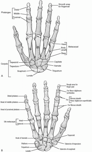
Osteology of the wrist and hand is complex, and designed to maximize manipulative ability. The intricacy of the articulations sacrifices weight-bearing power in favor of fine motor manipulation and prehensile strength. Twenty-nine bones articulate with each other to form the distal radioulnar joint DRUJ), radiocarpal joint, intercarpal joints, carpometacarpal (CMC) joints, metacarpophalangeal (MCP) joints, and interphalangeal joints (Fig. 19.1.1). Anatomy of the wrist and hand is presented in detail in major anatomic textbooks (1,2,3,4,5,6,7,8,9).
The distal radius and ulna articulate, forming the most proximal joint of the wrist, the DRUJ. Pronation and supination of the hand are permitted at this synovial joint. The distal ulna rests in the ulnar notch of the radius, allowing the radius to swivel about the ulna, which is fixed by its proximal articulation with the humerus by the triangular fibrocartilage complex (TFCC). The TFCC attaches medially to the ulnar styloid process and laterally just distal to the ulnar notch of the radius. This contributes to the joint capsule and allows the joint to pivot. The fibrocartilaginous disc of the TFCC fills a hollow in the distal ulna (ulnar fovea). This contributes to the ulnar proximal articular surface. A superior redundancy in the synovial membrane of the articular capsule forms the sacciform recess. This connective tissue tether stretches as the radius pivots about the ulna. Transverse bands of the palmar and dorsal radioulnar ligaments complete the articular capsule of the DRUJ (4).
The radiocarpal joint includes the articulation of the distal radius and articular disc of the DRUJ with the proximal carpal row. These bones include the scaphoid, lunate, and triquetrum, with the pisiform, a sesamoid bone, anterior to the triquetrum. This complex synovial condyloid joint permits flexion and extension, circumduction, and radial-ulnar deviation of the wrist. The proximal carpal row glides as a unit within the axial concavity of the DRUJ. The carpals translate anteriorly to permit wrist extension, and posteriorly to allow wrist flexion. Radial and ulnar deviation are permitted by lateral and medial glides. Circumduction is a combination of these motions. The articulations of the distal carpal row (trapezium, trapezoid, capitate, and hamate) combine anterior and posterior glide on the proximal carpal row to contribute to full wrist flexion and extension.
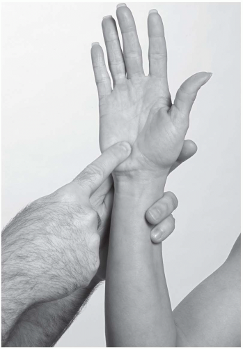
Palpation of each carpal bone is performed using surface landmarks. The pisiform carpal bone is most prominent, and can be palpated on the medial volar surface of the wrist at the proximal flexion crease. The hook of the hamate bone is less easily palpated just distal to the pisiform, deep to the hypothenar eminence.
Proceeding laterally on the volar wrist, a depression is noted between the pisiform and hamate. The carpal bones form the floor and sides of the carpal tunnel, an arch with a volar concavity that houses the tendons of the finger and thumb flexors and the median nerve (Fig. 19.1.2). The transverse carpal ligament
forms the roof by attaching to the pisiform and hamate medially and the scaphoid and trapezium laterally. The trapezium is palpated at the base of the thumb, distal to the distal volar flexion skin crease of the wrist.
forms the roof by attaching to the pisiform and hamate medially and the scaphoid and trapezium laterally. The trapezium is palpated at the base of the thumb, distal to the distal volar flexion skin crease of the wrist.
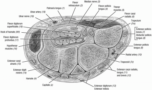 FIGURE 19.1.2. Section through the distal carpal tunnel. (From Agur AMR, Lee ML. Grant’s Atlas of Anatomy, 10th ed. Baltimore: Lippincott Williams & Wilkins, 1999.) |
Landmarks of the lateral wrist include the anatomic snuffbox, a small depression between two prominent tendons where the scaphoid bone can be palpated dorsolaterally (Fig. 19.1.3). The medial dorsum of the wrist is marked by a small, visible depression where the lunate can be palpated as it articulates with the radius. The capitate bone is palpated just distal to the lunate during wrist flexion.
Much of the stability of the wrist is provided through the numerous ligaments securing the carpal bones to the DRUJ. These include the palmar radiocarpal and ulnocarpal ligaments, dorsal radiocarpal and ulnocarpal ligaments, and radial and ulnar collateral ligaments. Intercarpal ligaments permit gliding of the carpals upon each other as described earlier (2).
Continuing distally, the CMC joint of the thumb can be palpated distal to the radial styloid. Because of their limited motion, the CMC joints of the fingers are less easily palpated, but are located across the midpalm. These condyloid joints allow very little motion, with the exception of the CMC joint of the thumb. This is a saddle joint, named for the concavity of the trapezium as it articulates with the first metacarpal bone. The saddle joint permits opposition, circumduction, flexion, and abduction of the thumb. Without this versatile articulation, prehension and fine manipulation would not be possible.
The MCP joints are the most proximal joints of the digits. They are formed by the articulation of the bases of the proximal phalanges with the metacarpal bones. These hinge joints allow flexion and extension of more than 90 degrees, and small medial and lateral movements that permit abduction of the digits away from the third digit, or middle finger. Radial (lateral) and ulnar (medial) collateral ligaments support the MCP joints.
Extrinsic muscles that are prime movers of the wrist and hand originate at the elbow. Muscles that act to move the wrist arise from the medial and lateral epicondyles of the humerus and forearm. Long tendons, with few exceptions, skip the carpals and attach these fusiform muscles to the proximal metacarpals (Fig. 19.1.4). The flexor carpi ulnaris (FCU) is the exception. It inserts on the pisiform bone, at the hook of the hamate bone, and finally on the base of the fifth metacarpal bone. The flexor carpi radialis (FCR) inserts on the anterior aspect of the base of the second metacarpal bone, the distal flexor retinaculum, and into the palmar aponeurosis. The extensor carpi radialis longus (ECRL) inserts on the posterior aspect of the base of the second metacarpal; the extensor carpi radialis brevis (ECRB) inserts onto the third metacarpal bone; and the extensor carpi ulnaris (ECU) inserts onto the base of the fifth metacarpal bone. More information on the compartments and proximal attachments of these muscles can be found in Chapter 18.
The five muscles that are primary movers of the wrist—the FCU, FCR, ECRB, ECRL, and ECU—work in synchrony. If all five muscles contract equally, they increase the stability of the wrist by compressing the intracarpal articulations, and the radiocarpal articulation (1). Wrist flexion is the action of both FCU and FCR together. Wrist extension is the action of the ECRL, ECRB, and ECU working evenly together. Radial and ulnar deviation require shortening of the corresponding pair of muscles, and circumduction of the wrist requires coordination of all five muscles shortening and lengthening progressively. The FCR has a close association with the carpal tunnel, but traverses the wrist in its own compartment along the radial side of the carpal tunnel. Guyon’s canal houses the ulnar nerve and is found in a groove formed along the medial aspect of the triquetrum-pisiform joint proximally, and medial to the hook of the hamate bone distally in the wrist.
The extrinsic forearm finger muscles and intrinsic hand muscles perform the motion of these joints. The tendons of the extrinsic finger flexors, the flexor digitorum superficialis (FDS) and the flexor digitorum profundus (FDP), traverse the wrist in close proximity through the
carpal tunnel (Fig. 19.1.5). The FDP lies deep, and the FDS more superficial, with the tendons to each digit encased in synovial sheaths that begin just proximal to the MCP joints, and run to the points of attachment. There is an interruption of the synovial sheath in the distal palm, and then it continues proximally through the carpal tunnel to the proximal borders of the flexor retinaculum. The sheath of the small finger is continuous with the more proximal common flexor sheath. The extrinsic finger extensors—the extensor digitorum communis (EDC), extensor indicis proprius, and extensor digit minimi—divide into the central and collateral slips or bands to form a complex extensor hood.
carpal tunnel (Fig. 19.1.5). The FDP lies deep, and the FDS more superficial, with the tendons to each digit encased in synovial sheaths that begin just proximal to the MCP joints, and run to the points of attachment. There is an interruption of the synovial sheath in the distal palm, and then it continues proximally through the carpal tunnel to the proximal borders of the flexor retinaculum. The sheath of the small finger is continuous with the more proximal common flexor sheath. The extrinsic finger extensors—the extensor digitorum communis (EDC), extensor indicis proprius, and extensor digit minimi—divide into the central and collateral slips or bands to form a complex extensor hood.
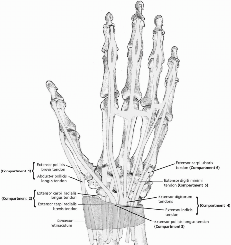 FIGURE 19.1.4. Extensor tendons of the hand and wrist, dorsal view. (From Stoller DW. MRI, Arthroscopy, and Surgical Anatomy of the Joints. Baltimore: Lippincott Williams & Wilkins, 1999.) |
 FIGURE 19.1.5. The flexor digitorum tendons in relation to the median nerve. (From Stoller DW. MRI, Arthroscopy, and Surgical Anatomy of the Joints. Baltimore: Lippincott Williams & Wilkins, 1999.) |
The intrinsic hand muscles are housed within the confines of the hand, arranged between the metacarpal bones. The interossei muscles insert laterally and medially onto the proximal dorsal hood to perform finger adduction and abduction. The lumbrical muscles lie parallel to the interossei along the metacarpal bones. The proximal attachment of these muscles is along the FDP tendon; the distal attachment is just distal to the proximal interphalangeal (PIP) joints on the lateral border of the middle phalanx of digits 2 through 5. These muscles act to assist in full extension of the fingers at the PIP joints. Innervation is split between the first and second lumbricals by the median nerve (C8, T1) and the deep branch of the ulnar nerve (C8, T1). The deep branch of the ulnar nerve innervates the interossei muscles.
The wrist and hand are supplied by the radial and ulnar arteries. The two arteries have both deep and superficial branches (7). The wrist receives its blood supply from the anterior and posterior interosseous arteries, which are distal continuations of the brachial artery. The superficial palmar arch is primarily supplied by the ulnar artery (5,7). The deep palmar arch is formed by the deep palmar branch of the ulnar artery and the radial artery and is also located at the midpalmar aspect of the wrist and is distal to the carpal tunnel.
Nerves that supply the DRUJ are the anterior branch of the median nerve (C6-C7) and
the posterior interosseous branch of the deep radial nerve (C7-C8) (6).
the posterior interosseous branch of the deep radial nerve (C7-C8) (6).
REFERENCES
1. Basmajian JV, Slonecker CE. Grant’s method of anatomy, 11th ed. Baltimore: Williams & Wilkins, 1989.
2. Buchler U, Kauer JMG, Garcia-Elias M, Hagert CG. Wrist instability. St. Louis: Mosby, 1996.
3. Clemente CD. Gray’s anatomy of the human body, American 30th ed. Philadelphia: Lea & Febiger, 1985.
4. Moore KL, Agur AM. Essential clinical anatomy. Baltimore: Williams & Wilkins, 1996.
5. Moore KL, Dalley AF II. Clinically oriented anatomy, 4th ed. Philadelphia: Lippincott Williams & Wilkins, 1999:1164.
6. Netter FH. Atlas of human anatomy, Summit, NJ: CIBA-Geigy Corporation, 1995.
7. Rosse C, Gaddum-Rosse P. Hollinshead’s textbook of anatomy, 5th ed. Philadelphia: Lippincott-Raven, 1997.
8. Williams PL, Warwick R, Dyson M, Bannister LH. Gray’s anatomy, British 37th ed. London: Churchill Livingstone, 1989.
9. Woodburne RT, Burkel WE. Essentials of human anatomy, 9th ed. New York: Oxford University Press, 1994.
19.2 Physical Examination
Sean M. Mcfadden
OBSERVATION
Evaluation of the wrist and hand starts when the examiner enters the room. It is important to note the manner in which the athlete is holding the hand and whether or not he or she can shake hands. Flexor tendon injuries are easily noted because the injured digit will not be maintained in its natural flexed position. Although rudimentary, it is always important to count all digits and note any previous amputations.
Palmar Surface
The palmar, or volar, aspect of the hand and wrist has multiple creases that are located where the underlying fascia attaches to the skin. The four important creases are as follows (Fig. 19.2.1):
The distal palmar crease marks the volar location of the metacarpophalangeal (MCP) joints.
The proximal palmar crease lies in the midmetacarpal area and is found just proximal to the distal palmar crease.
The proximal interphalangeal crease is located at the proximal interphalangeal (PIP) joint and marks the distal aspect of no man’s land.
The thenar crease denotes the thenar eminence.
Note the “attitude” of the hand, that is, its natural position when relaxed (Fig. 19.2.2). The MCP and interphalangeal (IP) joints should have slight flexion at rest, and the fingers should line up parallel to each other and pointing toward the thenar and hypothenar eminences. Any significant discrepancy could mean a tendon injury or a contracture held in flexion with the fingers directed toward the thenar eminence (1,2). The normal appearance of each eminence has a prominence, which is important to note because atrophy of either eminence is representative of nerve damage to the muscle
bellies that control the thumb (median nerve) and little finger (ulnar nerve) (1,3).
bellies that control the thumb (median nerve) and little finger (ulnar nerve) (1,3).
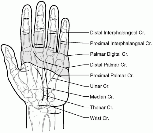 FIGURE 19.2.1. Palmar view of the hand with the four main creases: distal palmar, proximal palmar, proximal interphalangeal, and thenar crease. |
The skin of the volar surface is usually thicker than the dorsal side because it is the working side of the hand. The volar skin is fixed with fascia, which binds it with the structures beneath it at the creases. This fixation is important because it allows for objects to be held securely by the hand.
The skin on the fingers is connected to bone with septa and small ligaments running from skin to bone along the lateral and medial sides of the fingers (Cleland’s and Grayson’s ligaments) (4). This arrangement results in little rotatory movement of the skin around the fingers.
 FIGURE 19.2.2. Attitude of the hand, noting the natural flexion at the metacarpophalangeal and proximal interphalangeal joints. |
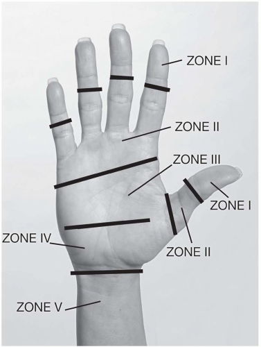
Flexor Zones (Fig. 19.2.3)
Zone I: The profundus tendon exists on its own and inserts at the proximal aspect of the distal phalanx.
Zone II: The profundus tendons become volar to the superficialis tendons. The profundus tendon will emerge through the superficialis tendons at the level of the MCP joint. The superficialis tendons bifurcate and then insert on the midaspect of the middle phalanx.
Zone III: The lumbricals originate from the profundus tendon in this zone.
Zone IV: The median nerve and the nine flexor tendons run through the carpal canal.
Zone V: The flexor tendons begin in the distal aspect of the forearm at the musculotendinous junction. The superficialis tendons lie volar to the profundus tendons.
Dorsal Surface
The skin on the dorsal aspect of the hand is more mobile, primarily to allow for MCP joint
flexion. The knuckles on the dorsal aspect of the hand are usually most prominent, the middle one more so. When an athlete clenches a fist, the fifth knuckle will become less prominent. The valleys between each knuckle should all be symmetrical. Any asymmetry, for example, as a result of swelling, is indicative of an injury (1,4).
flexion. The knuckles on the dorsal aspect of the hand are usually most prominent, the middle one more so. When an athlete clenches a fist, the fifth knuckle will become less prominent. The valleys between each knuckle should all be symmetrical. Any asymmetry, for example, as a result of swelling, is indicative of an injury (1,4).
Inspection of the nails is also important. The normal nail bed should have a white base (the lunula) with the remainder of the bed being pink. Observation of the nail is important because a nail bed that is lighter in color could be indicative of anemia. Nails that have ridges or are brittle could be a result of malnutrition. Clubbing of the fingertips and nails may indicate pulmonary disease.
Extensor Tendon Zones
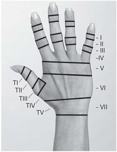
As with the volar aspect of the wrist and hand, the extensor side is subdivided into zones and compartments for the purpose of describing extensor tendon injuries (Fig. 19.2.4). The six dorsal extensor tendon compartments are as follows (Fig. 19.2.5) (1,4,5):
Compartment | |
|---|---|
1 | Abductor pollicis longus, extensor pollicis brevis |
2 | Extensor carpi radialis longus and brevis |
3 | Extensor pollicis longus |
4 | Extensor carpi digitorum communis, extensor indicis proprius |
5 | Extensor digiti minimi |
6 | Extensor carpi ulnaris |
PALPATION
Palpation of the wrist bones starts at the radius and ulna and proceeds distally. Note tenderness on the radial and ulnar styloids (Fig. 19.2.6). The indentation at the distal aspect of the radial
styloid process is the anatomic snuffbox. A branch of the radial artery can be palpated within the snuffbox, but any tenderness located in the area could represent a scaphoid fracture (1,4,6). The scaphoid is the most commonly fractured carpal bone, and depending on the location of the fracture, healing can be difficult due to recurrent blood flow. It is the most radial of the proximal carpal row. Palpation is facilitated by placing the wrist into ulnar deviation (Fig. 19.2.7).
styloid process is the anatomic snuffbox. A branch of the radial artery can be palpated within the snuffbox, but any tenderness located in the area could represent a scaphoid fracture (1,4,6). The scaphoid is the most commonly fractured carpal bone, and depending on the location of the fracture, healing can be difficult due to recurrent blood flow. It is the most radial of the proximal carpal row. Palpation is facilitated by placing the wrist into ulnar deviation (Fig. 19.2.7).
 FIGURE 19.2.5. The six dorsal extensor tendon compartments. (From Agur AMR, Lee ML. Grant’s Atlas of Anatomy, 10th ed. Baltimore: Lippincott Williams & Wilkins, 1999.) |
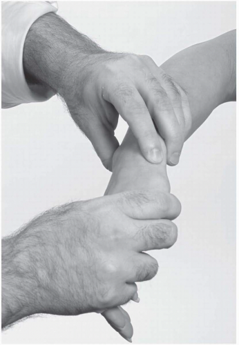
Examine the proximal carpal row carefully (scaphoid, lunate, triquetrum, pisiform). The distal row includes the trapezium, which can be easily palpated by moving distal to the anatomic snuffbox and asking the athlete to flex and extend the thumb. The trapezoid, capitate, and hamate round out the row. The hamate hook can be easily palpated by placing the IP joint of the thumb on the pisiform and then rolling the thumb forward (Fig. 19.2.8). The hamate is important because it forms the ulnar border of Guyon’s canal, which contains the ulnar nerve and artery (7).
RANGE OF MOTION
Wrist
The wrist range of motion (ROM) should be evaluated by assessing both passive and active ranges before continuing the examination. Significant loss of motion from trauma may need radiographs to rule out unstable fractures. Bilateral comparison is essential to evaluate for any unilateral restrictions. Ranges of motion move in three planes as follows:
Flexion and extension.
Ulnar and radial deviation.
Supination and pronation (this motion actually emanates from the elbow, not the wrist).
To assess passive motion, the examiner isolates the wrist joint by holding the forearm with one hand and using the other hand to manipulate the wrist. At the same time, he or she puts the wrist into radial and ulnar deviation and then supinates and pronates the wrist. Be aware of any discrepancies in (ROM) or restrictions that could reflect an injury to the wrist. Normal passive ranges of motion are listed in Table 19.2.1.
TABLE 19.2.1. RANGES OF MOTION (ROM) WRIST | ||||||||||||||
|---|---|---|---|---|---|---|---|---|---|---|---|---|---|---|
|
Finger
Finger motion involves three joints per digit, so the examiner should look at all of them. To grossly assess active finger flexion and extension, ask the athlete to make a fist while observing the finger motion. Make sure the fingers are all moving together throughout the range of motion. When all joints of the fingers are completely flexed, the fingers should be at the level of the distal flexion crease.
The specific ranges of motion that need to be evaluated are as follows:
Finger and thumb flexion and extension at the MCP joint.
Finger and thumb flexion and extension at the IP joints.
Finger and thumb abduction and adduction at the MCP joint.
Finger and thumb opposition.
Normal ranges of motion for the finger are listed in Table 19.2.2.
To examine the passive motion of the fingers, the examiner grasps the hand at the distal metacarpal using his or her index finger and thumb to stabilize each joint. The mobilizing hand then moves the digit through its ROM. To test MCP flexion and extension, the examiner grasps the metacarpal with the stabilizing
hand and the proximal phalanx with the mobilizing hand. The MCP joint has some lateral movement when in extension, but no lateral movement when in flexion because the collateral ligaments are loose in extension and tight in flexion. This becomes an important factor when the hand is placed into a cast that extends beyond the MCP joint. The cast should maintain MCP joint flexion in order to prevent collateral ligament tightness.
hand and the proximal phalanx with the mobilizing hand. The MCP joint has some lateral movement when in extension, but no lateral movement when in flexion because the collateral ligaments are loose in extension and tight in flexion. This becomes an important factor when the hand is placed into a cast that extends beyond the MCP joint. The cast should maintain MCP joint flexion in order to prevent collateral ligament tightness.
TABLE 19.2.2. ACTIVE AND PASSIVE FINGER RANGES OF MOTION (ROM) | ||||||||||||||||||||||||||||||||||||||||||||
|---|---|---|---|---|---|---|---|---|---|---|---|---|---|---|---|---|---|---|---|---|---|---|---|---|---|---|---|---|---|---|---|---|---|---|---|---|---|---|---|---|---|---|---|---|
|
Stay updated, free articles. Join our Telegram channel

Full access? Get Clinical Tree


