Myofascial Release


Myofascial Release


Cliff Stark

Wayne English


OVERVIEW

Myofascial release refers to a class of manual techniques that are used to relieve the abnormal constriction of tense fascia. Myofascial treatments are widely used by physical therapists, massage therapists, and osteopathic and allopathic clinicians. Myofascial release is especially useful in sports medicine, as it helps athletes to relieve pain, resolve structural dysfunction, restore function and mobility, release emotional trauma, and promote healing.

Myofascial release is of significant benefit in treating a wide range of conditions that exist in sports medicine. Fascia acts much like a flexible skeleton, moving in response to complex activities. The fascial system and musculoskeletal system are anatomically connected in a complex and intricate framework that has many implications in the overall well-being of the body.

The benefits of myofascial therapy are not limited to muscle and fascia, but extend also to bones, joints, ligaments, skin, blood, cartilage, tendons, and other collagenous tissue as well as soft tissues of the body. Because fascia exists in virtually every part of the body, myofascial treatment can be tailored to any limb or body part. Myofascial techniques can be extremely useful in treating any type of athlete, from novice to elite and in any sport, from ballet to bodybuilding.

Many variations of myofascial techniques continue to be developed; in addition to myofascial release, the technique is also referred to as myofascial unwinding, myofascial manipulation, active release technique, myofascial massage, and soft tissue manipulation. All of these techniques view a healthy fascial system as integral to good health. The art of myofascial technique lies in understanding its basic concept, and learning to apply it anywhere it is useful in the body, trunk, or extremity.

Because the fascial system is very comprehensive, myofascial release is a very broad topic with many indications beyond the scope of this chapter. Some osteopathic physicians consider the realm of myofascial release to be very extensive, including counterstrain, direct fascial release, cranial osteopathy, facilitated positional release, and visceral manipulation in its repertoire. The goal of this chapter is to address the basic general concepts of myofascial release and its application in sports medicine.


RATIONALE

The athlete’s body is subject to greater than normal biomechanical stress with the extremes of movement and repetition as well as the one-sided nature of many sports, and thus is prone to asymmetrical restrictions. The nature of the particular sport and the athlete’s dynamic posture within the sport dictate the most common injuries and causes of pain. Asymmetry causes the body to adopt compensatory patterns. The reason for compensatory patterns may be handedness, eye dominance, or foot preference. Postural imbalances such as leg-length discrepancies and eye-level imbalance may also play a role. The tendency to move in one direction results in the body’s attempt to maintain a center of gravity and a balance of all forces to attain equilibrium.

Problems in the myofascial system are recognized by loss of function, such as decreasing ability of the muscles to work properly, as well as nerve numbness, tingling, and pain. In addition,
myofascial problems may result in decreased range of motion, weakness, stiffness, soreness, and even in slower reaction time.

Myofascial pain is relatively quick and easy to assess, and myofascial techniques are usually quick and easy to perform. As long as the athlete is stationary, the treatment setting can be anywhere from the field or office, in the sitting or standing position. The benefits of myofascial release may be both immediate and long term. The technique has relatively few contraindications, and it does not aggravate hypermobility. Because myofascial release is noninvasive and safe with virtually no side effects, it comes with a record of very good results. Performance of any sport or task—ranging from basketball, golfing, bodybuilding, and running to administrative work—can be improved significantly with myofascial release.


CONCEPTS AND PRINCIPLES


Fascial Pain

Fascia is continuous from head to toe, surrounding every muscle, tendon, nerve, blood vessel, bone, and organ. It is interconnected in various sheaths or planes, holding structures together, giving them their characteristic shapes and support. The ability of fascia to absorb and redistribute forces makes it a compensatory organ.

Fascia is comprised of three layers, superficial, deep, and subserous. Superficial fascia is a continuous layer of connective tissue over the entire body between the skin and the deep fascia. Holding muscles and other structures in place are the connective sheets and bands of deep fascia. Lining the body cavities and viscera between deep fascia and the subserous membrane is a subserous fascia, addressed more commonly in visceral techniques. Its elastic and flexible properties afford fascia its dynamic capabilities and fluid movements. In addition, movement and warmth increase the elasticity of fascia as it stretches and moves freely to accommodate the biomechanical stresses of the body.

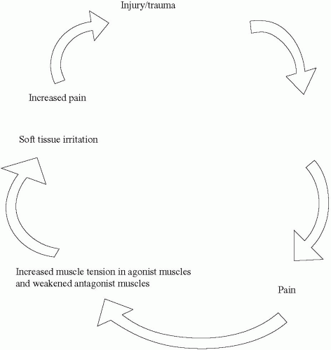
The repetitive stress and mechanical loading of tissues by athletic training cause increased strain on the fascial system. This increase in muscle tension commonly disrupts flow in the delivery of nutrients and removal of waste products in the tissues. The accumulation of waste products acts as an injurious stimulus, inducing further tissue irritation, pain, and inflammation. Up to this point, the process can be thought of as acute. Acute processes generally manifest as severe, sharp tenderness, edema, erythema, and bogginess in the soft tissues, with increased moisture and hypertonicity.

The continuation of this course results in a fibrous tissue reaction leading to a chronic process. Chronic changes are less likely to be reversible, making the soft tissue damage more permanent. Furthermore, the body experiences muscle shortening, decreasing stretch, and restricting movements. Manifestations of chronic processes are usually dull, achy, burning, ropy, cool, dry skin with fibrotic changes and slight tension.

Both acute and chronic tissue changes often present with compensation in other parts of the body. The pervasiveness and interconnectedness of fascia throughout the body create a scenario in which restriction in one part of the body will not only affect local, adjacent structures but may also affect areas distal to the site of injury. It then becomes necessary to address dysfunction in both the local area of injury as well as in distant areas.

Myofascial pain is sometimes associated with fibrositis, defined by fibrous tissue inflammation and connective tissue hyperplasia. Frequently, a hyperirritable region within a taut band of tissue with referred pain is known as a myofascial trigger point. Trigger points have a predictable distribution and are commonly found within areas of myofascial pain. The concept and principles of myofascial trigger points can be further studied in works by Janet Travell.

Because of the fascia’s integration with the neuromuscular system, many symptoms are mediated by the sympathetic nervous system. Myofascial release elicits neuroreflexive changes in the musculoskeletal system, from the skin to deep spinal joints. During myofascial release, the afferent stimulation caused by a stretch movement results in the sequential relaxation
of tight tissues by efferent inhibition, providing instant relief on many occasions.


FIGURE 6.1. Positive feedback cycle of myofascial pain. The initial injury or trauma causes soft tissue irritation. The body interprets the irritation as pain and reacts by increasing muscle tension in the agonist muscle and weakening the antagonist muscle, which increases pain and further soft tissue injury and irritation.


Positive Feedback Cycle of Myofascial Pain

The soft tissue irritation caused by injury or dysfunction is read by the body as pain, and it reacts by increasing the tension in the agonist muscle and weakening the antagonist muscle. An increase in muscle tension is interpreted as increasing pain leading to increased muscle injury, thus positively feeding back into the cycle as soft tissue irritation (Fig. 6.1). Some examples of the multiple causes of myofascial pain are included in Table 6.1. Myofascial therapies are useful in reversing this damage.


Tight-Loose Concept (Direct-Indirect Barriers) in Myofascial Release

Tightness creates asymmetry, and looseness permits asymmetry. Three main barriers are encountered in osteopathic manipulation: the physiologic, anatomic, and restrictive (pathologic) barriers. A
restrictive barrier causes asymmetry and inhibits movement in one direction. This is a basic concept of myofascial release, and looking for three-dimensional tightness and looseness is essential. Areas of myofascial injury can vary in size, pattern, and depth (superficial to deep), and they can be tight or loose in relation to one another.


TABLE 6.1. CAUSES OF MYOFASCIAL PAIN


Causes of Myofascial Pain


Examples


Poor posture


Chronic muscular strain, scoliosis, resting and exercise posture, leg-length discrepancies, anatomic variants, skating


Malpositioning/maladjustments


Bicycle seat, sleep position, weight training


Poor biomechanics


Improper techniques, shifting of center of gravity, jogging, running, snowboarding, skiing


Repetitive stresses, overuse injuries


Golf, tennis, soccer, swimming, baseball


Immobility


Sedentary lifestyle, prolonged rest


Steady contractions and position


Weight training, yoga, Pilates exercises, wrestling, prolonged sitting


Prolonged constriction of soft tissues


Ski and snowboard boots, rollerblades, ill-fitting sports bra or sportswear, tight shirts, collars, belts, shoes


Nutritional/vitamin deficiencies


Vitamin B12, folate, thiamine deficiency, calf cramps, paresthesias


Infections/other processes


Abscesses, local inflammations, allergies, edema, effusions


Psychological factors


Depression, sleep disturbance, anxiety, tension


Endocrine/metabolic


Hypothyroidism, hypoglycemia


Other


Lifestyle, accidents, trauma, drug use, tobacco, alcohol, physical fitness


The term “end-feel” describes the sensation one feels when a mobilized joint moves into a barrier, or the end of its range of motion. Asymmetrically perceived end-feels are commonly referred to as direct and indirect barriers. Direct barriers suggest tethering and tightness, whereas indirect barriers suggest tissue laxity and looseness. Movements are easier in some directions, less so in others. End-feels that are hard and terminate abruptly occur with direct barriers, whereas indirect barriers have soft and easy-tonavigate end-feels. Restrictions involving one side of the body frequently affect the opposite side.

Tight muscles are not always sources of pain and altered neuromusculoskeletal functioning. On the other hand, loosened sites are often chronic, painful, and susceptible to injury at relatively low thresholds of stress. Tight muscles can be self-limiting or they can steady an unstable region by increasing fibrotic tissue formation.


Physiologic Principles

DiGiovanna and Schiowitz (1) have described several principles underlying myofascial techniques. Their effects cross over to several types of tissues and systems, which is part of the reason why myofascial techniques are effective. The principles are as follows:


  • Increased circulation to the area of restriction delivers oxygenated blood and nutrients to the tissues and removes harmful metabolic waste products.


  • Increased venous and lymphatic drainage decreases local swelling and edema caused by tissue inflammation.


  • The elasticity and flexibility of connective tissues elongate connective tissues secondary to mechanical loading.


  • Increased temperature causes an increase in elasticity and stretch of the muscle.


  • The stretch reflex stimulates tone in hypotonic muscles in areas of looseness. Muscle stretching excites the muscle spindle causing reflex contraction of the muscle.


  • The stretch reflex is sustained by the muscle spindle reflex. The Golgi tendon organ can cause prompt relaxation of the muscle (via
    inhibition) when tension on the tendon becomes extreme. Relaxation of contracted muscles occurs decreasing the oxygen demand of the muscle, decreasing pain, and allowing normalized range of motion across a joint.


  • Reciprocal inhibition: The stretch reflex activates one muscle (e.g., quadriceps), while simultaneously inhibiting its antagonist muscle (the hamstrings).


  • Crossed extensor reflex: The stretch reflex stimulates one muscle (e.g., right quadriceps), while simultaneously activating the contralateral antagonist muscle (the left hamstrings), creating an “X” pattern.


MECHANICS OF MYOFASCIAL PATHOLOGY

The effects of multidirectional forces on both local and distant joints and soft tissues manifest in injury patterns. Mechanical loading, increasing strain, and repetitive stress on a soft tissue over time under a constant load are certain to cause deformation. All tissues are mechanically responsive, exhibiting stress-strain responses that affect the body neurologically and anatomically.

Two-handed palpation is generally required to interactively assess and modify dysfunctional patterns and the athlete’s ability to adapt. Searching out tight and loose end-feels allows assessment and simultaneous treatment of dysfunctional soft tissue patterns and joint-related movements. Using compression, traction, and twisting maneuvers to mechanically load areas of restriction can help release barriers. Athleteassisted release-enhancing maneuvers may be further integrated to complement the treatment process.

The fascia release phenomenon is also known as melting or quivering of the segment. As treatment is directed to an area of tissue injury using layer-by-layer palpation, the practitioner’s hands move further into the restrictive barrier, which begins to soften, allowing the muscle to relax into the fascia. By permitting the fascia to guide the practitioner, the tissue will ease up progressively from superficial to deep. The fascia responds by becoming more pliable and increases in elasticity. In addition, the heat imparted by the hands further increases the stretch and melting of the segment. Appropriate application of stress on the tissue results in both muscle and fascia tissue relaxation, as the tightness “melts” and gives way under the application of load.

Only gold members can continue reading. Log In or Register to continue

Stay updated, free articles. Join our Telegram channel

Aug 27, 2016 | Posted by in ORTHOPEDIC | Comments Off on Myofascial Release

Full access? Get Clinical Tree

Get Clinical Tree app for offline access