The Wafer Procedure
Paul Feldon
Indications and Contraindications
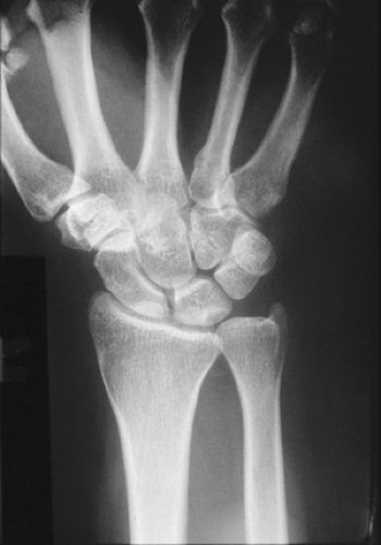
Partial resection of the distal portion (“pole”) of the ulna or wafer procedure was designed to treat ulna impaction syndrome and symptomatic tears of the triangular fibrocartilage complex (TFCC) (Fig. 42-1). The wafer procedure requires only a limited arthrotomy of the distal radioulnar joint (DRUJ) and allows direct inspection of the TFCC, débridement or repair of the TFCC, if necessary, as well as decompression of the ulnocarpal junction (1,2). It preserves the ligamentous attachments of the TFCC to the base of the ulna styloid process and preserves the function of the DRUJ. Patients with chronic ulnar-sided wrist pain may have one or more of the following: positive ulnar variance (long ulna relative to radius), sclerotic or cystic changes in the lunate and triquetrum, TFC perforations on arthrogram or arthroscopy, and disruption of proximal surface of the TFC. The wafer procedure can be used when any or all of these are present (2,3).
The advantages of the wafer procedure over other decompressive procedures, such as ulnar shortening osteotomy, are as follows:
Does not require bone healing
Does not require internal fixation
Requires limited surgical exposure
Provides direct access to TFCC
Allows ulnar shortening, decompression of the ulnocarpal junction, and TFCC debridement or repair in a single procedure
The wafer procedure can be used to treat ulna impaction syndrome in a patient who (a) refuses ulnar-shortening osteotomy, (b) has some contraindication for ulnar shortening, or (c) has not done well following a wafer procedure done arthroscopically.
The procedure does have some disadvantages:
Technically more difficult than osteotomy
Risk of cutaneous nerve damage
Amount of bone resection is limited
Long recovery time
Most of the contraindications to the wafer procedure relate to the anatomy and condition of the patient’s DRUJ. These include the following:
DRUJ/wrist (lunotriquetral) instability
DRUJ arthritis
Ulna impingement (ulna against radius) syndrome
Ulna minus variance
Limited articular surface of the ulna “seat”
Positive ulna variance >4 mm
Surgical Technique
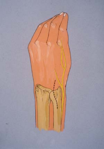
In the original description of the wafer procedure, the DRUJ is exposed through a slightly serpentine longitudinal incision on the dorsal and ulnar aspect of the wrist between the fifth and sixth compartments (Fig. 42-2).

Figure 42-1 Radiograph of a patient with ulna impaction syndrome. Note crescent-shaped subchondral cyst in proximal and ulnar corner of lunate.

Figure 42-2 Initially, the wafer procedure was done through a slightly serpentine incision. Now, a straight longitudinal incision is used.
Now, use a straight longitudinal incision.
It is important to mobilize and protect any branches of the dorsal sensory portion of the ulnar nerve.
Open the fifth dorsal compartment.
Preserve the sixth compartment intact to prevent both destabilizing the DRUJ by disrupting the subsheath and postoperative extensor carpi ulnaris (ECU) tendonitis by closing the sheath too tightly (Fig. 42-3).
Elevate a radially based U-shaped flap of capsule to expose the DRUJ, the vertical edge of the TFCC, and the articular surfaces of the lunate and triquetrum (Fig. 42-4).
With an osteotome, microsagittal saw or side-cutting burr remove the distal 2 to 4 mm of the head of the ulna (Fig. 42-5
Stay updated, free articles. Join our Telegram channel

Full access? Get Clinical Tree


