and Peter J. Millett2
(1)
Steadman Philippon Research Institute, Vail, CO, USA
(2)
The Steadman Clinic, Steadman Philippon Research Institute, Vail, CO, USA
Keywords
ShoulderSternoclavicularSC jointCostoclavicularMedial clavicleDislocationPhysical examClinical exam8.1 Introduction
The sternoclavicular (SC) joint is a complex articulation that can have significant anatomic variations within individuals, between individuals, and throughout a population. Nevertheless, the inherent stability and biomechanics of the SC joint remain relatively unchanged. While an acute traumatic injury can lead to an anterior or posterior SC joint dislocation, repetitive micromotion and shear can lead to degenerative changes which may produce chronic symptomatology. Clinicians should be familiar with the anatomy, biomechanics, and process of diagnostic evaluation in patients with pain or disability related to the SC joint such that an accurate diagnosis and effective treatment modality can be chosen.
8.2 Relevant Anatomy and Biomechanics
8.2.1 Osseous Anatomy
Although the clavicle is the first bone to ossify (fifth gestational week), the medial clavicular physis typically does not close until at least 25 years of age. In fact, some studies have shown that physeal closure may not actually occur until 31 years of age in some patients [1, 2]. Therefore, patients younger than 31 years of age who present with possible SC joint dislocations should be evaluated for both ligamentous integrity and patency of the medial clavicular physis.
The SC joint is a diarthrodial joint lined with synovium that primarily functions to connect the shoulder girdle to the axial skeleton (Fig. 8.1). Because the articular surface of the medial clavicle is highly incongruent with the manubrium, joint stability is maintained primarily by strong ligamentous attachments. Specifically, the articular surface of the medial clavicle is much larger than that of the manubrium and has been described as having a “saddle-like” configuration (i.e., concave in the axial plane and convex in the coronal plane) which biomechanically functions similarly to a ball-and-socket joint (Fig. 8.2) [4, 5].
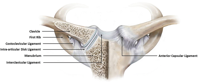
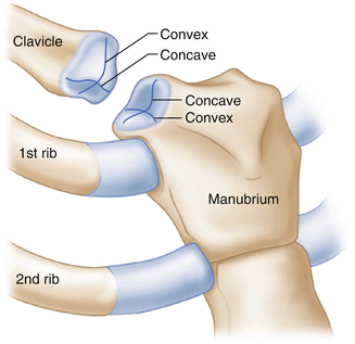
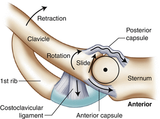
Fig. 8.1
Illustration highlighting the important structural components of the SC joint. (From Martetschläger et al. [3]; with permission).
Fig. 8.2
The articular surfaces of the sternum and medial clavicle are incongruent, although the medial clavicle typically exists in a “saddle” configuration (i.e., concave in the axial plane and convex in the coronal plane).
Both the manubrium and the medial clavicle can have a variety of different anatomic configurations which may vary across populations, between genders and, potentially, within the same patient [6, 7]. Tuscano et al. [7] quantified this asymmetry in a series of 104 patients who had previously undergone a computed tomography (CT) scan of the chest for other reasons. In their study, the investigators measured joint spaces and the maximum diameter of the medial clavicular head in each patient. Overall, joint spaces ranged from 0.2 to 1.37 cm across the study population and medial clavicular head diameters ranged from 1.2 to 3.7 cm. Interestingly, some patients displayed differences in medial clavicular head diameters between their right and left clavicles (range, 0.0–1.0 cm difference). Other authors have found that the manubrium may also be subject to significant anatomic variation [8]. As a result, osseous asymmetry of the SC joint should be expected in the clinical setting to prevent misdiagnoses and unnecessary surgery.
8.2.2 Chondral Surfaces
The articular surfaces of the medial clavicle and the manubrium are covered with hyaline cartilage that eventually become fibrocartilage with increasing age [9]. Recent dissections performed at this institution revealed that only approximately two-thirds of the medial clavicle was covered with articular cartilage: the majority of this cartilage was found anteriorly and inferiorly where the medial clavicle was devoid of capsuloligamentous attachments (Fig. 8.3). This finding has also been confirmed by others [10]. We also identified a previously undescribed ridge that traveled along the superior aspect of the clavicular head of the pectoralis major insertion site [11]. This “pectoralis ridge” may prove to be an important landmark for orientation during arthroscopy or rotational alignment during open reconstructive procedures (see Figs. 8.3 and 8.4).
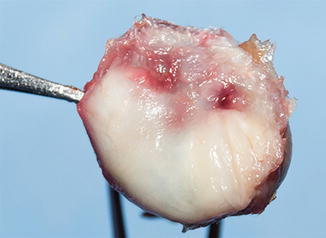
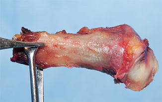
Fig. 8.3
Approximately two-thirds of the medial clavicle is covered with articular cartilage. The forceps point to the pectoralis ridge which may be an important landmark for surgical orientation. (From Warth RJ, Lee JT, Millett PJ. Anatomy and biomechanics of the sternoclavicular joint. Oper Tech Sports Med. 2014;22(3):248–52; with permission).
Fig. 8.4
The forceps point to the pectoralis ridge of the medial clavicle. The ridge extends from the tip of the forceps medially towards the articular surface.
8.2.3 Ligamentous Anatomy
As mentioned above, the articular surfaces of the SC joint are highly incongruent and rely primarily on the patency of static capsuloligamentous structures to maintain stability. These structures include the capsular ligaments, costoclavicular ligament, interclavicular ligament, and the intra-articular disc ligament.
8.2.3.1 Capsular Ligaments
Integrated within the SC joint capsule are discrete thickenings that represent the anterior and posterior capsular ligaments. The anterior capsular ligament runs from an area just superior to the articular cartilage of the medial clavicle to an area just superior to the articular cartilage of the manubrium. The posterior capsular ligament is essentially a thickening of the entire posterior capsule and spans between the posterosuperior aspect of the medial clavicle to the posterior aspect of the manubrium. Several studies have shown that the posterior capsule is most important for the maintenance of horizontal stability, thus preventing anterior and posterior translation of the medial clavicle [12, 13]. The anterior capsular ligament most likely plays a secondary role in the maintenance of horizontal stability.
8.2.3.2 Costoclavicular Ligament
The costoclavicular ligament is a thick, robust fibrous band that inserts across the costochondral junction of the first rib and travels towards the inferior aspect of the medial clavicle to insert on the costoclavicular tubercle (see Fig. 8.1) [11]. The ligament is commonly described as being composed of two separate fascicles (anterior and posterior) oriented in a “twisted” configuration with an interposed bursa spanning between the first rib and the medial clavicle [14]. However, our recent cadaveric dissections revealed that the costoclavicular ligament may actually exist as a single ligament since we were unable to identify or separate the previously described anterior and posterior fascicles [11]. The costoclavicular ligament may be the most important ligamentous stabilizer of the SC joint in both the vertical and the horizontal axes [14, 15].
8.2.3.3 Interclavicular Ligament
The interclavicular ligament is a thick, fibrous band that lies over the superior aspect of the manubrium as it runs between the superomedial aspect of each SC joint capsule (see Fig. 8.1) [11]. Despite its anatomic position and thickness, the interclavicular ligament actually does not significantly contribute to vertical stability of the SC joint. Although we were able to identify the ligament in all of our recent cadaveric dissections, its attachments to the manubrium and the SC joint capsules are quite weak and could potentially be removed during the reflection of overlying soft tissues [11].
8.2.3.4 Intra-Articular Disc Ligament
The intra-articular disc ligament attaches near the chondral junction of the first rib, passes through the SC joint (thus creating two separate joint spaces), and inserts along the superior margin of the articular cartilage of the medial clavicle (see Fig. 8.1). This ligament does not confer joint stability. Rather, it probably functions to diminish the force transmission between the manubrium and the medial clavicle which is thought to decrease the detrimental effects of bony incongruity on the health of the articular surfaces [9, 16, 17]. The morphology of the intra-articular disc has also been shown to vary considerably between individuals. Although DePalma [9] found the intra-articular disc to be complete in 97 % of his specimens, others have found that as many was 52 % of specimens may have disc degeneration which is often clinically associated with symptomatic osteoarthritis of the SC joint [10, 13, 17, 18].
8.2.4 Mediastinal Vessels
Traumatic injuries to the SC joint can result in severe complications due to the proximity of the mediastinal vessels, especially in those with posterior SC joint dislocations. These mediastinal vessels include the subclavian vessels, the right and left brachiocephalic veins, the brachiocephalic artery and the left carotid artery (Fig. 8.5). According to Ponce et al. [6], the closest vessel lies approximately 6.6 mm deep to the posterior SC joint capsule (left or right brachiocephalic vein). In some cases, the subclavian vein may actually cross over the first rib along the posterolateral aspect of the costoclavicular ligament before merging with the brachiocephalic vein [14].
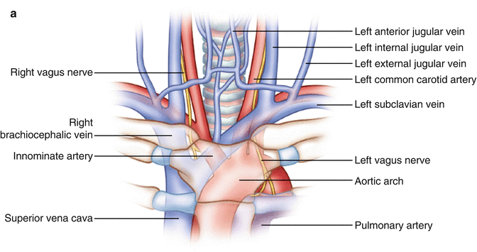
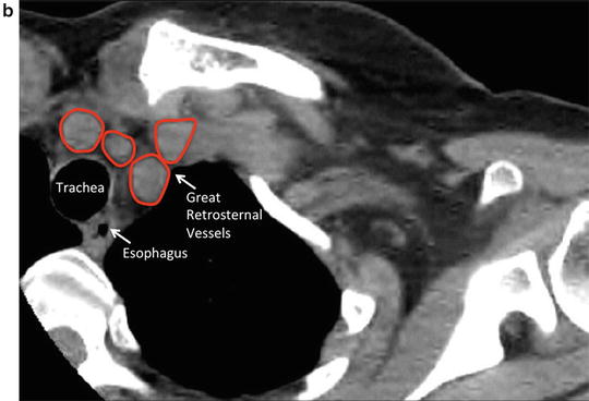
Fig. 8.5
(a) Illustration showing the important structures that are situated posterior to the SC joint. (b) CT scan showing the orientation of these structures in the axial plane.
In our cadaveric dissections, the fascial raphe between the sternohyoid and sternothyroid muscles was found to provide some protection to the mediastinal vessels since it forms a physical barrier between the medial clavicle and the mediastinal vessels (Fig. 8.6). We also noted a “safe zone” for posterior dissection in the interval between the sternothyroid muscle and area directly posterior to the manubrium and the medial clavicle [11]. However, in the event of posterior dislocation, the sternohyoid and sternothyroid muscle bellies may become disrupted, placing the posterior vessels at an increased risk for iatrogenic injury during posterior surgical dissection of the SC joint. Disruption of the sternohyoid and sternothyroid muscle bellies can easily be identified on diagnostic axial MRI scans.
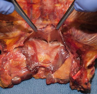
Fig. 8.6
Cadaveric dissection photograph showing the fascial raphe between the sternohyoid and sternothyroid muscles which is thought to provide some protection to the mediastinal vessels.
8.2.5 Biomechanics
For every 10° of humeral elevation, the SC joint contributes approximately 4° of rotation (Fig. 8.7) [19]. The SC joint also contributes approximately 50° of posterior rotation with every 35° of elevation, flexion and extension of the humerus (Fig. 8.8) [20, 21]. Therefore, surgical procedures involving rigid fixation of the SC joint have largely been abandoned due to the potential for poor functional outcomes in addition to the high reported incidence of hardware failure. Large resections of the medial clavicle have also been reported; however, this can result in uncontrollable scapulothoracic motion and, therefore, may require subsequent scapulothoracic fusion [22].
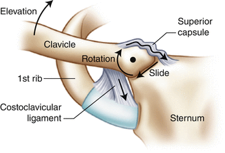
Fig. 8.7
Anterior view showing the motion across the SC joint as the humerus is elevated above 90º.