The Skeletal Dysplasias


The Skeletal Dysplasias


Paul D. Sponseller

Michael C. Ain


GENERAL PRINCIPLES

Osteochondral dysplasias are rare disorders of growth and development that affect cartilage and bone. Knowledge about these dysplasias is important to orthopaedic surgeons as an aid to understanding skeletal development. In the preface to the classic text, McKusick’s Heritable Disorders of Connective Tissue (1), the late Victor McKusick stated, “Nature is nowhere more openly to display her secret mysteries than in cases where she shows traces of her workings apart from the beaten path…” It may be true that there is a mutation and a disorder representing nearly each step of skeletal development. Although there is substantial overlap between conditions that primarily affect cartilage and those that primarily affect bone because of shared matrix elements, metabolic pathways, hormonal influences, and other processes (2), this chapter focuses on those that affect cartilage (for a summary, see Appendix 1).

A useful tool for diagnosis and additional research is the Online Mendelian Inheritance in Man. This web-based compendium is publicly available and readily accessible on the PubMed Web site of the National Library of Medicine (http://www.ncbi.nlm.nih.gov/omim). It allows a user to search by physical features or diagnosis and provides a compilation of applicable knowledge on each (3).


Terminology.

Most skeletal dysplasias result in short stature, defined as height more than 2 standard deviations below the mean for the population at a given age. The term “dwarfing condition” is used to refer to disproportionate short stature. The disproportion is commonly referred to as “short trunk” or “short limb.” The short-limb types are further subdivided into categories based on which segment of the limb is short. “Rhizomelic” refers to shortening of the root (proximal) portion of the limb; “mesomelic,” to the middle segment; and “acromelic,” to the distal segment. Achondroplasia is a classic example of rhizomelic involvement, with the femora and especially the humeri being most affected by shortening. Some of these disorders are named after the appearance of the skeleton (diastrophic means “to grow twisted,” camptomelic means “bent limbs,” and chondrodysplasia punctata refers to stippled cartilage). Eponyms such as Kneist, Morquio, and McKusick are used to name others.


Pathogenesis.

Although their pathogenesis is only slowly being investigated, a number of mechanisms have been discovered to lead to skeletal dysplasia. Some result from an alteration in transcription or in the intracellular or extracellular processing of structural molecules of the skeleton (Fig. 7-1). Others are caused by a defect in a receptor or signal transduction in pathways of skeletal differentiation and proliferation. These abnormalities tend to occur in the pathway of cartilage differentiation, growth, and development.

Abnormalities in the form of a structural macromolecule may occur, as in type-II collagen causing spondyloepiphyseal dysplasia (SED). In some cases, the effect may be magnified—a phenomenon termed a “dominant negative” effect. This phenomenon occurs as the defective gene product binds to normal copies of the product, leading to early destruction of normal and defective copies, as seen in osteogenesis imperfecta type II. Pseudoachondroplasia provides another example, with the abnormal cartilage oligomeric matrix protein (COMP) accumulating in the rough endoplasmic reticulum and causing secondary retention of type-IX collagen and other proteins. By contrast, models in which COMP is completely knocked out and not expressed display no disease.

Another pathway through which mutations may act is the alteration of the transport of structural molecules. One example of this mechanism is the group of conditions that includes diastrophic dysplasia (DD) and achondrogenesis type 1, the result of a defect in sulfate transport. This alteration disturbs proteoglycan assembly, leading to diffuse changes in the articular surface cartilage, growth plate, and other areas. An example of receptors gone awry is the family of disorders that includes achondroplasia, hypochondroplasia, and thanatophoric dysplasia. These disorders occur as a result of varying defects in fibroblast growth factor receptor protein. These mutations result in a constitutively active receptor
(gain of function). Because this receptor down-regulates endochondral growth, mutations result in decreased endochondral growth. Another example is Jansson metaphyseal dysplasia, which is the result of a constitutively active mutation in parathyroid hormone receptor protein. This protein inhibits the expression of the signaling factor Indian hedgehog, which is needed to stimulate terminal differentiation to hypertrophic chondrocytes and produce normal metaphyseal growth. Disorders of transcription may also cause skeletal dysplasia, as seen in cleidocranial dysplasia, a defect in core-binding factor 1. Because this transcription factor stimulates osteoblast differentiation, a defect in this factor leads to a cartilage model that is well formed but not normally ossified.


Classification.

The classification of skeletal dysplasias has traditionally been structured according to the pattern of bone involvement, as in the International Classification of Osteochondrodysplasias (4) (Table 7-1). Another approach, however, is to group them according to the specific causative gene defect for cases in which the defect is known (Table 7-2). A schematic representation of the effects of the known mutations on cartilage development is shown in Figure 7-1. It is also useful for the orthopaedic surgeon to classify the dysplasias into those that are free from spinal deformity [for instance, hypochondroplasia and multiple epiphyseal dysplasia (MED) rarely have significant spinal abnormalities] versus those for which spinal deformity is a frequent problem (such as SED, DD, and metatropic dysplasia). Which disorders are free from epiphyseal involvement and therefore from risk of subsequent degenerative joint disease (DJD)? Achondroplasia and hypochondroplasia, cleidocranial dysplasia, and diaphyseal aclasia rarely present these problems in adulthood, but SED, MED, DD, and others commonly do.


TABLE 7-1 International Nosology and Classification of Genetic Skeletal Disorders 2006 (Partial List)


FGFR3 group


Type-II collagen group


Sulfation disorder group


Perlecan group


Filman group


MED/pseudoachondroplasia group


Metaphyseal dysplasias


Spondylometaphyseal dysplasias


Spondyloepimetaphyseal dysplasias


Acromesomelic dysplasias


Mesomelic and rhizomelic dysplasias


Bent bone dysplasias


Slender bone dysplasias


Dysplasias with multiple joint dislocations


Chondrodysplasia punctata group


Increased bone density group


Decreased bone density group


Lysosomal storage diseases


Cleidocranial dysplasia group


TABLE 7-2 Classification of Skeletal Dysplasias Based on Pathogenesis (Partial List)


Defects in extracellular structural proteins


COL1 (OI)


COL2 (achondrogenesis, hypochondrogenesis, SEDC, SEDC, Stickler, Kneist)


COL9 (MED)


COL10 (Schmidt)


COL11 (Stickler variant)


COMP (pseudoachondroplasia, MED)


MATN3 (MED)


Defects in metabolic pathways


AP (hypophosphatasia)


DTDST (achondrogenesis B, DD, rMED)


Defects in processing and degradation of macromolecules


Sedlin (SED-X-linked type)


Lysosomal enzymes (mucopolysaccharidoses, mucolipidoses)


EXT1, EXT2 (MHE1,2)


Defects in hormones, growth factors, receptors, and signal transduction


FCGRs 1-3 (craniosynostoses, achondroplasia, thanatophoric)


PTH/PTHrP (Jansen metaphyseal dysplasia)


GNAS1 (McCune Albright, pseudohypoparathyroidism)


Defects in nuclear proteins and transcription factors


SOX1 (camptomelic dysplasia)


CBFA1 (cleidocranial dysplasia)


SHOX (Leri-Weill)


Defects in RNA processing and metabolism


RMRP (cartilage-hair hypoplasia)


Defects in cytoskeletal proteins


Filamins (Larsen syndrome, Melnick-Needles)


Prenatal Diagnosis

With the increasing availability of prenatal screening, many individuals with skeletal dysplasia are being diagnosed before birth. When ultrasound shows a fetus with shortening of the skeleton, femur length is the best biometric parameter to distinguish among the five most common possible conditions. In one study, fetuses with femur length <40% of the mean for gestational age most commonly had achondrogenesis, those with femur length between 40% and 60% most commonly had thanatophoric dysplasia or osteogenesis imperfecta type II, and those with femur length >80% most commonly had achondroplasia or osteogenesis imperfecta type III (5). Additional testing may be performed, if indicated, by chorionic villous sampling and mutation analysis.


Evaluation.

In evaluating for skeletal dysplasia in a patient with short stature or abnormal bone development, there are several aspects of the medical history that should be investigated as an aid to diagnosis and coordination of care. Birth length
is usually shorter than normal in patients with achondroplasia, SED, and most dysplasias but not in those with pseudoachondroplasia or storage disorders. Head circumference is usually larger than normal in patients with achondroplasia. Respiratory difficulty in infancy may occur as a result of restrictive problems in the syndromes with a small thorax, neurologic problems such as foramen magnum stenosis in achondroplasia, or upper airway obstruction in various conditions. A history of heart disease suggests the possibilities of chondroectodermal dysplasia, which may be associated with congenital heart malformations, or storage disorders, such as Hurler or Morquio syndromes, in which cardiac dysfunction may be acquired. A history of immune deficiency or malabsorption is common in cartilage-hair hypoplasia. Retinal detachment may occur with Kneist syndrome or SED. The clinician should elicit information about a family history of short stature or dysmorphism any previous skeletal surgery the patient may have had.


FIGURE 7-1. Schematic illustration of the sites and effects of the known cartilage defects in the skeletal dysplasias. Section of cartilage matrix of physis and epiphysis is simplified and enlarged; genetic abnormalities often affect both regions. DST, diastrophic sulfate transporter, deficiency of which leads to undersulfation of proteoglycans in epiphysis and physis of DD and achondrogenesis types 1B and 2; Col II, type-II collagen, which is defective in Kneist dysplasia and SED; COMP, cartilage oligomeric matrix protein, abnormal pseudoachondroplasia, and some forms of MED; Col IX, type-IX collagen, which is closely linked to type-II collagen and is abnormal in some forms of MED; FGFR3, fibroblast growth factor receptor 3, which inhibits chondrocyte proliferation in achondroplasia, hypochondroplasia, and thanatophoric dysplasia; Col X, type-X collagen, which is synthesized only by the hypertrophic cells of the growth plate and is abnormal in Schmidt-type metaphyseal chondrodysplasia.

The presence of unusual facial characteristics, a cleft palate, or extremity malformations should be noted. Height percentile for age should be determined using standard charts. Most skeletal dysplasias result in adult height of <60 in. Measurement of the upper:lower segment ratio may be helpful in distinguishing disproportion early. This value can be obtained by measuring the distance from the top of the pubic symphysis to the sole of the plantigrade foot and subtracting it from the overall length. The normal ratio is 1.6 at birth (given that extremities develop later than the trunk) and diminishes to 0.93 in adults and teens. If shortening of the extremities is noted, it is helpful to classify it as rhizomelic (shortest in the humerus and femur), as in achondroplasia, mesomelia (shortest in the forearms and the legs), or acromelia (shortest distally). The extremities should be examined for ligamentous laxity or contracture (6, 7).

A thorough neurologic examination is needed because of the frequent incidence of spinal compromise at the upper cervical level in SED, DD, Larsen syndrome, and metatropic dysplasia, or at any level in achondroplasia.

A skeletal survey should be ordered, including lateral radiographs of skull and neck and anteroposterior views of the
entire spine, pelvis, arms, hands, and legs. Much of this information can be gleaned from reviewing previous radiographs of the child’s chest and abdomen that may have been obtained. Sometimes, pathognomonic features will be revealed, such as the caudal narrowing of the interpediculate distances in achondroplasia, double-layered patella in MED, and the iliac horns in nail-patella syndrome. Flexion-extension radiographs of the cervical spine should be ordered if instability is suspected to be causing delay in reaching milestones, loss of strength, or loss of endurance. In many syndromes in which cervical instability is common, such as SED, such radiographs should be ordered as a matter of course. Magnetic resonance imaging (MRI) in flexion and extension may be helpful in some cases to determine if the instability is causing critical risk. However, the limitation of this test is that it often must be done under anesthesia or sedation and the degree of cervical movement is less. If conventional radiographs show substantial motion and a static MRI shows signal changes at the same location, then flexion and extension images are usually not needed.

Laboratory tests may include calcium, phosphate, alkaline phosphatase, and protein to rule out metabolic disorders such as hypophosphatemia or hypophosphatasia. If a progressive disorder is found, the patient’s urine should be screened for storage products (under the guidance of a geneticist). To rule out hypothyroidism, serum thyroxine should be measured if the fontanels in an infant are bulging and bone development is delayed. After the differential diagnosis is clinically focused, DNA testing for mutation analysis is increasingly being done in the clinical setting for patients with skeletal dysplasias. A geneticist should be consulted to help establish a diagnosis and a prognosis and to address medical problems. The geneticist sometimes functions as a primary physician for a patient with a genetic disorder because a geneticist has the best overview of the medical issues facing the patient.


ACHONDROPLASIA


Overview.

Achondroplasia, an abnormality of endochondral ossification, is the most common form of skeletal dysplasia and occurs in approximately 1 of 25,000 live births (14, 15).

Achondroplasia is caused by a gain of function in the mutation of a gene that encodes for fibroblast growth factor receptor 3 (FGFR3) (16, 17, 18 and 19). Achondroplasia arises from a point mutation on the short arm of chromosome 4 at nucleotide 1138 of the FGFR3 gene. The mutation is located on the distal short arm of chromosome 4. The result of this mutation is endochondral-ossification-engendered underdevelopment and shortening of the long bones that does not involve intramembranous or periosteal components.

Achondroplasia is inherited as a fully penetrant autosomal dominant trait, but more than 80% of such cases are sporadic, meaning both parents are unaffected (20). If one of the parents is affected, there is a 50/50 chance that the child will develop achondroplasia (14, 15, 20). However, because there is also an increased incidence when the parents are more than 33 years old at the time of conception, a de novo mutation is implied (21).


Etiology.

The cause of achondroplasia is a single-point mutation in the gene that encodes for FGFR3. FGFR3 mutations have also been found in individuals with thanatophoric dysplasia and hypochondroplasia. Almost all people with achondroplasia have the same recurrent G-380Rlocus mutation, which causes a change in a single amino acid. This mutation substitutes an arginine for a glycine residue in the transmembrane domain of the tyrosine-coupled transmembrane receptor in the physis (1, 22). 
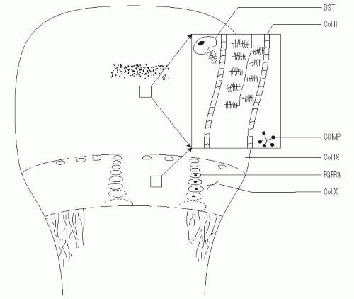
FGFR3 is expressed in the cartilaginous precursors of bone, where it is believed to decrease chondrocyte proliferation in the proliferative zone of the physis and to regulate growth by limiting endochondral ossification (23). However, in persons with achondroplasia, articular cartilage formation, articular cartilage development, and the intramembranous and periosteal ossification processes are unaffected (24).

It is not known why the proximal portions of the long bones (rhizomelic) are affected more than the distal aspects.


Clinical Features.

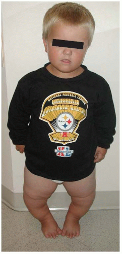
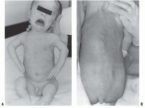
In the achondroplastic population, the extremities are most affected, that is, they are shorter than those in an unaffected individual. The most commonly affected bones are the humerus and femur, which present a rhizomelic appearance (25). The trunk length is within normal limits or at the lower end of normal limits. This combination typically results in the fingertips reaching only to the tops of the greater trochanter (26), a condition that can lead to possible difficulties in personal hygiene and care and that can worsen as decreasing amount of flexibility occurs in the normal aging process (Fig. 7-2).

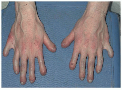
The hands are described as trident in nature, that is, the individual is unable to oppose the third and the fourth ray, leaving a space that cannot be closed. There are flexion contractures at the elbow and decreased ability to supinate, most often secondary to the fact that the radial head can be subluxed, as evidenced on radiographs. None of the above features are clinically important, but a nonknowledgeable physician might misdiagnose such a presentation as a “nursemaid’s elbow” and incorrectly attempt a reduction.

The facial appearance of patients with achondroplasia is characterized by an enlarged head, mandibular protrusion, frontal bossing (flattened or depressed nasal bridge), and midface hypoplasia. The bones in the midface are more affected than the other facial bones because of their endochondral origin (16).

Although the lower extremities are typically in varus secondary to knee and ankle morphology, the lower extremities can be straight or occasionally in valgus, and internal tibial torsion may also be seen. Typically, the knee and ankle joints have excessive laxity, although usually such patients do not develop premature arthritis. The femoral necks are often shortened, giving an appearance of coxa breva.

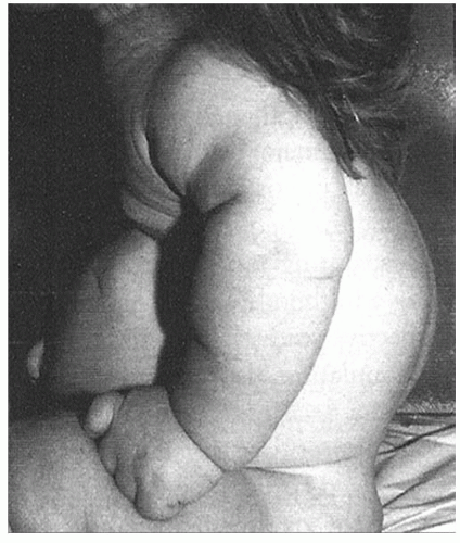
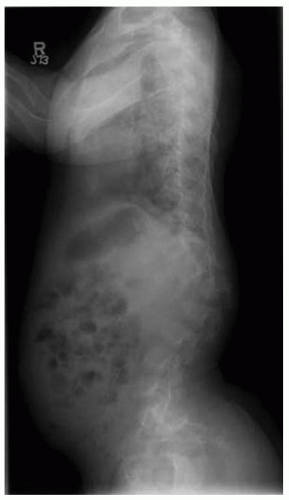
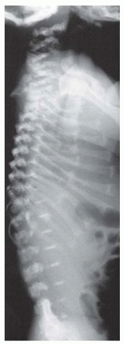
In terms of the spine, kyphosis at the thoracolumbar junction is very common and is typically seen in the first 1 to 2 years of life (Fig. 7-3). In most children, this condition will correct spontaneously within a few months of ambulation, although ambulation is often delayed in patients with achondroplasia (27). As kyphosis improves, lumbosacral lordosis may seem to progress.


FIGURE 7-2. Photograph of a 5-year-old male with achondroplasia. Note the typical bowing and fingertips reaching to the top of the hips.

One study found that life expectancy is not substantially diminished in individuals with achondroplasia (28), but a more recent report has indicated a higher mortality rate in 30- to 50-year-old people with achondroplasia compared with age-matched controls (29). This reported increased mortality is typically secondary to heart disease. One indicator for heart disease is abnormal blood pressure, but it is possible that standard blood pressure cuffs may underestimate the pressures in the achondroplastic population, leading to the nonidentification and nontreatment of a large number of patients with high blood pressure. New cuffs have been developed and their use for this population is being reviewed.


Growth and Development.

In most children with achondroplasia, growth and development fall behind those of unaffected children.

Widely available growth charts (25) indicate that the infant with achondroplasia is shorter than an unaffected infant, a
height deficit that increases markedly during the first few years of life and becomes even more marked during the growth spurt at puberty (30). The average height for an adult with achondroplasia is 132 cm for men and 125 cm for women (20).


FIGURE 7-3. Photograph of an 18-month-old female with achondroplasia sitting with typical postural kyphosis.

Children with achondroplasia also have delayed motor milestones (head control, 4 months; sitting up independently, 10 months; ambulation, 18 to 20 months) (31, 32), and threequarters of them have ventriculomegaly (33).

Historically, hydrocephalus was thought to be the cause, leading also to macrocephaly, but only a very small subset has been shown to have clinically significant hydrocephalus (34); standardized head circumference charts can help track such children (35). Ventricular peroneal shunting is indicated only for rapid progression of head circumference, or for signs and symptoms of increased intracranial pressure.

Mental development is typically normal in children with achondroplasia, but physical manifestations are often delayed, especially in the first 2 to 3 years of life (36). Typically, motor development normalizes by 3 years of age. There are standardized developmental charts that are available for monitoring such children.

Foramen magnum stenosis is one of the earliest serious health consequences faced by some children with achondroplasia. Its symptoms, which most commonly occur in the first 2 years of life but which may present later (37), include chronic brain stem compression, sleep apnea, lower cranial nerve dysfunction, difficulty in swallowing, hyperreflexia, hypotonia, weakness, paresis or clonus, and severe developmental delay, and are quantified in sleep studies (34, 38, 39, 40, 41 and 42). The most common presenting symptom of foramen magnum stenosis is respiratory difficulty with excessive snoring or apnea (43). Apnea can be central in nature (because of brainstem compression) or just obstructive because of the individual’s small midface. The American Academy of Pediatrics recommends screening for foramen magnum stenosis with polysonography and computed tomography (CT) or MRI in all infants with achondroplasia. Because CT and MRI in the first year of life require sedation, the child who is developing well, has no abnormal reflexes, and is alert, oriented, and meeting all milestones can typically just be followed clinically. If head circumference changes or if a patient is not reaching milestones, a sleep study should be ordered. If the sleep study is abnormal, then a CT or an MRI scan should be obtained. We prefer MRI because, in our opinion, it produces a better image of the brain stem and the upper cervical spinal cord.

Some studies have shown a high mortality rate (2% to 5%) in infants with achondroplasia and have indicated foramen magnum stenosis as the responsible factor (33).


Radiographic Characteristics.

There are several features typically seen on the radiographs of individuals with achondroplasia, but caution should be exercised in interpreting the absence of such findings: not all affected individuals exhibit such radiographic characteristics.

The key feature is the typical narrowing intrapedicular distance from L1 to L5 seen on the anteroposterior radiographs of affected individuals (44, 45); in the unaffected population, the intrapedicular distance from L1 to L5 increases. The presence of such narrowing is an absolute indicator of achondroplasia, but the lack of such narrowing does not rule out the presence of achondroplasia. In addition, pedicles in those with achondroplasia are approximately 30% to 40% thicker than those in unaffected individuals (46). In this patient population, the vertebral bodies have a scalloped appearance (20), lumbar lordosis increases to the sacrum segment and may even become horizontal, and severe scoliosis is rare but can be seen; however, the incidence of cervical instability is not higher than that in the unaffected population (20, 46).

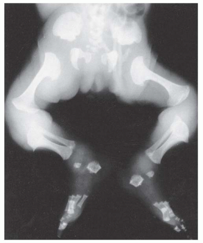
Other radiographic abnormalities include underdeveloped facial bones, skull base, and foramen magnum; square iliac rings; rhizomelic shortening; and flared metaphysis of the long bones. Affected individuals also have a pronounced, inverted “v” shape of the distal femoral physis with normal distal femoral epiphysis, and the metacarpals and metatarsals are almost all equal in length. The iliac wings have a squared appearance. The metaphysis of all long bones is flared in appearance. Despite being short, the diaphyses of all long bones are thick. The sites of major muscle insertions, such as the tibial tubercle, greater trochanter, and insertion of the deltoid, are more prominent than usual. The epiphysis throughout the skeleton is normal in appearance and development; consequently, degenerative joint arthritis or changes are rarely seen.


General Medical Treatment.

Although children with achondroplasia are typically healthier than those with other dysplasias, infants and young children with achondroplasia should be closely monitored and evaluated, especially during the first few years of life, for signs and symptoms of foramen magnum stenosis (see earlier). If the diagnosis is made clinically, an MRI should be ordered to show the stenosis. At this point, neurosurgery can enlarge the foramen magnum. Sometimes, surgeons may need to perform a durotomy or an expansion of the dura and a C1 laminectomy. Many of these children also have dilatation of the veins of their cranium secondary to venous distension, which can also be relieved by such surgery. As indicated earlier, children with achondroplasia have delayed motor milestones; for example, most unaffected children walk by 12 months, whereas most of those with achondroplasia do not walk until 18 months. Postsurgery, patients typically are able to start achieving milestones much more quickly and progress rapidly (38, 40). In addition, children with achondroplasia have a higher risk of respiratory complications than do unaffected children, not only because of midface hypoplasia and upper airway obstruction, but also because of a decreased respiratory drive that can be secondary to foramen magnum stenosis. Early brain stem decompression can decrease the risk (37).

Otolaryngeal problems are also prevalent: 90% of the patients with achondroplasia can experience otitis media before they are 2 years old (47), and many require ear tube placements. Adenoid and tonsil hypertrophy in the presence of midface hypoplasia can cause obstructive sleep apnea. The otitis media and adenotonsil hypertrophy may result in conductive hearing loss that can impair speech development and delay.

Achieving and maintaining an ideal body weight is also difficult and a lifelong struggle. Currently, because there are no standardized charts for size and weight, observing skin-fold thickness and noting general appearance may be the best clinical option (48, 49).

Children with achondroplasia are typically not deficient in growth hormone levels, but there is a substantial amount of research with regard to the administration of growth hormone to supplement height (18, 50, 51). Typically, in the first year of receiving growth hormone treatment, there is an increase in the growth height velocity, but it diminishes over the next 2 to 3 years, with a net result of no real increase. There has been speculation that too much growth hormone can hasten the development of spinal stenosis, which is one of the worst complications of achondroplasia (18, 50, 51).

There are several other otolaryngologic problems that are usually secondary to the underdevelopment of midface skeletal structures. Maxillary hypoplasia can lead to dental overcrowding and malocclusion (52). Many children with this condition require orthodontic attention. In such children, Eustachian tubes often do not function properly because the children are smaller than the unaffected population and more horizontally than vertically positioned, decreasing the ability to drain middle ear fluid (53).

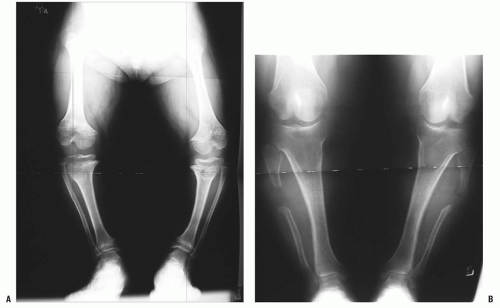
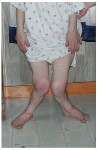
Orthopaedic problems include angular deformities of the lower extremities, genu varum at the knees, thoracolumbar kyphosis, and spinal stenosis (which can occur at any level of the spinal canal). Malalignment of the lower extremities is typically secondary to genu varum or ankle varus (24, 54). A very small percentage of patients have genu valgum, which rarely becomes severe enough to require treatment, but genu varum may progress to cause substantial pain and difficulty in ambulation (24, 55). Some clinicians have postulated that the longer fibula is the cause of this pain, but others have shown that the length of the fibula has no direct relationship on the amount of bowing on the knee (56, 57 and 58) (Fig. 7-4). Leg malalignment has been shown to be the result of ankle, distal femur, or proximal tibia deformity, or from a combination thereof. Incomplete ossification epiphysis often makes it quite challenging to elicit the source of this malalignment. Arthrograms are typically used at our institution to help identify the exact location of the deformity and are especially helpful in patients <8 years old (12). Although bracing has been used elsewhere to help control ligamentous laxity and to try to correct bowing, we have found that the short and often pendulous nature of the legs of patients with achondroplasia makes it difficult to provide a brace with proper fit and enough of a mechanical advantage to correct the malalignment. During the past 10 years of our practice, no brace has been used to control malalignment, and surgical decisions are not made until the child is at least 3 years old. The indication for surgery is persistent pain that is secondary to malalignment (not to spinal stenosis) deformity severe enough to cause a fibular thrust, resulting in a gap between the proximal tibia and the femur on ambulation (40, 57, 59). Again, in our practice, if the decision has been made for surgical intervention, an arthrogram is obtained to evaluate the optimal location of the osteotomy. Such arthrography also often identifies internal tibial torsion, which can then be corrected concurrently. Although fibular shortening has been advocated in the past (55), we and others (60, 61) do not think it is ever indicated. Treatment indications are difficult to define clearly because there are no natural history studies showing which degree of deformity causes early degeneration.


Short Stature and Limb Lengthening.

Infants with achondroplasia are shorter than other individuals and the deficit progresses until skeletal maturity.

Everyday difficulties as the result of short stature include using public restrooms, face washing in public restrooms, hair combing, engaging in hobbies involving physical activity, playing sports with average-statured individuals, conducting routine business transactions (often at countertop level), and driving a car. Nevertheless, the decision to augment stature is difficult and controversial because the procedure is time consuming, complicated (40, 62), and fraught with complications (38, 40).

First, surgical lengthening can achieve quite a bit of height if done safely and correctly (13, 63, 64), but because it is a time-consuming process, it removes these children from their normal activities of school and socialization. The psychologic impact can be tremendous, especially if the lengthening goals
are not achieved. At some centers, lengthening is performed at two separate time intervals, the first typically at the age of 7 years old and the second at preadolescence. The overall time frame for surgery and postoperative therapy may be up to 3 years. Some centers prefer to delay lengthening until early adolescence to increase the patient’s participation for the rehabilitation process and also the decision making as to whether or not the lengthening should be done. We know of only one child with achondroplasia who has had limb lengthening and whose parents were also affected.


FIGURE 7-4. Prefibulectomy (A) and postfibulectomy (B) (without change of alignment) radiographs of a 14-year-old male with achondroplasia and tibia vara.

Second, surgical limb lengthening is a very complicated endeavor (40, 62), and one that is fraught with complications (38, 40). In one study by Aldegheri and Dall’Oca (65), 43% of the patients who underwent limb lengthening had complications, including fracture, failure of premature consolidation, malunion, malalignment, joint stiffness, and infection. One report in the literature indicates increased symptoms of lumbar spinal stenosis (66). The effect of limb lengthening on spinal stenosis needs additional investigation. Humeral lengthening, often is combined with lower extremity limb lengthening, may be the most functionally appreciated because it makes it is easier to perform personal care, put on shoes and socks, and perform extended reaching. The procedure also has lower risks than lower limb lengthening.

Despite the fact that limb lengthening has been a procedure in frequent use for several decades, to our knowledge, the functional benefits after elective limb lengthening have never been studied.


Spinal Aspects

Thoracolumbar kyphosis develops in most infants with achondroplasia. A newborn with achondroplasia typically has a thoracolumbar kyphosis centered at approximately T12-L1. When sitting begins, the infant slumps forward because of trunk hypotonia, in combination with a relatively large head, flat chest, and protuberant abdomen. The apex of the vertebral deformity becomes wedge-shaped anteriorly, although it usually is a reversible phenomenon. This condition should not be confused with a diagnosis of congenital kyphosis. Most of these patients improve by the 2nd or the 3rd year of life, after walking begins and muscle strength increases (15, 27, 67, 68). However, persistent kyphosis can increase the risk of symptomatic stenosis, putting pressure on the conus (Fig. 7-5). To prevent persistent kyphosis, Pauli et al. (27) recommended early parental counseling (before the infant is 1 year old) for prohibition of unsupported sitting or sitting up at more than a 45-degree angle and for the use of the following measures: firm, back-seating devices; curling the infant into a “C” position; hand counterpressure when holding the infant; and bracing as needed. In the study by Pauli et al. (27), bracing is initiated for patients who develop kyphosis that does not correct to <30 degrees on prone lateral radiographs. Those authors initially used bracing but found it cumbersome
and not very helpful. In addition, the braced patient may be at an increased risk for falls because of the brace’s large size and the patient’s small body, poor trunk control, and developmental delay. Bracing may also have a detrimental effect on pulmonary function in children with small thoracic cages. In our practice, we have found that bracing has delayed the onset of walking. If wedging of the vertebrae persists beyond the ages of 4 or 5 years, and surgical intervention is not sought, we recommend a trial of using hyperextension casts to see if it will help with the wedging. Although some surgeons have used this technique with some benefit (69), we have seen several instances of such use that have resulted in numerous complications, including skin breakdowns, the inability to tolerate the casts, decreased ability to ambulate, and others. Currently, the indication for safe surgical intervention in our practice is kyphosis ≥50 degrees at 5 years of age with no sign of improvement (70). The key is twofold: (a) no hooks, wires, or any other hardware in the canal and (b) no overcorrection. Correction should limited to what is obtainable preoperatively with the awake child hyperextended laterally over a bolster. The threshold for performing an anterior arthrodesis has decreased with increased rigidity instrumentation by the placement of pedicle screws at every level (70, 71, 72, 73, 74, 75 and 76).


FIGURE 7-5. Radiograph of a 4-year-old patient with achondroplasia and thoracolumbar wedging and kyphosis.

For the child with achondroplasia, thoracolumbar kyphosis, and concurrent spinal stenosis symptoms, MRI will be obtained; if it shows anterior cord impingement, a corpectomy via an anterior approach will be performed. In this situation, we would not perform a vertebral body resection posteriorly because we think the achondroplastic spinal cord is not mobile enough to tolerate such a procedure. Currently, any child >3 years old who can accommodate pedicle screws, including cervical spine screws, is not placed in a cast or brace postoperatively. However, for a child <3 years old with pedicle screws, a bracing protocol is instituted for 3 months.

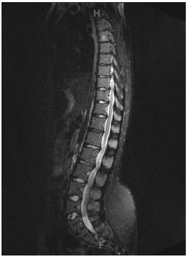
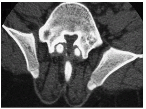
Lumbar stenosis typically can present during the second or third decade of life of an individual with achondroplasia, but it can be seen as early as 18 months (Fig. 7-6). Patients typically present with complaints of lower back pain, leg pain, progressive weakness of the extremities, numbness, and tingling, symptoms that often are decreased or alleviated completely by squatting or bending over—maneuvers that reduce the lumbar lordosis, increase the size of the canal, and relieve the pressure. Surgical indications include myelopathy, progressive signs and symptoms, inability to ambulate more than one or two city
blocks without having to stop or squat to relieve the pressure. Preoperative workup includes MRI and possibly a CT scan (Fig. 7-7). By correlating the physical examination and the MRI study, the clinician can identify the approximate level of the most severe stenosis. Usually, treatment is a wide decompression that extends at least two levels above the point of the most severe stenosis and down to the sacrum. In skeletally immature patients, a posterior spinal fusion with pedicle screw instrumentation needs to be done concurrently to prevent progressive kyphosis. If the patient is skeletally mature and has no underlying kyphosis, posterior decompression can be done alone, without a concurrent fusion (40, 46, 71, 72, 73 and 74, 77).


FIGURE 7-6. T2-weighted MRI of a 12-year-old patient with achondroplasia and lumbar stenosis.


FIGURE 7-7. CT myelogram in a patient with achondroplasia and severe lumbar/sacral stenosis.


HYPOCHONDROPLASIA


Etiology and Pathogenesis.

Hypochondroplasia is an autosomal dominant disorder, and the chance of passing it on to offspring is approximately 50% (1). Although hypochondroplasia and achondroplasia have similar names and are similar phenotypically (individuals with mild achondroplasia can appear similar to individuals with severe hypochondroplasia), they are two distinct disorders. The mutation that causes hypochondroplasia is located on the short arm of chromosome 4, in gene FGFR3, as it is in achondroplasia and thanatophoric dysplasia. However, the nucleotide change is in a different region, the tyrosine kinase domain. In hypochondroplasia, the mutation results in increased activation of factors that slow cell growth (16, 78, 79, 80 and 81).


Clinical Features.

Hypochondroplasia can usually be identified at birth, but it can also be unrecognized until early puberty if the individual is only mildly affected. The presentation is more varied than that of achondroplasia; foramen magnum stenosis and thoracolumbar stenosis are extremely rare in patients with hypochondroplasia.

Compared with the achondroplastic population, individuals with hypochondroplasia have less of a height discrepancy (118 to 160 cm) (20, 82); similar, but less pronounced, facial characteristics; limbs shorter than the trunk, but to a lesser extent; milder other features such as thoracolumbar kyphosis, spinal stenosis, and genu varum; and mesomelic rather than rhizomelic long-bone shortening. In addition, the need for surgical intervention for patients with hypochondroplasia is much lower than that for those with achondroplasia. In our practice, we have surgically treated several hundred patients who had achondroplasia with spinal stenosis and/or kyphosis, but only a few patients with hypochondroplasia have required surgical intervention. Unlike individuals with achondroplasia, in whom intelligence is normal, a small portion (<10%) of those with hypochondroplasia have been associated with mental retardation (83).


Radiographic Features.

Hall and Spranger (84) have proposed primary and secondary criteria for making this diagnosis. Primary criteria are narrowing of the pedicles in the lumbar spine, squaring of the iliac crest, broad femoral necks, mild metaphyseal flaring, and brachydactyly. Secondary criteria are shortening lumbar pedicles, mild posterior scalloping of the vertebral bodies, elongation of the distal fibula, and ulnar styloid. In patients with achondroplasia, the sciatic notches are narrow in nature; in patients with hypochondroplasia, the notches are unaffected and normal in appearance.


Differential Diagnosis.

Compared with achondroplasia, hypochondroplasia is a much milder form of skeletal dysplasia and has a much more variable presentation; it can also go unrecognized until early puberty. However, severe cases of hypochondroplasia can overlap mild forms of achondroplasia. Occasionally, hypochondroplasia can be confused with Schmidt metaphyseal dysplasia because both disorders have mild short stature, typically normal faces, and mild genu varum.


METATROPIC DYSPLASIA


Overview.

The term “metatropic dwarfism” comes from the Greek word metatropos, or “changing form,” because patients with this condition appear to have short-limb dwarfism early in life, but later develop a short-trunk pattern as spinal length is lost with the development of kyphosis and scoliosis. The condition has been likened to Morquio syndrome because of the enlarged appearance of the metaphyses and the contractures (87).


FIGURE 7-8. Histology of the growth plate in metatropic dysplasia, showing relatively normal columns of proliferating chondrocytes (C), but absence of the hypertrophic or the degenerating zones, as well as a “seal,” or bony end plate (EP), over the metaphysis. (From Boden SD, Kaplan FS, Fallon MD, et al. Metatropic dwarfism: uncoupling of endochondral and perichondral growth. J Bone Joint Surg Am 1987;69:174, with permission.)

It is a rare condition that may be inherited in an autosomal dominant or recessive manner (88). The cause of this dysplasia has not been elucidated. However, histologic abnormalities of the growth plate have been studied and appear to be characteristic, as shown in the study by Boden et al. (89). The physis shows relatively normal columns of proliferating chondrocytes. However, there is an abrupt arrest of further development, with absence of a zone of hypertrophic or degenerating chondrocytes. Instead, there is a mineralized seal of bone over the metaphyseal end of the growth plate (Fig. 7-8). The perichondral ring remains intact, and circumferential growth is preserved. This uncoupling of endochondral and perichondral growth appears to account for the characteristic “knobby” metaphyses and the platyspondyly. Additional understanding of the defect in this disorder will shed light on the normal maturation of the physis.


Clinical Features.

One of the most characteristic features of this condition is the presence of the “coccygeal tail,” a cartilaginous prolongation of the coccyx that is not present in other dysplasias (Fig. 7-9A,B). It is usually a few centimeters long and arises from the gluteal fold. The facial appearance includes a high forehead, and there may be a high arched palate. The sternum may display a pectus carinatum, the limbs have flexion contractures of up to 30 to 40 degrees from infancy, and other joints may have ligamentous laxity. The limbs appear relatively short with respect to the trunk. The metaphyses are enlarged, which, when combined with underdeveloped musculature, gives a “bulky” appearance to the limbs. Some patients have been reported to have ventriculomegaly or hydrocephalus (90) or to develop upper cervical spine instability and/or stenosis (90, 91). Scoliosis develops in early childhood and is progressive (92, 93). Some restrictive lung disease is usually present,
and it may cause death in infancy for the one-third of patients who are afflicted by the autosomal recessive form of the disease (88, 93). However, for those who survive into adulthood, height varies from 110 to 120 cm.


FIGURE 7-9. A 1-year-old infant with metatropic dysplasia, illustrating knee-flexion contractures, “bulky” metaphyses (A), and a coccygeal tail (B).


Radiographic Features.

Prenatal sonographic diagnosis may be possible in the first or second trimester, with the finding of substantial dwarfism, narrow thorax, and enlarged metaphyses (94, 95). Odontoid hypoplasia frequently exists in patients with this condition, as in many patients with skeletal dysplasia. In infancy, the vertebrae are markedly flattened throughout the spine, but normal in width. Kyphosis and scoliosis develop in most patients. The ribs are short and flared, with cupping at the costochondral junctions (Fig. 7-10).

The epiphyses and metaphyses are enlarged, giving the long bones an appearance that has been likened to that of a barbell (Fig. 7-11). The epiphyses have delayed and irregular ossification. Protrusio acetabuli has been reported (93). Genu varum of mild-to-moderate degree usually develops. Degenerative changes of major joints often occur in adulthood.


FIGURE 7-10. Newborn with metatropic dysplasia. Note platyspondyly with delayed vertebral ossification and flared ribs. (Courtesy of Judy Hall, Vancouver, BC.)


FIGURE 7-11. Newborn with metatropic dysplasia. The diaphyses are short and the metaphyses are broad and flared; their appearance has been likened to dumbbells. The iliac wings are flared, and the acetabulae are deep. (Courtesy of George S. Bassett, MD.)


CHONDROECTODERMAL DYSPLASIA


Overview.

Chondroectodermal dysplasia is an uncommon disorder. It is also known as Ellis-Van-Creveld syndrome and is prevalent among the Amish (96). It results in disproportional short stature and abnormalities in the teeth, limbs, and cardiac areas. The pathognomonic characteristic of this condition is severe flattening or wedging of the lateral proximal tibial physis, which leads to the severe genu valgum (Fig. 7-12).

It is a defect in EVC gene, or in the short arm of chromosome 4 (97, 98 and 99). It results in the defect of maturation of endochondral ossification. It is transmitted as an autosomal recessive condition.


Orthopaedic Treatment.

The first priority for patients with chondroectodermal dysplasia is stabilization of the heart. Approximately one-third of these infants die in the first few weeks of life (100). In the first year, most patients with this condition have polydactyly, which can be reconstructed (Fig. 7-13). Genu valgum frequently occurs and can be quite severe. If seen early, genu valgum can be treated with guided growth, such as a hemiepiphysiodesis with an 8-plate. In our practice, bracing has had no effect on this condition and does not help control the severe ligamentous laxity. If surgical intervention (i.e., osteotomies) is warranted because of severe valgus angulation, rotational malalignment should be considered along with any genu valgum. The distal femur is typically externally rotated, and the tibia is internally rotated. It appears as though there is a flexion contracture in the lower extremities, but after correcting the malrotation, the flexion contracture typically disappears and then the malalignment needs to be corrected (62, 101). Clinicians can correct the malalignment with external or internal fixation. In these children and young adults, there is an increased risk of patellar subluxation and dislocation. Many times, lateral release, medial reefing, and even tibial tubercle osteotomies are required. In the presence of genu valgum, after correcting the malrotation, lateral proximal tibial elevation can also be entertained. Before the plateau elevation, an external fixator across the knee can be placed to open the lateral joint line. Osteotomy is necessary in severe cases because there is a high rate of recurrence.


FIGURE 7-12. Photograph of a 16-year-old Amish male with Ellis-Van-Creveld syndrome and severe genu valgum.


FIGURE 7-13. Photograph of a 21-year-old male with Ellis-Van-Creveld syndrome who did not undergo polydactyly correction.


Clinical Features.

Approximately half of the children with chondroectodermal dysplasia have cardiac defects, most commonly atrial septal defects. One-third of children with this condition die during the neonatal period, most from cardiac abnormalities.

Patients with this disorder develop hypospadious and epispadious. They have narrow chests, abnormal dentition (with crooked, sparse, and sometimes lost teeth), abnormal nails, and postaxial polydactyly. This condition presents as acromesomelic shortening of the middle and distal segments
of the upper and lower extremities (102, 103, 104 and 105). The spine is typically uninvolved. The lower extremities have significant genu valgum secondary to a hypoplastic proximal laterotibial plateau and lax ligaments, and rotational abnormalities (such as external rotation of the femur or internal rotation of the tibia) are often present, as though there were a flexion contracture.


Radiographic Features.

The ribs are short, the chest is narrow, and there is uneven growth of the proximal tibial epiphysis laterally. Exostosis can develop from the proximal tibial epiphysis medially and acetabular spike of the medial and lateral edges. The greater trochanteric epiphyses are quite pronounced, and the wrists can display fusion of the capitates, hamate, and (sometimes) other carpal bones. Carpal bones typically have delayed maturation, in contrast to the accelerated maturation of the phalanges.


DIASTROPHIC DYSPLASIA


Overview.

DD is perhaps the dysplasia with the most numerous, disparate, and severe skeletal abnormalities. The term “diastrophic” comes from a Greek root meaning “distorted,” which aptly describes the ears, spine, long bones, and feet. Before the current level of understanding of the skeletal dysplasias was developed, early authorities referred to this condition as “achondroplasia with clubbed feet” (106, 107). Certainly, the skeletal abnormalities are much more extensive than that.

The disorder is autosomal recessive and is extremely rare, except in Finland, where between 1% and 2% of the population are carriers, and there are more than 160 people known to be affected because of an apparent founder effect (108). The defect is on chromosome 5 in the gene that codes for a sulfate transporter protein (aptly named “diastrophic dysplasia sulfate transporter” or DTDST) (109, 110). This protein is expressed in virtually all cell types. Decreased content of sulfate in cartilage from patients with DD has been shown (111). A defect in this gene leads to undersulfation of proteoglycan in the cartilage matrix. If one considers proteoglycans to be the “hydraulic jacks” of cartilage at the ultrastructural level, it is understandable that there should be such impairment of performance of physeal, epiphyseal, and articular cartilage throughout the body. Achondrogenesis types 1B and 2 are more serious disorders causing mutations on the same gene.

Histopathology reveals that chondrocytes appear to degenerate prematurely, and collagen is present in excess (112, 113). Tracheal cartilage has some of the same abnormalities seen in other cartilage types, but it still does not explain some of the specific focal malformations seen in DD, such as proximal interphalangeal joint fusion in the hands, short first metacarpal causing hitchhiker thumbs, or cervical spina bifida. Additional work on the role of this sulfate transporter on skeletal growth and development must be done to explain these curious findings.


Clinical Features.

Prominent cheeks gave rise to the previously used name “cherub dwarf” (Fig. 7-14). The nasal bridge is flattened. Up to one-half of patients have a cleft palate, which may contribute to aspiration pneumonia (112). The cartilage of the trachea is abnormally soft, and its diameter may be narrowed. The ear is normal at birth but develops a peculiar acute swelling of the pinna at 3 to 6 weeks in 80% to 85% of cases (114). The reasons for this event and this timing are not known. The cartilage hardens in a deformed shape—the “cauliflower ear,” which is one of the pathognomonic features of this dysplasia.

Patients with diastrophism have a slightly increased [approximately 5% (106, 107)] perinatal mortality as a result of respiratory problems, especially aspiration pneumonia and tracheomalacia. Motor milestones are delayed: sitting occurs at a mean age of 8 months, pulling up to a stand at 13 months, and walking at 24 months (115).

The skeleton displays abnormalities from the cervical spine down to the feet (6). The posterior arches of the lower cervical spine are often bifid. There are no external clues to this occult underlying abnormality. Cervical kyphosis is seen in one-third to one-half of patients (11, 116); it may be present in infancy, and its course is variable. Spontaneous resolution has been reported in a number of patients, even with curves of up to
80 degrees (117, 118) (Fig. 7-15A-C). However, others progress, and several reports of quadriparesis from this deformity exist (11, 119). Scoliosis develops in at least one-third of patients (116), but many curves do not exceed 50 degrees. Tolo (120) has stated that the scoliosis may be one of two types: idiopathic-like or sharply angular. The sharply angular type is usually characterized by kyphosis at the same level as the scoliosis. Spinal stenosis is not common, in contrast to achondroplasia. Most patients have substantial lumbar lordosis, likely to compensate for the hip flexion contractures in diastrophism.

Only gold members can continue reading. Log In or Register to continue

Stay updated, free articles. Join our Telegram channel

Jul 21, 2016 | Posted by in ORTHOPEDIC | Comments Off on The Skeletal Dysplasias

Full access? Get Clinical Tree

Get Clinical Tree app for offline access