The Shoulder
17.1 Anatomy
William M. Falls
Gail A. Shafer-Crane
The shoulder girdle is composed of three joints and one articulation, which work together to permit movement of the upper limb. These include the sternoclavicular joint, the acromioclavicular joint, the glenohumeral (shoulder) joint, and the scapulothoracic articulation (1,2,3,4,5,6).
Several bony structures can be palpated as one examines the shoulder girdle (Fig. 17.1.1). Beginning on the midline is the suprasternal notch situated along the superior margin of the manubrium of the sternum. Immediately lateral to the suprasternal notch is the sternoclavicular joint. The medial end of the clavicle participates in this joint and lies slightly superior to the manubrium. Moving laterally from the sternoclavicular joint one can palpate the smooth superior surface of the clavicle, which is devoid of muscle attachment and is covered by the thin platysma muscle. The medial two thirds of the clavicle are convex, while the lateral one third is concave. At the deepest point of the concavity of the clavicle and inferior and deep to the anterior edge, one can palpate the coracoid process of the scapula. The coracoid process faces anterolaterally, lies deep to the pectoralis minor muscle, and may be felt as one palpates into the deltopectoral triangle.
At the lateral end of the clavicle is the subcutaneous acromioclavicular joint. At this joint the clavicle, which has begun to flatten out, protrudes slightly above the acromion process of the scapula. The acromion is rectangular in shape, contributes to the contour of the shoulder, and can be palpated on its superior and anterior surfaces.
As one palpates laterally and inferiorly from the lateral lip of the acromion, the greater tubercle of the humerus can be felt. The bicipital groove of the humerus is situated anterior and medial to the greater tubercle. It is bordered laterally by the greater tubercle and medially by the lesser tubercle and is best palpated with the arm laterally rotated. Within the bicipital groove is the tendon of the long head of the biceps muscle. Inferior to the acromion is the shoulder joint, which cannot be palpated. This is a very mobile joint where the head of the humerus fits into a very shallow glenoid cavity. As a result, the humerus is suspended from the scapula by muscles, ligaments, and a loose joint capsule with very little bony support.
Posteriorly, the acromion tapers and continues as the spine of the scapula. The spine extends obliquely across the superior four fifths of the posterior surface of the scapula and ends as a flat triangle on the vertebral border of the scapula at the level of T3. The vertebral border, lying approximately 5 cm from the spinous processes of the thoracic vertebrae, terminates superiorly at the superior angle, which is covered by the levator scapulae muscle. The vertebral border ends inferiorly at the subcutaneous inferior angle. The lateral border, which is covered by the latissimus dorsi, teres major, and teres minor muscles, runs obliquely toward the glenoid.
The sternoclavicular joint is divided into two compartments by an articular cartilage. The medial end of the clavicle articulates with the manubrium of the sternum and the first costal cartilage, reinforced by the anterior and
posterior sternoclavicular ligaments. The costoclavicular ligament, medial to the joint, attaches the inferior surface of the clavicle to the first rib and costal cartilage. The sternoclavicular joint moves in anterior, posterior, superior, and inferior directions. The joint is innervated by the medial supraclavicular nerve and the nerve to the subclavius.
posterior sternoclavicular ligaments. The costoclavicular ligament, medial to the joint, attaches the inferior surface of the clavicle to the first rib and costal cartilage. The sternoclavicular joint moves in anterior, posterior, superior, and inferior directions. The joint is innervated by the medial supraclavicular nerve and the nerve to the subclavius.
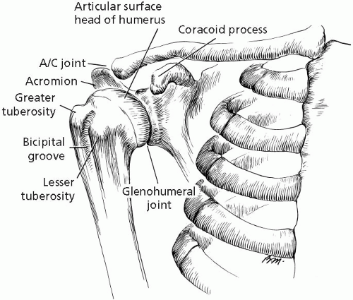 FIGURE 17.1.1. Bony anatomy of the shoulder. (From Rockwood, Green. Fractures in Adults, 4th ed., vol 2. Baltimore: Lippincott Williams & Wilkins, 1996.) |
The acromioclavicular joint is a synovial joint, surrounded by an articular capsule, where the lateral end of the clavicle articulates with the acromion of the scapula. The superior and inferior acromioclavicular ligaments strengthen the joint superiorly and inferiorly, and the coracoclavicular ligament attaches the clavicle to the underlying coracoid process of the scapula. The acromion rotates on the clavicle from scapulothoracic movements. The acromioclavicular joint is innervated by the supraclavicular, lateral pectoral, and axillary nerves.
The glenohumeral joint is a synovial joint where the head of the humerus articulates with the shallow glenoid cavity of the scapula, which is deepened by the fibrocartilaginous glenoid labrum (Fig. 17.1.2). The loose joint capsule is attached medially to the margin of the glenoid cavity and laterally to the anatomic neck of the humerus. The synovial lining of the capsule forms a sheath for the tendon of the long head of the biceps muscle, which runs through the joint cavity.
The three glenohumeral ligaments strengthen the anterior aspect of the joint capsule, and the coracohumeral ligaments strengthen the joint superiorly. The capsule is also strengthened by the transverse humeral ligament, which helps to hold the tendon of the long head of the biceps muscle in the bicipital groove. The coracoacromial ligament extends across the superior aspect of the joint, separated from the joint capsule, and together with the coracoid process and the acromion forms the strong coracoacromial arch, which protects the joint superiorly. The shoulder joint receives its blood supply from the suprascapular artery and branches of the anterior and posterior humeral circumflex arteries from the axillary artery and is innervated by the suprascapular, axillary, and lateral pectoral nerves.
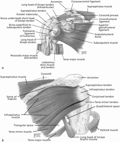
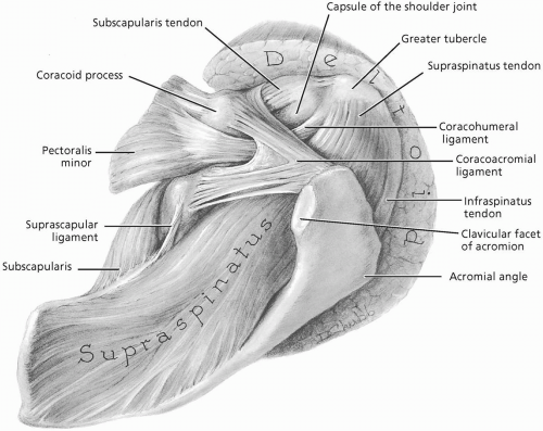
The rotator cuff muscles attach the scapula to the humerus (Fig. 17.1.3A and B). The supraspinatus, infraspinatus, and teres minor are palpable at their attachments onto the greater tubercle of the humerus. The supraspinatus is an abductor of the humerus, while the infraspinatus and teres minor are lateral rotators. With the arm hanging at the side, the supraspinatus
lies directly over the head of the humerus deep to the acromion (Fig. 17.1.4). The infraspinatus is posterior and inferior to the supraspinatus, and the teres minor is posterior and inferior to the infraspinatus. The fourth cuff muscle, the subscapularis, lies anterior to the humeral head, attaches to the lesser tubercle of the humerus, and acts as a medial rotator of the humerus. The supraspinatus is innervated by the suprascapular nerve (C4, C5, C6); the infraspinatus by the suprascapular nerve (C5, C6); the teres minor by the axillary nerve (C5, C6); and the subscapularis by the upper and lower subscapular nerves (C5, C6, C7).
lies directly over the head of the humerus deep to the acromion (Fig. 17.1.4). The infraspinatus is posterior and inferior to the supraspinatus, and the teres minor is posterior and inferior to the infraspinatus. The fourth cuff muscle, the subscapularis, lies anterior to the humeral head, attaches to the lesser tubercle of the humerus, and acts as a medial rotator of the humerus. The supraspinatus is innervated by the suprascapular nerve (C4, C5, C6); the infraspinatus by the suprascapular nerve (C5, C6); the teres minor by the axillary nerve (C5, C6); and the subscapularis by the upper and lower subscapular nerves (C5, C6, C7).
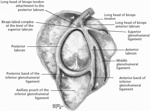 FIGURE 17.1.2. Glenohumeral ligaments and labrum, lateral view. (From Stoller DW. MRI, arthroscopy, and surgical anatomy of the joints. Baltimore: Lippincott Williams & Wilkins, 1999.) |
Bursae surround the shoulder lying between the tendons of the rotator cuff muscles and the shoulder joint capsule. They reduce friction on the tendons passing over bones and other areas of resistance. The subacromial bursa lies between the tendon of the supraspinatus muscle and the overlying acromion process of the scapula. Several portions of this bursa are palpable. A lateral extension of the subacromial bursa, the subdeltoid bursa, extends deep to the deltoid muscle, separating it from the rotator cuff below and allowing each to move more freely.
Several other muscles attach to the shoulder girdle and can be palpated. The sternocleidomastoid muscle arises from the manubrium of the sternum and the medial third of the clavicle, bisects the neck obliquely, and attaches superiorly to the mastoid process of the temporal bone. This muscle laterally flexes the neck and rotates the head to the opposite side and is innervated by the spinal accessory nerve (CN XI).
The fan-shaped pectoralis major muscle forms the anterior wall of the axilla, arises from the medial two thirds of the clavicle and the anterior surface of the sternum, and inserts into the lateral lip of the bicipital groove of the humerus. It adducts and medially rotates the humerus, and draws the scapula anteriorly and inferiorly. The pectoralis major receives innervation from the lateral and medial pectoral
nerves (clavicular head [C5, C6] and sternal head [C7, C8, T1]).
nerves (clavicular head [C5, C6] and sternal head [C7, C8, T1]).
The biceps muscle, located in the anterior compartment of the arm, is composed of two heads, the long head and short head. The long head runs through the bicipital groove of the humerus and the shoulder joint cavity to attach to the supraglenoid tubercle of the scapula. The short head attaches to the coracoid process. Both heads attach distally to the radial tuberosity in the forearm. The muscle supinates the forearm and flexes a supinated forearm. It receives its innervation from the musculocutaneous nerve (C5, C6).
The deltoid muscle contributes to the full contour of the shoulder. It has a broad, curved origin from the lateral one third of the clavicle, acromion, and spine of the scapula. The fibers converge to attach halfway down the lateral surface of the humeral shaft on the deltoid tuberosity. The anterior part of the muscle flexes and medially rotates the arm, the middle part abducts the arm, and the posterior part extends and laterally rotates the arm. The deltoid muscle is innervated by the axillary nerve (C5, C6).
The trapezius muscle is prominent in the cervical region. It arises from the medial one third of the superior nuchal line, external occipital protuberance, nuchal ligament, and the spinous process of the C7-T12 vertebrae. Its fibers attach onto the shoulder girdle at the lateral one third of the clavicle, acromion, and spine of the scapula. The main actions of this muscle are scapular elevation-depression, retraction, and rotation. The trapezius muscle is innervated by the spinal accessory nerve (CN XI).
The rhomboid (major and minor) muscles are postural muscles, that originate from the nuchal ligament and the spinous processes of the C7-T5 vertebrae. They extend laterally and attach to the vertebral border of the scapula from the spine to the inferior angle. These muscles retract the scapula and rotate it to depress the glenoid cavity as well as fixing the scapula to the thoracic wall. The rhomboid muscles are innervated by the dorsal scapular nerve (C4, C5).
The latissimus dorsi muscle has a broad origin from the spinous processes of the lower six thoracic vertebrae, thoracolumbar fascia, iliac crest, and the lower three or four ribs. The muscle is
directed toward the shoulder where its tendon twists upon itself before attaching into the floor of the bicipital groove of the humerus. The muscle forms the posterior wall of the axilla. The latissimus dorsi extends, adducts, and medially rotates the humerus and is innervated by the thoracodorsal nerve (C6, C7, C8).
directed toward the shoulder where its tendon twists upon itself before attaching into the floor of the bicipital groove of the humerus. The muscle forms the posterior wall of the axilla. The latissimus dorsi extends, adducts, and medially rotates the humerus and is innervated by the thoracodorsal nerve (C6, C7, C8).
The serratus anterior muscle can be palpated along the medial wall of the axilla. It originates from the external surfaces of the lateral aspects of the first to eighth ribs and attaches along the length of the anterior surface of the vertebral border of the scapula. The serratus anterior muscle is important in holding the scapula against the thoracic wall as well as in helping in scapular protraction and rotation. This muscle is innervated by the long thoracic nerve (C5, C6, C7). The attachments of the rhomboid and serratus anterior muscles from thoracic spinous processes and ribs, respectively, to the vertebral border of the scapula form the scapulothoracic articulation.
The axilla is a pyramidal area at the junction of the arm and the thorax. The apex lies between the first rib, clavicle, and superior edge of the subscapularis muscle. Arteries, veins, lymphatics, and nerves pass through the apex connecting the arm and the base of the neck. The pectoralis major muscle forms the anterior wall of the axilla; the posterior wall is formed chiefly by the latissimus dorsi muscle. The second through sixth ribs and the overlying serratus anterior muscle define the medial wall. The lateral wall is the bicipital groove of the humerus containing the tendon of the long head of the biceps muscle. The brachial plexus arises from C5-T1 nerve roots and forms the origin of the major nerves to the upper limb. It runs in the axilla along with the axillary artery and its branches, the axillary vein and its tributaries, and axillary lymph nodes.
REFERENCES
1. Basmajian JV, Slonecker CE. Grant’s method of anatomy, 11th ed. Baltimore: Williams & Wilkins, 1989.
2. Clemente CD. Gray’s anatomy of the human body, American 30th ed. Philadelphia: Lea & Febiger, 1985.
3. Moore KL, Agur AM. Essential clinical anatomy. Baltimore: Williams & Wilkins, 1996.
4. Rosse C, Gaddum-Rosse P. Hollinshead’s textbook of anatomy, 5th ed. Philadelphia: Lippincott-Raven, 1997.
5. Williams PL, Warwick R, Dyson M, Bannister LH. Gray’s anatomy, British 37th ed. London: Churchill Livingstone, 1989.
6. Woodburne RT, Burkel WE. Essentials of human anatomy, 9th ed. New York: Oxford University Press, 1994.
17.2 Physical Examination
Alan Stockard
Steven J. Karageanes
The examination always begins with a thorough history, which leads to specific physical examination findings and provocative tests/maneuvers for the suspected condition. Shoulder pain can be primary or referred from the cervical or thoracic areas, even from the heart, as with angina or myocardial infarction. The history leads the clinician to areas that require evaluation.
Therefore, the history should include questions about the onset of the pain. Was it acute or gradual? If the pain was acute, was there an associated pop? If so, inquire about hearing more than one pop (a popping-out and popping-backin sensation) that would suggest subluxation or dislocation. Ask about associated numbness, tingling, or burning pain distally along the upper
extremity. Also ask about falls or trauma onto the tip of the shoulder or onto a flexed elbow or an outstretched arm.
extremity. Also ask about falls or trauma onto the tip of the shoulder or onto a flexed elbow or an outstretched arm.
If the athlete is involved in a throwing or racquet sport, learn the phase of each activity when the pain occurs, such as the cocking phase, acceleration phase, release phase, or follow-through. Ask about any crepitance or a feeling of looseness since the injury. The clinician should assess the quality of pain: sharp or dull, radiating or localized, intermittent or constant, worse with certain ranges of motion or activities such as throwing or raising the arms.
When starting the physical examination of the shoulder, always examine the cervical spine as well. Even if the history is classic for an isolated shoulder injury, cervical pathology may be implicated, especially if the shoulder is targeted for treatment and the neck is not.
OBSERVATION
Observe the athlete entering the examination room. Observe the athlete’s movement of the arms as he or she walks, sits on the examination table, or possibly removes a jacket or other outer clothing. Many times, the examiner’s first encounter with the athlete is when he or she is seated on the examination table. Watch how the athlete extends his or her hand to shake and disrobe.
Exposing both shoulders, the examiner should observe the anterior view with close attention to the acromion heights, the sternoclavicular and acromioclavicular joints, and the deltoid, pectoral, and anterior trapezius muscles (Fig. 17.2.1). The bicipital groove and biceps muscle belly should be observed for deformity and swelling. In a lateral view, look for an increase in thoracic kyphosis, uneven scapulae, and/or winging of the scapulae. Also check proper alignment of the head with the acromion process to rule out a cervical process.
Posteriorly, look for a low shoulder or uneven scapulae by checking the acromion processes and the inferior angles of the scapulae. Note any hypertrophy or atrophy of the posterior shoulder girdle musculature. It is not uncommon to find hypertrophy in the dominant extremity, especially in throwing or racquet sport athletes. Examine the trapezius, levator scapulae, and rhomboid muscle groups for symmetry and strength, particularly for symmetry with the scapula. Note any scapular winging.
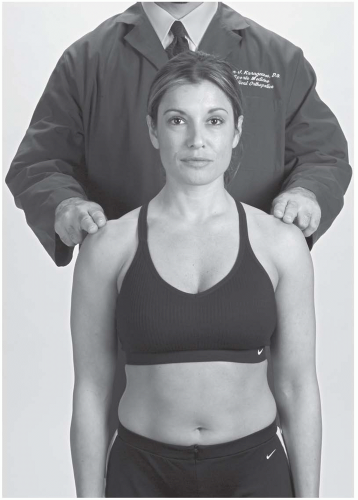 FIGURE 17.2.1. The clinician stands behind the athlete with fingers on the acromion to evaluate shoulder heights. |
On inspection, also note indentations, nodules, or skin changes such as discoloration, hematomas, abrasions, and erythema. Scars, either postsurgical or otherwise, should be noted. Look for deformities from such conditions as glenohumeral dislocations, clavicular fractures, and acromioclavicular sprains. The scapulothoracic relationship should be noted. The scapula may be either retracted, protracted, or exhibit uneven heights.
PALPATION
Begin by systematically palpating the bony prominences, and the muscles and tendons of the shoulder and the scapulothoracic region
(Table 17.2.1). Palpation should be aimed at eliciting tenderness. Be sure to palpate deeply enough where the athlete complains of pain and test bilaterally so the athlete has a comparison.
(Table 17.2.1). Palpation should be aimed at eliciting tenderness. Be sure to palpate deeply enough where the athlete complains of pain and test bilaterally so the athlete has a comparison.
TABLE 17.2.1. BONY PROMINENCES TO PALPATE | ||||||||||||||||||
|---|---|---|---|---|---|---|---|---|---|---|---|---|---|---|---|---|---|---|
| ||||||||||||||||||
An indentation posterior to the glenohumeral joint may suggest an anterior glenohumeral dislocation. Significant palpable tenderness on bony prominences should cause the clinician to order radiographic studies to rule out fracture. The greater tuberosity of the humerus should be easily palpated (Fig. 17.2.2).
Palpation of the muscles should target the pectoralis, biceps brachii, deltoid (anterior portion, midportion, and posterior portion), supraspinatus, infraspinatus, teres minor, trapezius, and rhomboid posteriorly, and the latissimus dorsi, which comprises the posterior axilla. The sternocleidomastoid muscle should also be evaluated, as it is the most superior muscle involved in shoulder motion.
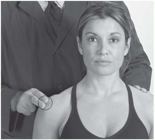
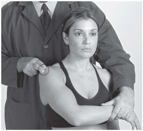
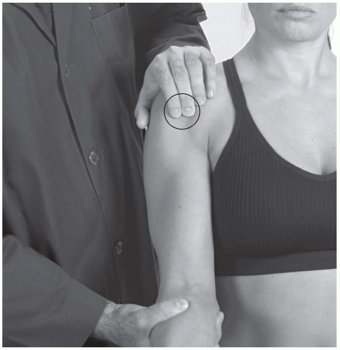
Palpate the tendons of the shoulder, specifically, the supraspinatus and the bicipital tendons. The subacromial bursa can also be palpated in most athletes. This can be done seated or standing. To palpate the supraspinatus tendon at the greater tubercle, internally rotate the arm with the humerus at the side and elbow flexed at 90 degrees (Fig. 17.2.3). The biceps tendon can be palpated between the greater and lesser tubercles of the humerus (Fig. 17.2.4). External rotation of the humerus allows the subscapularis to be palpated at the lesser tubercle (Fig. 17.2.5). Palpate all muscle bellies and attachments at the scapula, acromion, crest of the scapula, and thoracic wall.
RANGE OF MOTION
The shoulder should be actively and passively assessed in the following ranges:
Abduction—arms at side, out at side, and above the head
Adduction—arm at 90-degree elevation, brought across the chest
Flexion—arms at side, lifted in front of the athlete over the head
Extension—arms at side, brought straight back behind the athlete
Internal rotation—arm at side, elbow flexed 90 degrees, rotate arm in and bring behind back
External rotation—arm at side, elbow flexed 90 degrees, rotate arms out
Scapular retraction—squeeze scapula together
Scapular protraction—reach forward with both arms while observing scapular motion
Scapular elevation—shoulder shrug
ABER (ABduction, External Rotation)—same position as for the apprehension sign (see Fig. 17.2.18A).
Note range of motion and end-feel, as well as symptoms of instability, apprehension, and pain. If the active range of motion is limited, the examiner should assess the passive ranges of motion carefully. For instance, “sticking points” in a passive range of motion may identify a mechanical barrier such as a labral tear, rotator cuff tear, or adhesion formation. Pain can limit an athlete’s active range of motion, but if there is no mechanical barrier, then the examiner should be able to passively move the shoulder past that end point. Make sure the athlete is relaxed during passive range of motion. Note any popping, clicking, or crepitus perceived by the athlete or noted by the examiner.
There is quite a range of normal variance in the population. Some athletes may have flexibility that would suggest possible capsular laxity but is normal for that particular athlete. As with most “loose-jointed” individuals, this could predispose the athlete to muscular injury. Always compare both shoulders to distinguish general laxity from specific instability.
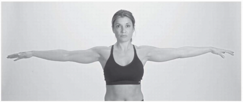
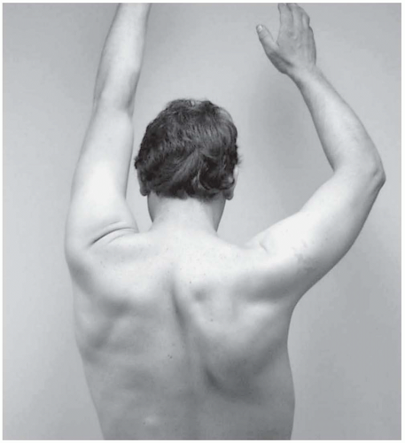
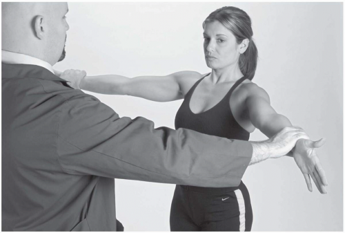
Look for glenohumeral motion isolated from scapulothoracic motion. Test the first 30 to 40 degrees of movement by having the athlete raise both arms from his or her side overhead with straight elbows. Normally, the scapula will not start to rotate until the arm is abducted beyond 30 degrees. If abduction occurs with the shoulders shrugging, the shoulder is using scapulothoracic motion to compensate for glenohumeral restriction. If abduction is not noted, glenohumeral motion is likely independent and normal (Fig. 17.2.6).
Glenohumeral restriction is best seen in cases of adhesive capsulitis. Usually the scapula rotates 1 degree for every 2 degrees of abduction at the glenohumeral joint. In a frozen shoulder, glenohumeral motion is markedly reduced, so abduction comes almost exclusively from scapular motion.
Apley’s Scratch Test
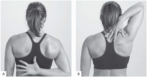
There are two Apley’s scratch maneuvers, lower and upper. The lower test grossly checks internal rotation, adduction, and extension. The upper test checks for external rotation, abduction, and flexion. It helps to determine range of motion early in rehabilitation and then to continually assess range of motion as rehabilitation progresses. It should be noted that most athletes have inequality with this maneuver, for most athletes normally have one shoulder that is somewhat tighter than the other.
Lower test. The athlete puts the arm behind his or her back with the palm facing outward and the thumb pointed cephalad. The athlete should then elevate the thumb as high as possible on the back while the examiner notes the level on the spine at which the thumb can reach (Fig. 17.2.7A). From here, the liftoff test can be done.
Upper test. The athlete abducts the arm, placing the palm of the hand behind the neck with palm facing toward his or her body. The athlete should then attempt to scratch the lowest possible vertebrae with the index finger (Fig. 17.2.7B).
NEUROVASCULAR EXAMINATION
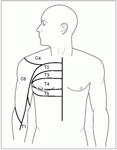
Sensorium. The dermatomes of the upper extremity are shown in (Fig. 17.2.8). Sensation is tested by pinprick and followed by a small brush. The two-point discrimination test is also helpful.
Reflexes
Biceps (C5 nerve root): The examiner places his or her thumb on the insertion of the biceps tendon. A small hammer then strikes the thumb of the examiner.
Triceps (C6 nerve root): The athlete’s arm is at 90 degrees of vertical abduction and the elbow is flexed to 90 degrees with 90 degrees of internal rotation. The examiner then holds the upper arm in one hand and taps the triceps tendon, which provokes elbow extension.
The brachial plexus traverses the glenohumeral joint on its way down the arm, and shoulder pain can emanate from any plexopathy. Any neurologic deficits can cause pain radiating down the nerves themselves, or create muscle weakness that can lead to shoulder pathology such as rotator cuff impingement and scapulothoracic dysfunction. These nerves can be tested through manual muscle testing.
Manual Muscle Testing
The list below demonstrates the maneuvers used to test specific muscles for strength. Table 17.2.2 shows the vertebral levels comprising upper extremity strength(1,2).
Specific Strength Testing
Deltoid (C5 and C6—axillary nerve):
Anterior deltoid: arm at 90 degrees of abduction, full external rotation and supination
Middle deltoid: arm as for anterior deltoid, but with some internal rotation and palm facing down
Posterior deltoid: arm as for anterior deltoid, but with full internal rotation and palm facing backward
Teres minor: arm at side, elbow at 90 degrees of flexion, athlete pushes hands out
Infraspinatus: arm at 90 degrees of abduction, elbow partially bent, resistance applied downward
Subscapularis: arm at 90 degrees of abduction, resistance directed upward
Supraspinatus: empty can or full can test
Trapezius: shrug shoulders against inferior resistance
Serratus anterior: arm flexed 90 degrees, palm down, resistance downward
Rhomboids: hand on hip with elbow at 90 degrees, resistance anterior against elbow
TABLE 17.2.2. UPPER EXTREMITY STRENGTH | |||||||||
|---|---|---|---|---|---|---|---|---|---|
|
Supraspinatus (C4-C6, Suprascapular). Set up athlete with arms abducted 90 degrees and flexed 45 degrees with the thumbs up (full can test). If the arm does not drop, then the examiner can assess strength by the athlete’s resistance against inferior directed force.
Subscapularis (C5-C7, Upper and Lower Subscapular). The athlete rests the arms at the side with elbows flexed at 90 degrees. The examiner places his or her hands inside the athlete’s wrists to resist internal rotation.
Infraspinatus (C5-6, Suprascapular). The athlete sets up as for the subscapularis, and the examiner resists external rotation by placing
the hands on the outside of the wrist, grading strength (+1 to +5) and noting any elicited pain.
the hands on the outside of the wrist, grading strength (+1 to +5) and noting any elicited pain.
Teres Minor (C5-6, Suprascapular). Same as for infraspinatus.
Levator Scapulae (C5, Dorsal Scapula; C3-4, Cervical). Either sitting or standing, the athlete elevates the scapula while the examiner resists with the hands on top of the shoulder, pushing inferiorly.
Serratus Anterior (C5-C7, Long Thoracic). Use the wall push-up test for scapular winging (see Fig. 17.2.12). Another way to test scapulothoracic strength is to ask the athlete to put both hands on the hips with the examiner behind him or her. The examiner then offers pressure to the medial aspect of the elbow and asks the athlete to push the elbows together posteriorly against the examiner’s resistance, noting weakness or scapular winging.
PROVOCATIVE TESTS AND MANEUVERS
Cervical Spine
Spurling’s Test. This is the first test to perform when evaluating the cervical intervertebral discs as a source of shoulder pain. The examiner extends the athlete’s neck and rotates it to one side with corresponding axial compression on the athlete’s head (see Fig. 16.2.9).
Positive sign: pain elicited down the ipsilateral arm from the neck.
Indicates: cervical disc disease.
Acromioclavicular Joint
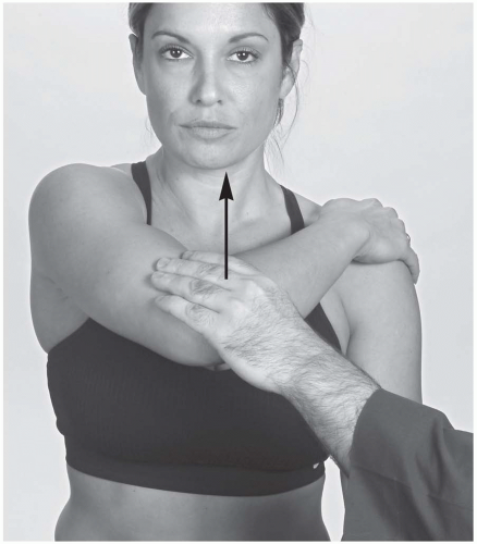
Cross-arm Test. The examiner passively adducts the athlete’s arm across the chest wall with the humerus parallel to the ground so that the free hand of the examined elbow rests on the opposite shoulder (Fig. 17.2.9). The athlete then pushes the elbow superiorly against the clinician’s resistance.
Positive sign: Pain with end-range adduction or with pushing arm up.
Indicates: Acromioclavicular joint pathology (3).
Active Compression Test. With the examiner behind the athlete, the athlete vertically abducts the arm to 90 degrees and adducts 10 to 15 degrees with internal rotation (thumbs down) (Fig. 17.2.10A). The athlete resists the examiner’s downward force. This maneuver is then repeated with the arms supinated (Fig. 17.2.10B).
Positive test: Pain that improves or resolves with the second maneuver.
Indicates: Acromioclavicular (AC) pathology if the pain is in the AC joint, or labral pathology if the pain feels more internal in the shoulder. (4)
Scapulothoracic Motion
Range of Motion. The examiner observes scapular rhythm as the athlete abducts the shoulder over the head. Note any difference between the scapulae in elevation and depression, external and internal rotation, retraction, and protraction.
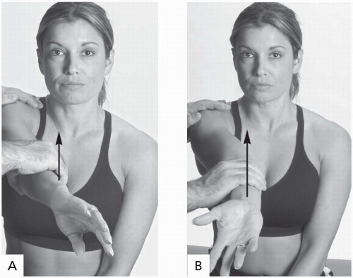 FIGURE 17.2.10. Active compression test. A, The athlete’s arm is internally rotated. B, Then the arm is externally rotated. |
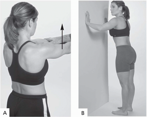
Scapular Winging. This is often related to weakness of the serrratus anterior, which is innervated by the long thoracic nerve (Fig. 17.2.11).
Test 2: The athlete stands in a relaxed position or with the hands on the hips. Have the athlete hang the arms at the side with palms out, then bring them up to a wall or door and perform a push-up off the wall. Observe the scapular behavior (Fig. 17.2.12B).
Test 1: The athlete forward flexes the arms at 90 degrees against clinician’s or tubing resistance (Fig. 17.2.12A).
Bicipital Tendon
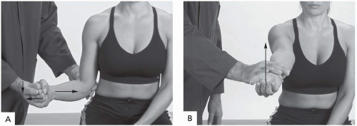
Yergason’s Test. The athlete is in the seated position with the elbow at his or her side and flexed to 90 degrees. The examiner holds the elbow with one hand while holding the wrist with the other hand. The examiner then tests the stability of the biceps tendon by externally rotating the athlete’s arm against resistance while simultaneously pushing downward on the elbow (Fig. 17.2.13A).
Positive test: Tendon will pop out of the groove and cause significant pain (1).
Indicates: Unstable bicipital tendon and subluxation, subscapularis damage.
Speed’s Test. This test also looks for bicipital groove pathology. The athlete’s shoulder is in 90 degrees of forward flexion, elbow extended and hand supinated, with applied resistance to cephalad motion (Fig. 17.2.13B) (5).
Positive test: Pain in the bicipital groove.
Indicates: Bicipital tendon pathology, usually tendinitis.
Rotator Cuff Impingement
Full Can Test. The athlete abducts both arms to 90 degrees and forward flexed 45 degrees with the thumbs pointing to the ceiling (Fig. 17.2.14). The athlete resists downward pressure.
Positive test: Weakness, pain, or dropping of the arm, which occurs in significant tears of the supraspinatus muscle with even a gentle tap to the forearm (6).
Indicates: Supraspinatus tendon tear.
Note: Recent studies suggest that the full can test is more sensitive for supraspinatus pathology than the empty can test, which has been the standard for years (5).
Empty Can Test. Same as the full can test, but with thumbs down.
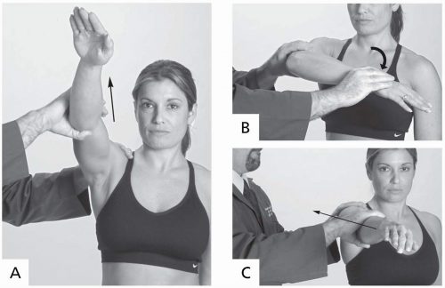
Hawkins Test (Coracoid Impingement Test). The examiner passively rotates the humerus into internal rotation while forward flexing to 90 degrees in the sagittal plane. This opposes the rotator cuff against the coracoacromial ligament and acromion (Fig. 17.2.15B).
Positive test: The maneuver produces pain.
Indicates: Rotator cuff pathology (usually supraspinatus) (3).
Classic Impingement Sign. The athlete is seated or standing. The examiner passively stresses the affected arm into forward elevation of the humerus (Fig. 17.2.15C).
Positive test: Pain is elicited as the impinged rotator cuff opposes the anterior acromial arch.
Indicates: Rotator cuff pathology (3).
Neer Impingement Test. The examiner stabilizes the athlete’s shoulder on the top with his or her off hand, forward flexes the humerus to 90 degrees, then abducts the arm to about 80 degrees with the arm internally rotated (Fig. 17.2.15A).
Positive test: Provokes pain.
Indicates: Rotator cuff impingement pathology (8). If pain is provoked with forward flexion to 90 degrees, it is primary impingement. Pain that is provoked when the arm is moved into abduction is secondary impingement (3).
Injection Test. The examiner injects the subacromial space with 1% lidocaine (approximately 5-10 mL) and then repeats the impingement test. If no pain is produced, the examiner knows that the problem is in the subacromial area.
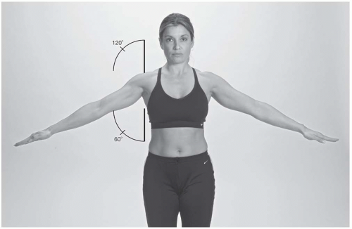
Painful Arc. The athlete abducts the arms overhead as far as they can go, bringing them out laterally (Fig. 17.2.16).
Positive test: Pain with shoulder abduction between 80 and 100 degrees in the coronal plane.
Indicates: Rotator cuff impingment.
Note: Pain starting after 100 degrees of abduction often suggests acromioclavicular joint
pathology, while pain immediately with abduction may indicate adhesive capsulitis, shoulder trauma, or neurapraxia.
pathology, while pain immediately with abduction may indicate adhesive capsulitis, shoulder trauma, or neurapraxia.
Subscapularis Injury
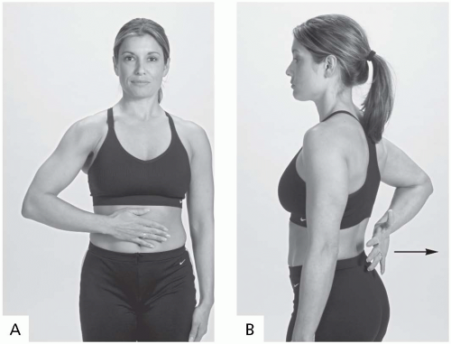
Napoleon Sign. If the athlete cannot fully internally rotate, place the athlete’s hand on his or her stomach and have the athlete push against it (Fig. 17.2.17A).
Positive test: The elbow will drop backward.
Indicates: Subscapularis injury or weakness.
Gerber’s (Liftoff) Test. This evaluates subscapularis muscle strength by limiting pectoralis major firing. The athlete puts his or her hand behind the lumbar spine and attempts to lift the hand away from the back (Fig. 17.2.17B).
Positive test: If the athlete cannot accomplish the liftoff (3).
Indicates: Subscapularis injury or weakness.
Glenohumeral Instability
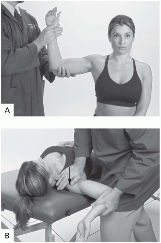
Apprehension Sign (Crank Test). The athlete is in the sitting or supine position, with the arm vertically abducted to 90 degrees and the elbow flexed to 90 degrees. The forearm is then forced into external rotation past 90 degrees (Fig. 17.2.18A).
Positive test: The athlete will be very apprehensive and ask the examiner to stop for fear of a repeat dislocation.
Indicates: Glenohumeral instability, previous glenohumeral dislocation or subluxation.
Stay updated, free articles. Join our Telegram channel

Full access? Get Clinical Tree


