8 The sacroiliac joint and pelvis
CHAPTER CONTENTS
Introduction
The pelvis has a curious place in the history of manual therapy. Perhaps more than any other joint complex in the body there is a mystique about the relevance of disorders of the sacroiliac joint (SIJ) and pubis, and confusion about their management. However, there is a growing body of evidence to support the notion that disorders of the pelvis form a significant pain subgroup and increasing insight into appropriate approaches to management. The SIJ is a well-documented source of buttock and leg pain in cases of chronic low back pain, pregnancy-related pain, post-partum pain, and chronic groin pain in athletes. Furthermore, there is evidence that SIJ pain and dysfunction are associated with a disruption of normal neuromuscular control of the trunk, the hip, and the knee, and this may be related to the pathogenesis of symptoms in these areas.
Clinical relevance
The SIJ is a relatively common cause of pain. Fluoroscopically guided anaesthetic injections suggest that 15–25% of chronic low back pain emanates from the SIJ (Maigne et al 1996; Schwarzer et al 1995). The incidence of pelvic-mediated pain is probably higher in the population with pregnancy-related low back and pelvic pain. Half or more of all pregnant women develop low back and pelvic pain (Bjorklund & Bergström 2000; Kristiansson et al 1996; Ostgaard et al 1991), and based on their clinical presentation, it has been estimated that around 50% of these individuals have symptoms emanating from the SIJ and pubis (Ostgaard et al 1991). Anecdotal suggestions that pelvis-related pain accounts for a proportion of groin and proximal lower-limb presentations in the sporting population have been strengthened by a recent study on groin pain by Mens et al (2006). Some 26% of the athletes in this study had a reduction of symptoms on the application of an SIJ stabilization belt when provoked using manually resisted adduction. Adduction force also improved, suggesting that either the pelvis is the source of pain or the mechanism of pain production is related in some other way to SIJ stability (Mens et al 2006).
As well as being clinically relevant because of its capacity to produce pain, dysfunction of movement and control of the pelvis may be clinically relevant to the development or maintenance of symptoms elsewhere. SIJ motion is considered to be important for shock absorbance during weight-bearing activities (Adams et al 2002), and therefore, a disruption to normal movement may mechanically and adversely affect adjacent structures. There also appears to be neuromuscular relationships. Hungerford et al (2003) identified disrupted neuromuscular control of the trunk and hip in a group of subjects with possible SIJ pain, and O’Sullivan et al (2002) identified disrupted respiratory and pelvic floor function in a similar population. Marshall and Murphy (2006) showed that manipulation of the SIJ can reverse timing deficits in the anterolateral abdominals, and Suter et al (1999) showed that it can improve the electromyographic activity of the vastii and extensor strength at the knee in anterior knee pain patients. These are intriguing results that help to support anecdotal evidence of a relationship between pelvic dysfunction and an array of other pain patterns.
The clinical picture
The availability of motion at the SIJ is well established (Jacob & Kissling 1995; Sturesson et al 1989; Vleeming et al 1992a). It is a synovial joint, but it is also surrounded by a strong capsuloligamentous complex so movement is limited. The best estimates of motion come from studies that have radiographically tracked the movement of implanted metalwork. In weight-bearing, these studies identify only small amounts of movement, on average 2° of rotation and 2 mm of translation (Jacob & Kissling 1995; Sturesson et al 1989, 2000; Vleeming et al 1992a). Studies of passive movement in fresh cadavers reveal more movement and a greater variation of movement, i.e. 3° to 17° of rotation (Smidt et al 1997).
Doppler studies have investigated the stiffness of the SIJ by measuring the conduction of vibration across the joint; if the vibration is conducted intact the joint is stiffer than if it attenuates as it crosses the joint (Buyruk et al 1999). The most interesting work has looked at the characteristics of the postpartum pelvic pain population. Contrary to popular conception, the difference between women in pain and those that are not is not that they are more mobile, but that they have asymmetrical stiffness values side to side (Damen et al 2001, 2002).
There are several features of the emerging understanding of the SIJ’s stability mechanisms that are relevant to clinical practice. Arguably the most important amongst them is that the joint’s stability is under dynamic muscular control. A number of muscles have the capacity to compressively stabilize the joint, from the inner core and pelvic floor to more superficial muscles, such as the gluteus maximus, the long head of biceps femoris, and the latissimus dorsi (Pool-Goudzwaard et al 2004; Richardson et al 2002; Snijders et al 1993a, b, 1998, 2006; van Wingerden et al 2004; Vleeming et al 1990a, 1992b). Deficits in neuromuscular control are implicated strongly in the pathogenesis of pain and dysfunction of this area.
Diagnosis
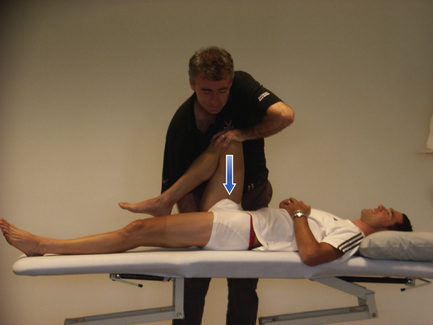
A clinical protocol for diagnosing pain of SIJ origin has also become clearer in the past few years. Recent injection studies have shown that a clinical examination incorporating provocation testing (Fig. 8.1) of the SIJ can accurately identify individuals with SIJ-mediated pain (Laslett et al 2003, 2005a,b; Petersen et al 2004; Young et al 2003). The clinical picture of SIJ-mediated pain that has arisen from these studies is one of unilateral pain with no referral up into the lumbar spine. Pain is often focused over the involved joint, and the sacral sulcus is often tender. Pain may refer down the lower limb and into the foot (Dreyfuss et al 1996; Fortin et al 1994a, b, Maigne et al 1996; Schwarzer et al 1995; Slipman et al 2000; van der Wurff et al 2006; Young et al 2003). Whilst it is very common for SIJ-mediated pain to be centred over the joint, it is worth noting that both this and tenderness of the sacral sulcus have low specificity to SIJ pain involvement (Dreyfuss et al 1996).
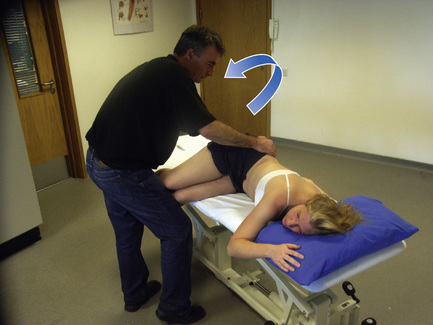
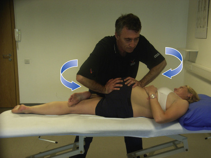
Traditionally, the diagnosis of SIJ dysfunction has been made by a palpation assessment of movement at the joint (Bourdillon et al 1992; DiGiovanna & Schiowitz 1999; Fowler 1986; Lee 1999; Mitchell & Mitchell 1999) (Figs. 8.2 and 8.3). Both the reliability and validity of this assessment have been questioned (Dreyfuss et al 1994; Egan et al 1996; Sturesson et al 2000; van der Wurff et al 2000a, b). Several studies have shown that the individual tests have poor reliability, but perhaps there may be more promise in using a composite of tests in assessment, as happens clinically (Cibulka et al 1988; Cibulka & Koldehoff 1999; Fritz et al 1992; Tong et al 2006). The validity of these tests has been questioned on two fronts. Some investigators query the specificity of the tests because a high proportion of the pain-free population test positive, but this may simply be an indication that dysfunction can occur with or without pain. Others point to the fact that very little joint motion has been identified in movement studies of weight-bearing active movement, and suggest that the therapist’s impression of joint movement is an illusion (Sturesson et al 2000). Unfortunately, the studies on motion have not looked at the tests as they are performed clinically, and therefore, it is not clear that the results apply. Further work is warranted since these are commonly used clinical tools.
Recently, another test of SIJ dysfunction has evolved, the active straight leg raise test (ASLR). This test involves asking the patient to report on the effort involved in lifting each leg 5 to 20 cm off the bed from a relaxed supine position (Mens et al 1999). The ASLR is considered positive if the subject’s perceived effort is altered when a compressive force is applied to the pelvis to stabilize the SIJ (Mens et al 1999; O’Sullivan & Beales 2007a). The test has been shown to be reliable and valid in discriminating between those with pregnancy-related pelvic pain and those without pain (Mens et al 2001). Moreover the perceived effort correlates well with the severity of symptoms and the degree of disability (Mens et al 2002), and it has been shown to correspond to hip flexion force output in that group of patients (de Groot et al 2006). It has been proposed that the ASLR identifies deficits in local muscle control, a proposition supported by the fact that aberrant muscle recruitment strategies have been identified in subjects with SIJ pain who test positive on these tests resolve on manual compression of the pelvis, and evidence that motor control rehabilitation strategies can resolve both the aberrant muscle activity and the effort of the ASLR manoeuvre (O’Sullivan et al 2002; O’Sullivan & Beales 2007a). It has been suggested that the ASLR may be a valid tool with which to monitor the improvement of patients through treatment and rehabilitation (O’Sullivan & Beales 2007a; Stuge et al 2004a, b).
Manual therapy
Manual therapy may involve manipulation or mobilization techniques to resolve movement restrictions and soft tissue techniques to improve muscle function. Whilst widely accepted as being beneficial, at least in the short term (O’Sullivan & Beales 2007b; Stuge et al 2003; Tullberg et al 1998), the nature of the effect of manual therapy is the subject of some debate. Traditional descriptions suggest that mobilization can correct the alignment of the joint if it is applied in a direction to oppose asymmetries of position (Bourdillon et al 1992; DiGiovanna & Schiowitz 1997; Fowler 1986; Lee 1999; Mitchell & Mitchell 1999). This concept that the effect of treatment will be direction-specific, i.e. that it will vary depending on the direction of the applied manual force, is not without merit. For example, it is known that stability of the pelvis is direction-specific. The ligaments connecting the innominate to the sacrum are arranged in such a way that movement of the joint in one direction serves to compress and stabilize the pelvis, and the opposite movement disengages joint compression (Snijders et al 1993a, b; Vleeming et al 1990a, b). Specifically, a relative posterior rotation of the innominate or nutation of the sacrum increases joint compression and posterior rotation decompresses the joint. Research on the ASLR has shown that joint compression can alter the recruitment of the lumbopelvic musculature (O’Sullivan et al 2002; O’Sullivan & Beales 2007a), and therefore it seems reasonable to propose that, if manual techniques can indeed alter the alignment and orientation of the joint, they may create changes in the activation of the surrounding musculature, to the potential benefit of the patient.
However, there is no evidence that manipulation and mobilization can change the position of the SIJ. In fact, X-ray imaging of implanted metalwork has demonstrated the opposite, i.e. no change in joint position after treatment, as measured in standing (Tullberg et al 1998). Interestingly, a palpation-detectable change in the position of the bony landmarks of the pelvis has been demonstrated when subjects have been reassessed in non-weight-bearing positions (Ellis et al 2003). One possible explanation for these apparently conflicting results is that, rather than altering the position of the joint per se, manual therapy may create a change in the directional strain upon the pelvis that is associated with changes in the activity of the surrounding trunk and pelvic musculature. The directional strain may be what is detected as asymmetries of pelvic position on clinical assessment (O’Sullivan & Beales 2007b).
Neuromuscular effects such as this have been demonstrated in recent research on manipulation. Manipulation of the SIJ has been shown to improve the feed-forward activation of the anterolateral abdominal muscles in an asymptomatic group (Marshall & Murphy 2006) and to improve the activation of the vastii and knee extensor torque in a group of patients with anterior knee (Suter et al 1999). These are intriguing results, but unfortunately both studies included only immediate post-intervention measures so there is no indication of the longevity of these effects.
The mechanism of these neuromuscular responses may be explained by a study on the porcine SIJ. Stimulation of the joint capsule and joint produced a response in the surrounding musculature and the muscles involved in the response varied, depending on the location of the stimulus (Indahl et al 1999). This suggests that the SIJ and its capsule play a role in the regulation of the activity of the surrounding musculature. Indahl et al (1999) suggested that abnormal loading on these structures in the dysfunctional pelvis may mediate the aberrant patterns of neuromuscular control seen in patients and that manual therapy may normalize the loads on the joint, capsule, and surrounding ligaments. The challenge to the therapist is to choose the treatment most likely to benefit the patient. Traditionally this has been done by a manual evaluation of the pelvis, an assessment that has, by and large, been shown to have poor inter-tester reliability (Potter & Rothstein 1985; van der Wurff et al 2000a). A recently suggested alternative is to perform techniques in a trial-and-error fashion, and to be guided by the patient’s response (Horton & Franz 2007).
Rehabilitation
There is a growing body of literature to guide rehabilitation of the painful pelvis, although it focuses almost exclusively on pregnancy-related and postpartum pelvic pain. Various exercise protocols have been investigated. More general and strengthening exercise has not been shown to be of benefit. In pregnancy-related pain for example, exercise regimes incorporating strengthening exercises for the abdominals and gluteal muscles (Elden et al 2007), a home exercise regime of exercises performed with a ball between the knees in sitting, standing, and 4-point kneeling position with movements of the arms and legs (Nilsson-Wikmar et al 2005), and submaximal lateral pull-downs, standing leg-press, sit-down rowing, and curl-ups (Nilsson-Wikmar et al 2005), have been investigated with no measurable benefit. A general exercise class was also shown to provide no benefit with regard to function or pain (Dumas et al 1995). In postpartum pain, the efficacy of an exercise programme incorporating trunk-curl exercises and bridging, and one incorporating diagonal trunk-curls and diagonal extension (lifting one shoulder and the opposite leg off the supporting surface from a prone lying starting position), have been assessed with no measurable benefit compared to no exercise (Mens et al 2000). However, more specific exercise programmes that focus on the initiation of pelvic floor and anterolateral abdominal muscle activation do show promise. In a study of postpartum pelvic pain, Stuge et al (2004b) showed that a 20-week intervention that initially focused on specific activation of the transverse abdominal muscles produced significant benefits with respect to pain, functional status, and health-related quality of life compared to an intervention that did not include such specific stabilizing exercises. The group who performed the specific stabilizing exercises maintained their improvement, and were significantly better at both the 1- and 2-year follow-ups. An improvement in pain with a specific stabilizing exercise intervention has also been demonstrated in pelvic pain during pregnancy, but there is no indication of the longevity of that improvement (Elden et al 2005).
Conclusion
Whilst there is a general acceptance that manual therapy to the pelvis can be of benefit, there is little consensus on the nature of its effect, and as yet, no evidence of long-term benefit. The improvements in neuromuscular function that have been noted with manual therapy interventions may indicate that it can provide a window of opportunity for the restoration of more normal neuromuscular function when combined with rehabilitation. The ASLR appears to be an appropriate test for these changes in neuromuscular function.
8.1 Acupuncture in pelvic dysfunction
Within the sporting world, a staggering 58% of UK professional soccer players have reported a history of sports-related groin injury (Karlssonn et al 1994). Much of the pain experienced in such cases is referred from adjacent or even remote myofascial and articular structures, and involves extensive release, muscle re-education, and functional restoration of the entire complex of shortened muscles. It must also be considered that myofascial trigger points (MTrPts) in the region of the abdominal muscles and pelvis can cause abnormal function in the visceral organs that has a somatovisceral effect, and that may mimic gynaecological conditions or symptoms presented to general surgeons, such as vomiting and diarrhoea. King et al (1991) found that 70% of subjects with pelvic pain reported complete or significant relief of their symptoms when the musculoskeletal dysfunction found during physiotherapy assessment was evaluated and treated. MTrPts may have a profound effect on urinary dysfunction, where those along the suprapubic rim involving the insertions of rectus abdominus, internal oblique, and transversus muscles can cause increased sensitivity, and spasms of the urinary bladder and sphincter, resulting in urgency, frequency, urinary retention, and pain. How many male patients are given the diagnosis of prostatitis without adequate attention to and assessment of the myofascial component before more invasive medical testing is offered? Both MTrPt needling and muscle energy techniques may be effective in relieving pain and discomfort, restoring normal muscle length, and facilitating rehabilitation. This comprehensive clinical reasoning approach to pain with myofascial origins may make it possible to provide relief and management of the pelvic region without surgical or diagnostic intervention.
Although athletic injuries around the hip and groin occur less commonly than injuries in the extremities, they can result in extensive rehabilitation time and considerable cost (Anderson et al 2001). Accurate diagnosis and treatment plans are essential, together with adequate management of pain-propagating structures in order to facilitate re-education and rehabilitation. Pelvic anatomical, biomechanical, and pain-propagating structures are amongst the most complex in the musculoskeletal system, offering many challenges to management protocols. A multidisciplinary approach is often necessary for optimal management of complex athletic injuries (Anderson et al 2001) (Table 8.1).
Table 8.1 Common disorders of hip and groin region
| Acute injuries | Treatment priority |
|---|---|
| Muscle strain | Prevention |
| Trigger point dysfunction | Pain modification Muscle imbalance re-education |
| Contusions | Minimize bruising and muscle spasm Prevention of haematoma formation Rest and NWB Rehabilitation |
| Avulsions and apophyseal Injuries | More common in skeletal immaturity Reduce tenderness and swelling Rehabilitation |
| Hip dislocations and subluxations | Pain relief PWB 6–8 weeks Rehabilitation |
| Acetabular labral tears and loose bodies | Pain modification PWB 4 weeks Local anaesthetic injection Surgical option |
| Proximal femur fractures | Surgical management Rehabilitation |
| Insidious Onset | |
| Sports hernia and athletic pubalgia | Pain modification Address pelvic imbalance Rehabilitation |
| Osteitis Pubis/Bursitis | Pain modification Address instability of Pubic Symphysis SIJs Rehabilitation |
| Snapping hip syndrome | Pain modification Rest Trigger point deactivation ITB, TFL |
| Osteoarthritis Lumbar and SI disorders Entrapment of nerve structures | Treatment involving pain propagating structures L1-L3 Address any nerve entrapment/compression of nerves from trigger points |
Adapted from Anderson et al (2001).
Related posts:

Stay updated, free articles. Join our Telegram channel

Full access? Get Clinical Tree


