Fig. 13.1
AP X-ray of previous lumbosacral instrumented fusions
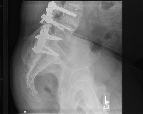
Fig. 13.2
Lateral X-ray of previous instrumented lumbosacral fusions
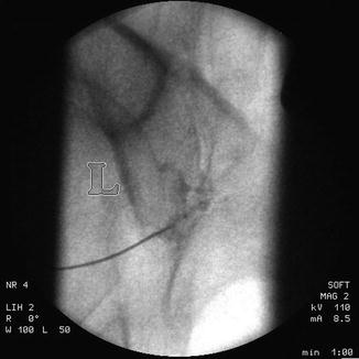
Fig. 13.3
Image guided diagnostic injection verified diagnosis
The patient noted significant pain relief after his SI joint fusions; however, he had persisting symptoms consistent with flat back syndrome, also known as positive sagittal imbalance (Figs. 13.4, 13.5, and 13.6).
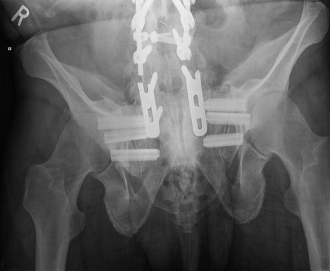
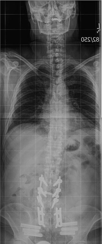
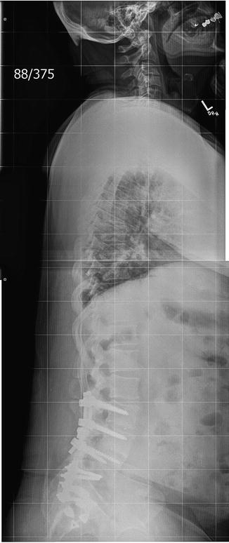

Fig. 13.4
Postoperative AP X-ray showing bilateral SIJ instrumentation

Fig. 13.5
PA scoliosis film

Fig. 13.6
Lateral scoliosis film showing “flat back” syndrome
The patient then underwent spinal reconstruction with a lumbar pedicle subtraction osteotomy. Distal fixation was challenging with the SI joint implants in place. The patient is quite satisfied with his clinical improvement (Figs. 13.7, 13.8, 13.9, and 13.10).
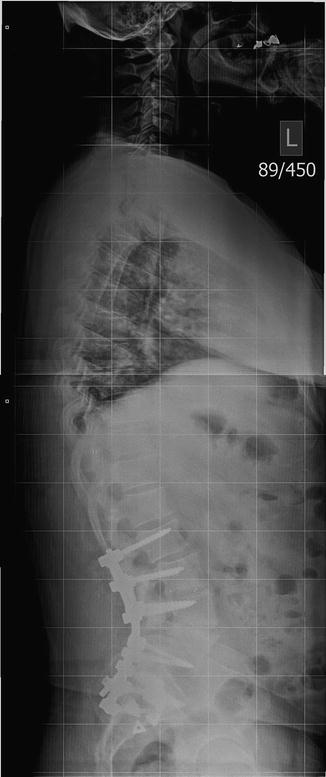
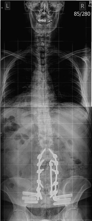
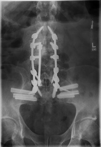
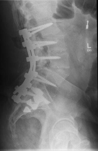

Fig. 13.7
Postoperative lateral scoliosis film after subtraction osteotomy

Fig. 13.8
Postoperative PA scoliosis film after subtraction osteotomy

Fig. 13.9
AP X-ray after subtraction osteotomy

Fig. 13.10
Lateral X-ray after subtraction osteotomy
Conclusion
It is now recognized that adjacent segment degenerative disease does affect the SIJ when a long lumbosacral fusion is performed. If the SIJ is fixated when performing a long lumbosacral fusion, certain pitfalls such as over abundant iliac bone graft harvesting, with its associated potential effects on the SIJ and the ramifications of positive sagittal balance, especially in the elderly, must be considered by the spine surgeon proactively to avoid potential long-term complications. When both positive sagittal balance and the SIJs are symptomatic and require surgery, the spine surgeon must be cognizant of the best methods to use and in what sequence to use them in order to achieve the best overall result for the patient.
References
1.
Ghiselli G, Wang JC, Bhatia NN, et al. Adjacent segment degeneration in the lumbar spine. J Bone Joint Surg Am. 2004;86-A(7):1497–503.PubMed
Stay updated, free articles. Join our Telegram channel

Full access? Get Clinical Tree


