The use of wrist arthroscopy is a valuable adjunct in the management of displaced intra-articular distal radius fractures as it has the advantage of viewing the articular surface under bright light and magnified conditions with minimal surgical morbidity. Fracture hematoma and debris removed arthroscopically may potentially improve the patient’s final range of motion. In addition, any associated soft tissue intra-articular injuries may be managed at the same sitting. Often, pathology—not readily identifiable on plain radiographs—is discovered during arthroscopic-assisted reduction of internal fixation distal radius fractures. It is much easier to manage an acute soft tissue injury that occurs with a fracture of the distal radius than a chronic injury.
Displaced intra-articular fractures of the distal radius are a unique subset of distal radius fractures. These fractures usually develop high-energy injury and are less amenable to traditional closed manipulation and casting. The prognosis for intra-articular fractures of the distal radius has been shown to depend on numerous factors. These include the amount of radial shortening, residual extraarticular angulation, articular reduction of both the radial carpal and the radial ulnar joints, and associated soft tissue injuries.
Lafontaine has described several radiographic features that signify when a fracture of the distal radius is unstable (
1). These include initial dorsal angulation greater than 20°, extensive dorsal comminution, associated ulnar styloid fractures, significant intra-articular involvement, and patients above the age of 60.
Edwards et al. (
2) described the advantage of viewing the articular reduction by wrist arthroscopy compared with monitoring reduction under fluoroscopy alone. In their series, 15 patients underwent arthroscopic evaluation of the articular surface of the distal radius following reduction and stabilization under fluoroscopy. They noted that 33% of the patients had articular step-off of 1 mm or more as viewed arthroscopically. Often, the fragment was rotated. Wrist arthroscopy is particularly useful in judging the rotation of fracture fragments, which is not readily identifiable under fluoroscopic guidance alone. Edwards et al. concluded that utilizing wrist arthroscopy is a useful adjunct and may detect residual gapping not previously identified under fluoroscopy.
Two millimeters or less of articular displacement for reduction has been the well-established threshold over the past several years. Knirk and Jupiter (
3), in their classic article, demonstrated the importance of articular reduction within 2 mm or less. Bradway et al. (
4) further substantiated their findings in their previously published study. Fernandez and Geissler, in their series of 40 patients, noted that the critical threshold may be as low as 1 mm or even less. They found the incidence of complications was substantially lower when articular reduction was within 1 mm or less (
5).
A high incidence of associated intra-articular soft tissue injuries involving the triangular fibrocartilage complex and the interosseous ligaments has been shown by several studies as displaced intra-articular fractures of the distal radius. Mohanti and Kar (
6) and Fontes et al. (
7), in two separate wrist arthrogram studies, noted a high incidence of tears to the triangular fibrocartilage complex in associated distal radius fractures. Mohanti and Kar (
6) noted the triangular fibrocartilage complex injury in 45% of 60 patients in their study. Similarly, Fontes et al. (
7) noted a 66% incidence of tears to the triangular fibrocartilage complex in 58 patients with fractures of the distal radius.
Several arthroscopic studies have documented the incidence of associated intracarpal soft tissue injuries associated with fractures of the distal radius. In three recent studies, tears to the triangular fibrocartilage complex are the most commonly associated intra-articular soft tissue injury (
8,
9 and
10). Geissler et al. (
8) reported their experience with 60 patients with displaced intra-articular fractures of the distal radius undergoing arthroscopic-assisted reduction. In Geissler et al. series, 49% of the patients had a tear of the triangular fibrocartilage complex. However, tears to the interosseous ligament were less common. Injuries to
the scapholunate interosseous ligament and the lunotriquetral interosseous ligament were identified in 32% and 15% of the patients, respectively.
Hanker (
9), in a study of 65 patients, noted that tears of the triangular fibrocartilage complex were present in 55% of the patients in his series of arthroscopic-assisted reduction of distal radius fractures. Lindau (
10), in an arthroscopic study of 50 patients, noted that injury to the triangular fibrocartilage complex was identified in 78% of patients, injury to the scapholunate interosseous ligament was identified in 54% of patients, whereas tears of the lunotriquetral interosseous ligament were far less common and were seen in only 16% of patients.
Geissler et al. (
8) noted that there was a spectrum of injuries that occurred to the interosseous ligaments, based on his findings of associated soft tissue injuries with fractures of the distal radius (
8). An arthroscopic classification of interosseous ligament injuries was described. He noted that the ligament attenuates and eventually tears usually in a volar to dorsal direction. This arthroscopic classification of carpal instability is based on observations of the interosseous ligaments from both the radiocarpal and the midcarpal spaces (
Table 43.1).
The normal scapholunate and lunotriquetral interosseous ligaments have a concave appearance between the carpal bones when viewed from the radiocarpal space. The scapholunate interosseous ligament is best seen with the arthroscope in the 3-4 portal, and the lunotriquetral interosseous ligament is best observed with the arthroscope in either the 4-5 or 6– portal. In the midcarpal space, the scapholunate interval should be tight and congruent without any articular gap or step-off. Similarly, the lunotriquetral interval may be congruent, but usually a 1-mm step-off or increased play may normally be seen between the lunate and the triquetrum when viewed from the radial midcarpal space.
In Geissler grade 1 injuries, there is a loss of the normal concave appearance between the carpal bones as the interosseous ligament attenuates to become convex as seen with the arthroscope in the radiocarpal space. Hemorrhage may be seen within the interosseous ligament itself. However, in the midcarpal space, there is no rotation between the carpal bones, and the carpal interval is tight and congruent.
In Geissler grade 2 injuries, the interosseous ligament continues to stretch and becomes attenuated with a convex appearance as seen with the arthroscope in the radiocarpal space. In the midcarpal space, the carpal bones are no longer congruent, and a step-off is present. With scapholunate instability, there is slight palmar flexion of the dorsal edge of the scaphoid in relation to the lunate. With lunotriquetral instability, increased play will be seen between the triquetrum and the lunate when palpated with a probe in the ulnar midcarpal portal.
In Geissler grade 3 injuries, the interosseous ligament starts to tear usually in a volar to dorsal direction as seen with the arthroscope in the radiocarpal space. A gap is seen between the carpal bones. In the midcarpal space, a probe may be inserted between the carpal bones. A portion of the interosseous ligament is still intact, and a complete separation of the carpal bones is not seen.
In Geissler grade 4 injuries, the interosseous ligament is completely detached and the arthroscope may be passed freely between the radiocarpal and the midcarpal spaces. (This is the so-called drive-through sign.)
Large joint instrumentation is not appropriate for arthroscopic-assisted reduction of distal radius fractures. Smaller joint instrumentation is essential. A smaller joint arthroscope of 2.7 mm or less is recommended. When the arthroscope is initially placed in the wrist, fracture debris and hematoma often obscure vision. It is important to irrigate out the fracture debris to improve utilization. A 3.5-mm or smaller shaver is used to help clear fracture hematoma and debris.
A traction tower is quite useful in the management of arthroscopic-assisted reduction of distal radius fractures. The tower allows continuous traction to the fracture fragments. It also allows the surgeon to flex, extend, and ulnar and radial deviate the wrist to help reduce the fracture fragments while maintaining constant traction. It is generally easier to insert the arthroscope with the wrist in slight flexion. The tower allows for slight flexion of the wrist during initial introduction of the arthroscope and cannulae.
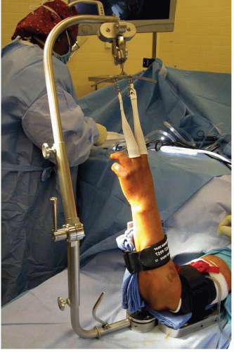
A new traction tower is designed to allow the surgeon to simultaneously evaluate the wrist arthroscopically to manipulate the articular reduction and monitor the reduction under fluoroscopy (
Fig. 43.1). The traction bar is uniquely placed at the side of the forearm and wrist so that it does not block fluoroscopic evaluation and the surgeon does not need to work around a central bar. In addition, the new traction tower allows the surgeon to perform arthroscopic-assisted fixation in the vertical or horizontal position, depending on the surgeon’s preference. If a traction tower is not available, the wrist may be suspended with a finger trap attached to weights suspended over the end of a horizontal hand table or suspended with a shoulder holder in the vertical position. A small bump placed under the wrist is useful if weights are being utilized over the end of the table to maintain the wrist in slight flexion to ease entry on reduction of instrumentation.
Patients who present with a fracture of the distal radius often have a swollen wrist. Because of this, it may be difficult to palpate the traditional extensor tendon landmarks for wrist arthroscopy. However, the bony landmarks are usually still palpable. These bony landmarks include the bases of the metacarpals, the dorsal lip of the radius, and the ulnar head. The 3-4 portal is made in line with the radial border of the long finger. The 4-5 portal is made at the mid-axial line to the ring finger. Precise portal placement is mandatory for arthroscopic-assisted reduction of distal radius fractures. If the portals are placed too proximally, the arthroscope may be placed within the fracture itself, or if placed too distally can injure the articular cartilage of the carpus. It is very important to place an 18G needle into the proposed location of a portal before making a skin incision. The needle should enter into the joint easily without interference. The portals are made upon the skin with the surgeon’s thumb against the tip of a no. 11 blade to avoid injury to the cutaneous nerves.
Blunt dissection is continued with the hemostat to the level of the joint capsule, and the arthroscope with a blunt trocar is introduced into the 3-4 portal. The 3-4 portal is the primary viewing portal in wrist arthroscopy. Fracture hematoma and debris are washed out to improve visualization through the 6-U portal. It is helpful to have a separate inflow provided with a 14G needle through the 6-U portal. Outflow is provided through the arthroscopic cannula. It is felt that a separate inflow and outflow are important to improve irrigation of the joint rather than inflow through the arthroscopic cannula alone. The small joint cannula in wrist arthroscopy does not allow much space between the cannula and the arthroscope itself and limits the amount of fluid irrigation into the joint. In addition, a separate outflow limits fluid extravasation into the soft tissues of the forearm and hand.
The ideal timing for arthroscopic-assisted reduction of intra-articular distal radius fractures appears to be between 3 and 10 days. Earlier attempts at arthroscopic fixation may result in troublesome bleeding obscuring visualization. Fractures that are stabilized after 10 days postinjury may be difficult to disimpact and elevate with arthroscopic techniques and manipulation.
Fractures without extensive comminution are most ideal for arthroscopic-assisted management. Radial styloid fractures, die-punch fractures, three-part T fractures, and four-part fractures are all amenable to arthroscopicassisted reduction and internal fixation. Radial styloid fractures are particularly amenable to arthroscopic reduction and are an ideal fracture pattern to gain experience in arthroscopic management of these fractures.
Three-part and four-part fractures with metaphyseal comminution are managed with a combination of open reduction and arthroscopic-assisted fixation. In these instances, the fracture is stabilized by a volar plate inserted through a volar approach, and the joint capsule is not opened. The articular reduction is provisionally pinned as viewed under fluoroscopy. The wrist is then suspended in traction, and articular reduction may be fine-tuned arthroscopically. Distal screws are then used to stabilize the fracture. Any associated soft tissue injuries are detected and managed in the same sitting.
 Acromioclavicular Separations: Arthroscopic Reconstruction of The Acromioclavicular Joint


