Arthroscopic Treatment of Dorsal and Volar Ganglions


Arthroscopic Treatment of Dorsal and Volar Ganglions


Scott G. Edwards

Evan Argintar


DORSAL WRIST GANGLION CYSTS


Introduction

Arthroscopic wrist ganglion resection offers several theoretical advantages over open techniques, including improved recovery, better joint visualization, lower complication and recurrence rates, and more satisfying cosmetic results. Initial outcomes of dorsal wrist ganglia resected arthroscopically have been favorable (1, 2 and 3). Although arthroscopic resection of dorsal wrist ganglion cysts is a procedure that is becoming more accepted, several questions still remain unanswered. Based on a critical review of the sparse literature on the subject and on clinical observations, this chapter attempts to clarify the ambiguity surrounding arthroscopic dorsal wrist ganglion resection and determine whether this is a useful technique to add to the arsenal or a triumph of technology over reason.


Anatomy and Pathoanatomy


Intra-articular Cystic Stalks

Cystic stalks, which may appear to be pedunculated or sessile protuberances, may be viewed in the radiocarpal joint at the interval between the dorsal scapholunate ligament and the capsule inflection that separates the radiocarpal and midcarpal joints (Fig. 41.1). In reviewing the current literature, the exact roles of intra-articular cystic stalks are somewhat ambiguous. According to previous reports, although not specifically stated, it has been implied that the identification and surgical excision of the stalk is paramount when using standard arthroscopic technique for ganglion excision. However, the presence of this important structure has been variable in the literature. Osterman and Raphael (1) identified a stalk in twothirds of their patients undergoing arthroscopic ganglion excision. Despite the fact that one-third of their patients had no identifiable stalk, ganglions were successfully excised with no recurrences. Other studies have reported a stalk incidence as low as 10% (2, 3 and 4). Despite vastly different reports on stalk identification, the importance of such pathology must be questioned. Rather than a cystic stalk, Edwards and Johansen (4) described intra-articular cystic material and redundant capsular tissue in the vast majority of their patients with ganglion cysts (Fig. 41.2). This finding, which was more consistently evident, was the focus of their resection, rather than the stalk.


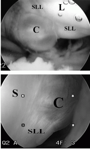
FIGURE 41.1. Intra-articular cystic stalk as viewed from the radiocarpal joint compartment. Cysts may be (A) pedunculated or (B) sessile in appearance. C, cyst; S, scaphoid; L, lunate; SLL, scapholunate ligament.


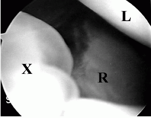
FIGURE 41.2. Diffuse cystic material and redundant capsule often occur more commonly than a discreet cystic stalk. X, cystic material and redundant capsule; R, radius; L, lunate.

The intra-articular limitations of arthroscopic viewing may explain the paucity of stalk identification. The radiocarpal and midcarpal joints are separated by the extrinsic capsular ligaments. At this separation, the dorsal capsular reflection is adherent to the interosseous scapholunate ligament. It is possible that a ganglion stalk travels toward the scapholunate ligament within the substance of the dorsal capsular reflection, rather than through the radiocarpal or midcarpal spaces, and the stalk may never be visualized by arthroscopy. Certain observations during arthroscopic resections may support this theory. On several occasions, extravasations of cystic fluid can be noted during the debridement of the dorsal capsular reflection between the radiocarpal and the midcarpal joints when stalks had not been visualized in either compartment. In other words, the stalk may have been hidden within the dorsal capsular extrinsic ligaments.


Intra-articular Associations

The dorsal ganglion may be an overt sign of intra-articular pathology. Povlsen and Puckett (5) found intra-articular abnormalities in 75% of patients with painful ganglia. They concluded that, like popliteal cysts in the knee, the dorsal ganglion was a marker of joint abnormality. Osterman and Raphael (1) found abnormalities in 42% predominated by findings at the scapholunate ligament (24%), triangular fibrocartilage (8%), lunatotriquetral ligament (3%), and significant chondromalacia. Despite only the ganglion being treated, wrist pain resolved in all cases. Edwards and Johansen (4) elaborated on this notion by showing that most ganglia are associated with type II and III scapholunate and type III lunatotriquetral laxities, as determined by the Geissler grading system (Table 41.1). Although it is reasonable to propose that increased intercarpal laxity may contribute to ganglion formation, the actual significance is unclear given that the natural incidence of these ligamentous laxities in the general population is not known. Cadaveric studies have suggested that type II and III laxities are actually within normal physiologic ranges (7).


Table 41.1. Arthroscopic classification of interosseous ligament tears (6)


Grade


Description


1


Attenuation and/or hemorrhage of interosseous ligament as observed from the radiocarpal joint. No incongruence of carpal alignment in midcarpal space


2


Attenuation and/or hemorrhage of interosseous ligament as observed from the radiocarpal joint. Incongruence and/or step-off as observed from midcarpal space. A slight gap (less than the width of a 2-mm probe) between carpals may be present


3


Incongruence and/or step-off of carpal alignment are observed in both the radiocarpal and the midcarpal space. The width of a 2-mm probe may be passed through gap between carpals


4


Incongruence and/or step-off of carpal alignment are observed in both the radiocarpal and the midcarpal space. Gross instability with manipulation is noted. A 2.7-mm arthroscope may be passed through the gap between carpals


Clinical Evaluation


History and Physical Examination

The first question to answer when a patient presents with a mass is whether it is a cyst or a tumor. Many elements of the history and physical are not conclusive. Occurrence, progression, size, shape, texture, the presence or absence of pain, and association with traumatic or repetitive activities provide little more than suggestions either way. One element of history, however, can be quite helpful in determining whether the lesion is cystic. Both cysts and tumors get larger, but only cysts get smaller. There are rare exceptions to this rule, such as some vascular tumors that involute over a period of months to years. Cysts, on the other hand, may decrease in size as quickly as overnight. On physical exam, transillumination can be helpful in differentiating a cyst from a tumor. This is performed by holding a penlight up against the lesion. A cystic lesion will allow the light to transmit through its fluid medium. The solid tissue of a tumor, however, will prevent any propagation of light.

Occasionally, cysts may herald a more dubious underlying pathology, such as a scapholunate ligament injury. The history and physical should focus on any recent or remote trauma. Oftentimes, patients may have incompetent scapholunate ligaments that remain clinically unapparent until the manifestation of an associated ganglion
cyst. Palpation of the dorsal portion of the scapholunate ligament, a positive Watson scaphoid shift test, or positive straight finger resistance test may be suggestive of scapholunate ligament pathology. Cysts may resemble other pathologies such as gouty tophus, tenosynovitis, and rheumatoid pannus. A careful history and physical should be able to differentiate these conditions.


Diagnostic Imaging

MRI and ultrasound remain the most commonly used imagery to differentiate fluid-filled cysts from solid tumors. Although both differentiate with equal reliability, MRI may suggest an etiology of a solid tumor, whereas ultrasound cannot. Even given this difference, there has been more of a shift toward using ultrasound as the preferred technique given its lesser comparable cost. Very small ganglions, though clinically significant, may be readily overlooked by both ultrasound and MRI. Surgeons should keep a high index of suspicion for these lesions despite a negative reading.


Treatment Options


Nonoperative Treatments

Although there is no consensus about the best nonoperative treatment for ganglion cysts, restriction of wrist activity seems to be well accepted. The efficacy of antiinflammatory medications is more controversial. Some believe that reducing inflammation help painful cysts, whereas others believe that the medications may make the cystic fluid less viscous and possibly more likely to spontaneously decompress. Neither belief has been substantiated in the literature. Needle aspiration seems to be relatively safe for dorsal ganglion cysts, but volar ganglions place neurovascular structures at particular risk with blind aspiration. Patients need to understand that recurrences after aspiration can be high. In summary, nonoperative treatments for dorsal ganglion cysts are unpredictable and the evidence to support such measures is largely anecdotal. Most surgeons, however, will attempt a trial of nonoperative treatment for some duration before committing the patient to surgical excision.


Surgical Indications and Contraindications

Indications and contraindications for arthroscopic dorsal wrist ganglion resection are still evolving. Ho et al. (2) reported two recurrences following resection of ganglia originating from the midcarpal joint. They concluded that arthroscopic resection was not indicated for cysts originating from the midcarpal joint. Many would agree that most dorsal wrist ganglia originate from the scapholunate interval. Given the capsular limitation in the wrist, however, this interval is only partially visualized from the radiocarpal joint. One study (4) observed that cysts communicated with the midcarpal joint in 75% of cases. In the same report, 25% of cysts were accessed exclusively through the midcarpal joint, which suggests that evaluation of the midcarpal joint is not only indicated, but also mandatory for successful resection. Although most cysts may be resected successfully through an isolated radiocarpal portal, some cysts may need supplemental debridement from the midcarpal joint.

Regarding recurrent cysts, one group of investigators has suggested that recurrent cysts following previous open surgical excision should be considered a contraindication for arthroscopic resection (8). Appropriate concerns are the risk of extensor tendon injury due to their potential displacement by the scar from the previous surgery. Thus, most previous studies have used recurrence as an exclusion criterion. One significant exception was a series in which 15% of the patients included recurrent ganglia (4). Patients with recurrent cysts had comparable outcomes with primary cyst resections. Therefore, the authors believe that arthroscopic resection of recurrent cysts is not contraindicated. In fact, arthroscopic resection may be helpful in identifying a potential cause of the recurrence. Previous studies have identified intra-articular abnormalities such as ligament tears, excessive intercarpal laxities, chondromalacia, and triangular fibrocartilage tears as being associated with ganglion cysts (1, 4). It is unclear whether these findings contribute to cystic development, but to the degree they have a role, arthroscopy is more effective at identifying and addressing these abnormalities compared with open excision. Given a recurrent cyst, an arthroscopic evaluation may identify a partial scapholunate ligament tear, which could be debrided, thus lowering the probability of further recurrence. An open technique may not identify the cause as easily and thus doom the recurrent excision to another recurrence.

Cosmetic reasons sometime drive decisions to pursue any endoscopic technique. Although an open incision across the dorsum of the wrist may not seem excessive to a surgeon, the patient may have another perspective. One study reported a very high postoperative satisfaction rate despite having 17% of patients be asymptomatic preoperatively and only opting for surgery for cosmetic reasons (4). There is no similar report for open resections. This implies that it would be reasonable to offer arthroscopic ganglion resections for patients primarily interested in the cosmetic appearance of their hands.


Surgical Technique for Arthroscopic Resection of Dorsal Wrist Ganglions

A tourniquet was placed on every patient as a precaution, and inflated in the event that intra-articular bleeding obscured visualization. While suspending the patient’s arm in a traction tower with 5 to 10 lb of traction applied, a 6R or 6U portal is created as a visualization portal. The more radial 3-4 or 4-5 portals are avoided at this time to prevent inadvertently decompressing the cyst (Fig. 41.3). Once the 2.7-mm arthroscopic camera is directed toward the dorsal compartment of the wrist, the capsule adjacent to the scapholunate ligament is visualized. Occasionally, either a sessile or pedunculated protrusion into the joint
can be seen in the area where the extrinsic capsule joins the distal portion of the dorsal scapholunate ligament. This capsular reflection serves as part of the barrier between the radiocarpal and the midcarpal joints, and the protrusion located here has been termed the cystic stalk (Fig. 41.1). More often, however, one may be impressed with the amount of synovitis and redundant capsule in this area instead of an actual cystic stalk (Fig. 41.2).

Only gold members can continue reading. Log In or Register to continue

Stay updated, free articles. Join our Telegram channel

Jul 22, 2016 | Posted by in ORTHOPEDIC | Comments Off on Arthroscopic Treatment of Dorsal and Volar Ganglions

Full access? Get Clinical Tree

Get Clinical Tree app for offline access