Fig. 5.1
Pubic-bone joint. The pubic-bone joint is made up of the anterior bony structures of the pelvis and all of the soft tissue attachments to the pubic bones. This diagram shows the many structures which attach to the anterior pelvis and the forces that are placed on the pubic-bone joint. From Meyers WC, Greenleaf R, Saad A: Anatomic basis for evaluation of abdominal and groin pain in athletes. Oper Tech Sports Med. 2007;15:165–77. Reprinted with kind permission from Elsevier Limited
The pubic-bone joint has multiple muscular attachments from both the abdomen and pelvis. The abdominal muscles surrounding the pubic symphysis consist of the external and internal oblique muscles, transversus abdominis, and rectus abdominis. The thigh adductors surrounding to the pelvis include the pectineus, gracilis, adductor longus, brevis, and magnus. The most robust and important muscles for maintaining stability of the anterior pelvis are the rectus abdominis and the adductor longus muscles (Fig. 5.2). These muscle groups pull the pelvis in opposite directions and function as antagonists to one another during flexion, extension, and rotation of the pelvis. Normally, contraction of the rectus abdominis muscle places both a posterior and superior force on the pubis leading to an upward tilt of the pelvis, while the adductor longus muscles pull the pelvis in an anterior and inferior direction (Fig. 5.3) [9]. The counter pull of the adductors and rectus muscles leads to an equilibrium of the two forces stabilizing the pelvis. The fibers of the rectus and adductor muscles mesh together forming one common attachment point on the pubic bone [16]. Injury to one of the muscles or its tendinous attachments can significantly affect its antagonist counterpart in addition to disrupting other muscles of the pubic-bone joint. Transection of the rectus abdominis muscle at its insertion has been shown to cause an excessive downward tilt of the anterior pelvis in cadaveric studies [9]. By removing the opposing force of the rectus muscle, increased pressure and stress is applied on the adductor compartment. A study conducted using cadavers, where the attachments of the rectus abdominis were cut to varying degrees, resulted in the increases in pressure of 30–100 cm of water within the adductor longus muscle compartments [15]. In fact, they described that when observers inserted their fingers behind the adductors while the rectus was being cut, bony projections from the anterior edge of the inferior pubic ramus caused significant pain to the observer as the force of the adductor compartment increased and pressed on the finger. Maintaining the balance between the superior pulling rectus muscles and the inferior pulling adductor muscles is important in preventing groin pain in athletes. Loss of function in one of these structures can lead to dysfunction in other surrounding structures, and it may be the beginning of a cascade of events leading to athletic pubalgia.
Fig. 5.2
Rectus abdominis and adductor attachments. The most important muscles in maintaining pelvis stability are the rectus abdominis and adductor muscles. These structures have a common attachment site on the anterior pubis and pull the pelvis in opposite directions. From Meyers WC, Greenleaf R, Saad A: Anatomic basis for evaluation of abdominal and groin pain in athletes. Oper Tech Sports Med. 2007;15:165–77. Reprinted with kind permission from Elsevier Limited
Fig. 5.3
Sagittal view of the pelvis showing the antagonistic pull of the rectus and adductor muscles. Injury to one of these structures can lead to muscle imbalance of the pelvis leading to pain. From Meyers WC, Greenleaf R, Saad A: Anatomic basis for evaluation of abdominal and groin pain in athletes. Oper Tech Sports Med. 2007;15:165–77. Reprinted with kind permission from Elsevier Limited
A tremendous amount of torque is created at the level of the pelvis in athletes participating in sports requiring twisting and cutting. The cutting and twisting activities require the use of the abdominal and pelvic muscles, which creates significant force through the pelvis and stress on the tendinous insertions. A majority of the force placed on the pelvis during high-level activities pulls the pelvis in an anterior direction leading to an anteromedial tilt of the pubic-bone joint that stresses the rectus tendon at its insertion on the pubic symphysis [17–20]. The net forces remain in equilibrium until one of the force vectors changes due to injury or fatigue of the contracting muscle or tendon. Overuse injuries due to repetitive hip hyperextension and truncal rotational movements lead to wear and tear of the tendon insertions culminating in partial or full tearing of these structures. When one muscle weakens or its associated tendon is torn, the result is an unequal distribution of pelvic forces and over-pulling of one of the muscles, leading to more anterior or posterior pelvic tilt depending on which muscle or tendon is injured. An injury to either the rectus abdominis or adductor longus tendon predisposes the opposing tendon to injury. In addition to the injury to the antagonist tendon, the loss of muscle balance can lead to changes of the surrounding pelvic structures causing the pelvis to be less stable. These injuries lead to the pain associated with athletic pubalgia.
The pubic symphysis is composed of opposing hyaline cartilage covered pubic bones with an intervening fibrocartilage disc and is the key structure in maintaining pelvic stability. The articulation is supported superiorly by the suprapubic ligament and inferiorly by the arcuate ligament [21]. The muscle imbalance associated with rectus or adductor dysfunction leads to increased stress in the pubic symphysis causing repetitive microtrauma across the joint and leading to periosteal inflammation, osseous resorption, or osteolysis along the symphyseal margin of the inferior pubic ramus [22, 23]. The breakdown of the pubic symphysis can lead to decreased stability and increased motion of the joint. The excess motion causes further instability of the pelvis, which places greater stress and strain on the soft tissues surrounding the pelvis. This cycle of increased motion and instability leads to further soft tissue injury, eventually leading to the pain associated with athletic pubalgia.
Most injuries leading to chronic groin pain occur due to repetitive trauma to the soft tissues surrounding the pelvis. However, a sudden forceful movement can also initiate symptoms of athletic pubalgia through a traumatic tear of the abdominal fascia. Dr. Tandy Freeman in Dallas described the development of traumatic groin injuries in bull riders [15]. The positioning necessary to ride bulls highlights the classic mechanism of injury to the pelvic musculature, including a combination of hyperextension of the abdominal muscles and hyperabduction of the thigh adductors. The forceful nature of bull riding associated with the specific body positioning often leads to complete avulsions of the rectus abdominis muscles and possible injury to the adductors. Whether it is from chronic overuse or a traumatic event, injury to the rectus abdominis and/or the adductor tendons appears to be the initial inciting event in the development of athletic pubalgia.
Not everyone agrees with all of the details as to how athletic pubalgia develops. Variations to the muscle imbalance theory do exist. Muschaweck describes sports hernias as a weakness in the posterior wall of the inguinal canal resulting in the irritation of the surrounding nerves and insertional tendon pain on the bone [24]. She theorized that the transversalis fascia dilates at its weakest point and widens the inguinal triangle, leading the rectus abdominis muscle to retract cranially and medially, producing increased tension on the pubis. The retraction may be associated with partial or complete tearing of the rectus abdominis muscles. The bulging of the posterior wall of the canal can lead to compression on the nearby genital branch of the genitofemoral nerve that may also play a major factor in creating chronic groin pain. Although there is some variation to the ideas of what leads to athletic pubalgia, a common thread of muscle imbalance and dysfunction of opposing structures is present in most theories.
The Role of FAI in Athletic Pubalgia
Femoroacetabular impingement (FAI) has become a very common topic in the orthopedic literature. FAI is the abnormal contact of the femoral neck with the acetabular rim in near physiologic ranges of motion. The exact etiology of FAI is not well understood, but appears to be associated with the developmental changes in the growing hip. The primary forms of FAI include cam lesions, pincer lesions, and combined cam-pincer lesions. Cam lesions represent a decreased head–neck offset due to an osseous-chondral overgrowth being present at the anterior–superior aspect of the femoral head–neck junction (Fig. 5.4). The cam lesion abuts the edge of the acetabulum during activities requiring flexion, adduction, and internal rotation of the hip. These positions move the cam lesion into appropriate position to impinge on the acetabular rim. A pincer lesion is associated with acetabular over-coverage of the femoral head causing abnormal contact of the normal femoral neck with the overhanging acetabulum (Fig. 5.5). Patients may also have a combined cam-pincer lesion where both the cam lesion and pincer lesion are present. The combined cam-pincer lesion is considered to be the most common form of FAI in the overall population [25]. However, isolated cam lesions are the most common type of FAI found in the young athletic male population [26]. FAI is known to lead to several intra-articular injuries including labral tears and cartilage damage (Fig. 5.6) [27–29]. These injuries often cause the same symptoms seen in athletic pubalgia, making it difficult to differentiate between the two disease processes. In addition, FAI and athletic pubalgia are common in the same patient populations, namely those participating in sports requiring significant cutting, twisting, and accelerating.
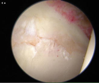
Fig. 5.4
Cam lesion. Cam lesions are an osteochondral growth on the anterior superior aspect of the femoral head–neck junction. As the leg is brought into a flexed, adducted, and internally rotated position (curved arrows), the bump strikes the chondral–labral junction causing articular cartilage injury. From Byrd JWT, Jones KS: Arthroscopic management of femoroacetabular impingement: minimum 2-year follow-up. Arthroscopy. 2011; 27(10):1379–1388. Reprinted with kind permission from Elsevier Limited
Fig. 5.5
Pincer lesion. Pincer lesions represent over coverage of the femoral head leading the normal femoral neck to impinge on the overhanging acetabular rim when the hip is flexed, adducted, and internally rotated (curved arrows). This type of FAI causes damage to the labrum and preserves the underlying cartilage. From Byrd JWT, Jones KS: Arthroscopic management of femoroacetabular impingement: minimum 2-year follow-up. Arthroscopy. 2011;27(10):1379–1388. Reprinted with kind permission from Elsevier Limited
Fig. 5.6
Intraoperative picture of chondral injury at the chondral–labral junction caused by the cam lesion striking the area
Several studies have explored the prevalence of FAI in high-risk sports populations. Gerhardt studied 95 elite male and female soccer players and identified a high percentage of these players to have at least one radiographic sign of FAI [26]. The study reported 72 % of the elite male soccer players and 50 % of the female soccer players, which showed at least one radiographic sign of FAI. Cam lesions were the most common form of FAI with 68 % of the males and 50 % of the females being identified with this morphology. The prevalence of FAI has also been examined in college football players. Kapron et al. [30] evaluated 67 Division-I collegiate football players with AP and frog-leg lateral radiographs to determine the prevalence of FAI in this group of athletes. The study found that 95 % of the 134 hips had at least one radiographic sign of cam or pincer impingement and 77 % had more than one radiographic sign of FAI. In a similar study, collegiate and professional hockey players were evaluated with MRI imaging of their hips to determine the prevalence of FAI [31]. Of the 39 athletes, 25(64 %) had intra-articular pathology of the hip with 56 % presenting with labral tears and 39 % of the participants having an alpha-angles greater than 55°. These studies confirm the high prevalence of FAI and intra-articular pathology in athletes participating in cutting and twisting activities. The fact that FAI occurs in the same patient population as athletic pubalgia points toward an association between the two processes. It is possible that the same forces that cause FAI also cause athletic pubalgia. Alternatively, the biomechanical changes associated with FAI may lead to athletic pubalgia.
Several studies have identified an association between chronic groin pain and lack of hip range of motion. The lack of ROM described in many of these studies most likely represented undiagnosed FAI, and it represented the first link between FAI and athletic pubalgia. The earliest study to link chronic groin and pubic pain with limited hip range of motion was published in the 1970s by Williams [23]. The study reviewed 12 athletes with osteitis pubis and described loss of internal rotation in all of these patients. The exact cause of the loss of hip ROM was not specifically identified in the study, but an abnormality of the femoral head–neck junction was described. The study found an increased femoral head ratio where the superior hemisphere of the femoral head measured an average 1.35 times the size of the lower hemisphere. It appears that Williams was describing the loss of femoral head roundness, which now represents a cam lesion. Further studies have attempted to link decreased hip ROM and chronic groin pain. Verrall studied 89 Australian Rules football players and found decreased internal and external rotation and total hip range of motion in patients with chronic groin pain as compared to those patients without groin pain [32]. The decreased ROM was felt to be caused by a soft tissue reaction to the stresses placed on the hip by the heavy activity associated with Australian Rules football. The author was unsure whether the decreased ROM preceded the chronic groin pain or was a result of the chronic groin pain, but favored the loss of hip ROM preceding the chronic groin pain. Radiographs of the hips were not performed in the study, so the presence of FAI bone morphology could not be determined; however, the limited hip ROM is consistent with the diagnosis of FAI. Verrall et al. [33] completed a follow-up to his first study confirming that the decreased hip IR and ER was associated with chronic groin pain.
These early papers showed an association between decreased hip ROM and chronic groin pain, but did not specifically identify FAI as the cause of decreased hip motion. More recent studies have specifically looked at FAI and its association with chronic groin pain. Weir evaluated the prevalence of FAI in patients with chronic adductor-related groin pain [34]. The study identified 94 % of the 34 athletes with long standing adductor-related groin pain had radiographic signs of FAI. In addition, all patients with chronic groin pain had significantly decreased internal rotation of the hip compared to those athletes without groin pain. The study was unable to specifically identify the reason for the decreased hip ROM, but did state a possible cause could be the associated FAI. Feeley identified a common theme of labral tears with adductor and rectus strains in NFL players and termed the findings the “sports hip triad” [35]. The study described an association between intra-articular pathology and FAI with adductor and rectus strains that further strengthened the relationship between FAI and athletic pubalgia.
The most direct evidence between athletic pubalgia and FAI was described by Larson et al. [25]. The study evaluated 1,033 hip arthroscopies in their practice and identified 31 patients with 37 hips presenting with both symptomatic intra-articular hip pathology and athletic pubalgia. In their study, 95 % of the 37 hips were diagnosed with FAI. Two of the hips had isolated cam lesions, while 35 had mixed cam-pincer lesions. With respect to extra-articular pathology, 26 hips were diagnosed with osteitis pubis, 21 had rectus abdominis pathology, 19 had adductor pathology, 27 had transversalis fascia/external oblique tears, and 4 had psoas impingement. Sixteen hips had athletic pubalgia surgery as their index procedure, and only 4 of the 16 athletes were able to return to unrestricted sports after their surgery. At a mean of 20 months after their index procedure, 11 of 16 patients went on to have hip arthroscopy due to persistent groin pain. Of the 11 patients that underwent hip arthroscopy, 10 returned to sports without limitations. In the same study, eight patients had hip arthroscopy as their index procedure. After hip arthroscopy, four of these patients were able to return to unrestricted sports. Three of the eight hips went on to have athletic pubalgia surgery due to continued disability and pain at a mean of 6 months after their initial hip arthroscopy. All three patients who underwent the second surgery were able to return to sports without limitations. Thirteen other patients in the study underwent concurrent hip arthroscopy and athletic pubalgia surgery. This cohort of patients undergoing concurrent surgery had the best return to sport with 11 of the 13 patients returning to unrestricted sports without further intervention. This study was the first to directly relate FAI and athletic pubalgia.
Related posts:
 Surgical Treatment of Sports Hernia: Laparoscopic Approach
Sports Hernia—Anatomy: What Is a Sports Hernia?
Rehabilitation and Return to Activity Following Sports Hernia Surgery
Sportsman’s Groin and the Inguinal Ligament Release Procedure
Surgical Treatment of Sports Hernia: Open Mesh Approach
The Role of the Team Physician and Athletic Trainer, Including Non-operative Management
Surgical Treatment of Sports Hernia: Laparoscopic Approach
Sports Hernia—Anatomy: What Is a Sports Hernia?
Rehabilitation and Return to Activity Following Sports Hernia Surgery
Sportsman’s Groin and the Inguinal Ligament Release Procedure
Surgical Treatment of Sports Hernia: Open Mesh Approach
The Role of the Team Physician and Athletic Trainer, Including Non-operative Management
Stay updated, free articles. Join our Telegram channel

Full access? Get Clinical Tree


