Diabetic foot ulcerations are a costly and common public health challenge. Although several organizations have emphasized the need to increase awareness of this problem and called health care providers to action to decrease the incidence of ulceration and amputation, there is limited evidence regarding what interventions are best suited to accomplish this goal. This article reviews the pathogenesis, risk factors, and current interventions that have been studied for the prevention of foot ulceration. Preventive measures with evidence for decreasing incidence of ulceration include patient education, offloading abnormal pressures with foot orthotics, and thermal monitoring.
The prevalence of diabetes in the United States and worldwide is growing steadily, and with it the impact of diabetes-related morbidity is likewise growing and presenting new challenges for public health systems. In 2007, there were an estimated 23.6 million people in the United States living with diabetes mellitus, representing a 13.5% increase since 2005. This trend is not limited to the United States alone, because the prevalence of type 2 diabetes mellitus and the economic stress of its associated complications is growing at a rapid rate globally. The growing burden of chronic disease is changing the face of health care and most profoundly affecting developing nations, where it is estimated that 80% of the world’s 250 million persons with diabetes reside.
Diabetic foot ulcerations and amputations are dreaded complications related to diabetes and have a severe impact on the individual and society. On an individual level, foot ulcers often represent a chronic disorder that may severely limit function, work capacity, and quality of life. On a public health scale, foot disorders represent a costly burden on the medical system as one of the leading causes for hospitalization in persons with diabetes. The rate of amputation in individuals with diabetes is 10 times higher than in persons without diabetes. Diabetes is associated with 60% of nontraumatic lower-limb amputations; in the United States, this represented 71 000 amputations in 2004. Even if an ulcer heals with medical therapy, the recurrence rate in patients with diabetes remains high at nearly 70% within 5 years.
Reducing the incidence of diabetic ulcerations and amputations has been pronounced a main public health goal for many years in the United States and abroad; however, despite the acknowledgment of this serious public health challenge, the number of amputations in persons with diabetes continues to rise, increasing 30% from 1980 to 1990. One of the main challenges in reducing the incidence of diabetic foot ulcerations is in determining what interventions are most effective toward this end. Unfortunately, research in prevention is still somewhat sparse in comparison to the body of evidence for treatment. Many of the current practice guidelines are based on consensus and tradition rather than research and evidence-based medicine. The aim of this article is to outline the current understanding of the pathologic process underlying diabetic foot ulceration, the risk factors associated with the development of ulcers, and interventions that are in use or have been studied to modify this risk.
Pathogenesis of diabetic foot ulcers
The feet of patients with diabetes are at increased risk for ulceration because of the damaging effects of diabetic peripheral neuropathy. Ulceration is usually the end result of interplay between several component factors, including poor sensation, structural foot abnormalities, and local trauma. The course leading from hyperglycemia to neuropathy is not entirely understood, although neural hypoxia secondary to metabolic abnormalities of hyperglycemia and dyslipidemia may be a contributing factor. Diabetic peripheral neuropathy is a length-dependent, mixed sensorimotor, demyelinating, and axonal process affecting multiple nerve fiber subtypes. The most well-known and common of these is sensory neuropathy, which leads to loss of protective sensation. Motor neuropathy also may produce intrinsic foot muscle atrophy and subsequent anatomic foot deformity, such as clawfoot, hammertoes, or Charcot foot. Range-of-motion limitations are also thought to result from direct glycosylation of tendons in the lower extremity.
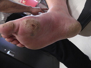
The result of these foot deformities is abnormal weight-bearing distribution in the foot, which places high-pressure areas at risk for skin breakdown ( Fig. 1 ). Finally, autonomic neuropathy decreases normal temperature and secretion regulation, which impairs the effectiveness of the skin barrier. Despite the extensive repercussions of diabetic neuropathy, ulcerations generally do not occur spontaneously in the foot of a patient with diabetes but occur as a result of local trauma in a predisposed foot. This may occur from local trauma caused by poor footwear or other external insult, such as trauma from nail clipping, falls, or repetitive trauma.
Once an ulcer is present, healing may be impaired by the same factors that contributed to the initial ulceration, including elevated blood glucose and abnormal pressure distribution. Poor circulation and infection may further impede healing in patients with foot ulcerations. Multiple classification systems exist to describe diabetic ulcerations, the most common of which include the Warner classification system and the University of Texas system, which reports the depth and width and presence or absence of ischemia and infection.
Risk factors for foot ulceration in patients with diabetes
To design interventions for the prevention of diabetic foot ulceration and estimate a patient’s risk of ulceration, multiple epidemiologic studies have evaluated cohorts of patients with diabetes to quantify the individual contributions of suspected clinical risk factors to lower extremity ulceration and amputation. The most consistently cited risk factors supported by evidence from prospective cross-sectional and case-control studies mirror the key elements in the pathogenesis of foot ulceration. This is witnessed by the systematic review and meta-analysis by Crawford and colleagues in 2007, which cited clinical indicators of diabetic neuropathy (high vibratory perception threshold, impaired sensation to monofilament testing, absent ankle jerk reflexes, high plantar pressures), foot deformities (limited subtalar joint and first metatarsal-phalangeal joint range of motion), or established vascular disease (by history of prior ulceration or amputation) as significant predictors of ulceration ( Table 1 ). Other proposed risk factors that predict ulceration with less consistent support from population studies include ankle-brachial index, HbA 1C , visual acuity, and duration of diabetes mellitus.
| Study, Publication Year | N | Design | Risk Factors Identified |
|---|---|---|---|
| Boyko, 2006 | 1285 | Observational cohort study, mean follow-up 3.38 years |
|
| Frykberg, 1998 | 251 | Cross-sectional |
|
| Cowley, 2008 | 2939 | Prospective |
|
Identified risk factors for amputation also include indicators of peripheral neuropathy (insensitivity to monofilament testing) and peripheral vascular disease (by history of prior lower extremity ulcers or lower extremity amputation). Other risk factors include peripheral vascular disease, which is measured by decreased transcutaneous oxygen pressure measurement (TcPO 2 < 50 mm Hg), and smoking, duration of diabetes, and treatment with insulin ( Table 2 ).
| Study | N | Design | Risk Factors Identified |
|---|---|---|---|
| Reiber, 1992 | 236 | Case-control |
|
| Adler, 1999 | 776 | Observational cohort study, mean follow-up 3.3 years |
|
| Samaan, 2008 | 4778 | Cross-sectional study |
|
Interventions for the prevention of diabetic foot ulcerations
Glycemic Control
Given the connection among hyperglycemia, peripheral neuropathy, and ulceration, it calls to question whether improving blood glucose control may decrease the risk of ulcer formation. Epidemiologic studies have revealed correlation between glycemic control (measured as HbA 1C values) and the risk of developing foot ulcers and lower extremity amputations. A prospective, observational study by Stratton and colleagues in 2000 revealed a linear association between HbA 1C values and incidence of amputation or death from peripheral vascular disease. Evidence indicates that aggressive glycemic control reduces the risk of developing peripheral neuropathy. Unfortunately, no studies have specifically evaluated interventions designed to gauge the effect of improved glycemic control on incidence of ulceration; however, this intervention has been included in combined/complex interventions, which is discussed later in this article.
Evaluation and Treatment of Peripheral Arterial Disease
A common comorbidity that contributes to the risk of amputation in patients with diabetes is peripheral arterial disease (PAD). Risk factors for PAD include smoking, hypertension, hyperlipidemia, and longstanding diabetes. Although arterial insufficiency is rarely the sole cause of ulcer formation, it often delays or prevents ulcer healing in patients with diabetes. The diagnosis of PAD is based on clinical, physiologic, or radiographic evaluation. Clinic-based evaluation for PAD begins with palpation of peripheral pulses. Unfortunately, wide variation in clinical techniques leads to a low rate of interexaminer reliability. Peripheral pulses also may be impalpable in a small proportion of patients without vascular compromise. An ankle-brachial index is another appropriate and more reproducible screening tool to detect PAD; values of less than 0.9 suggest arterial insufficiency in the lower extremities. Unfortunately, atherosclerosis may cause a false elevation of the ankle-brachial index. Another quantitative tool for assessing peripheral circulation is transcutaneous oxygen pressure measurement (TcPO 2 ); a value of more than 40 to 50 mm Hg is considered normal. Radiographically, peripheral arterial supply may be further assessed with angiography, MR angiography, CT angiography, or ultrasound.
Treatment for PAD is well described in the literature and may include smoking cessation, medications, or revascularization surgery. Although the treatment of PAD seems to aid wound healing in dysvascular patients, the role of this intervention in prevention of ulceration is unclear. No studies have evaluated the effect of revascularization alone on the incidence of ulceration.
Evaluation and Treatment of Peripheral Neuropathy
Diabetic neuropathy is one of the leading causes of foot ulceration, and it is estimated that 80% of patients with diabetes who have foot lesions have known peripheral neuropathy. Numerous clinical tools are used to evaluate patients for peripheral neuropathy. Currently, the gold standard for clinic-based evaluation of sensory neuropathy in patients with diabetes is examination with a 10-g Semmes-Weinstein monofilament. Other useful clinical evaluation techniques include biothesiometry to determine vibratory perception threshold, ankle reflex testing, and clinical history and using a subjective neuropathy scoring system, such as the University of Texas Subjective Peripheral Neuropathy verbal questionnaire. All of these screening techniques seem to be sensitive for the determination of neuropathy and prediction of ulceration risk; the use of multiple screening techniques seems to increase specificity. Electrodiagnosis can further confirm this condition and evaluate for peripheral nerve entrapment, such as tarsal tunnel syndrome, as a mimicking or coexisting condition.
Several medical therapies are under evaluation for treatment of diabetic neuropathy, although their impact on diabetic complications such as diabetic foot ulcers is unclear. Aggressive glycemic control has been associated with significant decreases in the incidence of peripheral neuropathy but has not been found to reverse the effects of neuropathy. Various other pharmacologic interventions, such as the use of cilostazol, have been studied as possible means of treating diabetic neuropathy; however, the research in this area is still in preclinical stages. Another class of medication currently being evaluated for effect on diabetic neuropathy is the statins. In a study of rouvastatin on a diabetic rat model, use of this medication was associated with improvements in nerve conduction velocities. These interventions are currently in preclinical stages for treatment of diabetic neuropathy, and the impact on diabetic complications also remains to be seen.
Peripheral nerve decompressive surgery has been proposed as a measure to improve protective sensation and decrease the risk of foot ulcer formation. The theory underlying this intervention is that persons with diabetes are more prone to peripheral nerve entrapment than patients without diabetes; peripheral nerve decompression may restore protective sensation and reduce the risk of ulceration. Commonly targeted nerves for surgical decompression in the lower extremities include the tibial nerve at the ankle, calcaneal, medial plantar, and lateral plantar nerves (tarsal tunnel release), and peroneal nerve at the ankle and knee. Unfortunately, there is no conclusive evidence that peripheral nerve decompression provides any significant benefit for peripheral neuropathy, as concluded in a recent systematic review by Chaudhry and colleagues. This study reviewed 6 observational studies detailing treatment in a total of 218 patients; however, the included studies were heterogeneous in the preoperative objective diagnosis of peripheral neuropathy and the postoperative improvement measurement tools. A retrospective study by Aszmann and colleagues followed 50 patients for a mean 4.5 years after peripheral nerve decompression surgery and reported decreased incidence of ulceration and amputation in the treated limb versus nontreated limb, with no ulcers or amputations in the treated limb, but 12 ulcers and 3 amputations in nontreated contralateral limbs.
Screening Foot Examination
The current standard of care for patients with diabetes includes an annual foot examination, during which the practitioner may evaluate for peripheral neuropathy, callus formation, onchomycosis, structural foot deformities, circulatory disturbances, wounds, and appropriateness of footwear. Specific foot deformities that have been found to be predictive of lower extremity ulceration include hammer/claw foot deformities and structural deformities, such as Charcot foot and drop foot. Regular foot examinations alone have not been found to decrease the incidence of lower extremity ulceration or amputation, however. Regular foot examination may help identify patients at risk for ulceration, which allows the practitioner to stratify patients according to risk of ulceration to guide interventions and may reveal modifiable risk factors that can decrease the likelihood of ulceration. Patients often fail to identify early-stage wounds, causing delay in appropriate treatment. This is supported by a recent prospective, observational study, which determined that a significant percentage (39.7%) of foot ulcers was first identified by a health care professional rather than the patient or caregiver/relative. The optimal frequency of screening foot examinations has not been established.
Evaluation and Treatment of Abnormal Plantar Pressure Distribution
Given that abnormal plantar pressure distribution is a known contributing factor to ulceration, monitoring and quantification of plantar pressures have been investigated as means of identifying patients at risk of ulceration and monitoring the effects of pressure-reducing interventions. Plantar pressure monitoring takes many forms, from qualitative podoscopy to digital plantar pressure monitoring. Pressure measurements may be performed on a static platform or mat or be measured with in-shoe inserts. Specific plantar pressure variables that have been used for predicting risk of ulceration include peak plantar pressure, pressure beneath the metatarsal heads, and forefoot-to-rearfoot plantar pressure ratio. In all cases, higher pressures are predictive of ulceration. In a prospective study by Caselli and colleagues, an elevated forefoot-to-rearfoot ratio was seen only in severe peripheral neuropathy (as measured by the neuropathy disability score) and was associated with an increased likelihood of ulceration (OR 1.37; 1.16–1.61, P < .0001).
Once a patient has been found to be at increased risk of ulceration because of abnormal plantar pressure distribution, plantar pressure monitoring can be used to assess the efficacy of measures designed to redistribute plantar pressures. This is most commonly performed with specialized footwear, such as custom insoles (discussed later). Surgical correction has been evaluated for this purpose. For example, Achilles tendon lengthening has been proposed as an intervention to decrease forefoot pressures in patients with diabetic neuropathy and possibly diminish the risk of forefoot ulceration.
Specialized Footwear
Custom footwear, including in-shoe orthotics and custom shoes, is the most common intervention for correction of abnormal pressure distribution in the feet of patients with diabetes and is a common clinical intervention used in an attempt to decrease the risk of ulcer formation. Despite the common usage of this intervention, however, the breadth of evidence relating therapeutic footwear for prevention rather than treatment of diabetic foot ulcers is surprisingly sparse.
The literature regarding primary ulcer prevention is limited, although several small studies do suggest that specialized footwear does confer benefit. A small, randomized trial by Colagiuri and colleagues in 1995 found that custom-made foot orthotics improved the rates of resolution of callus, an indicator of abnormal pressure distribution in the foot. Although the primary outcome of this study was callus resolution and not ulcer prevention, this does demonstrate modification of risk factors in normalizing plantar pressure distribution. A systematic review by Bus and colleagues in 2008 found no experimental studies on the role of footwear and offloading in primary ulcer prevention and mixed findings regarding the role of therapeutic shoes in secondary ulcer prevention. Another systematic review by Spencer in 2000 revealed callus resolution with offloading interventions but no studies demonstrating evidence of benefit of custom foot orthotics specifically for primary prevention of ulceration.
Custom footwear does seem to decrease the risk of re-ulceration. A randomized, controlled trial by Uccioli and colleagues in 1995 revealed that manufactured shoes significantly reduced the risk of re-ulceration in comparison to standard footwear. Another study by Busch and colleagues in 2003 reported similar benefits of specialized footwear in decreasing the incidence of re-ulceration. Although the evidence behind custom footwear for prevention of diabetic ulceration is limited, it remains a common intervention for patients deemed to be at higher risk of ulceration because of peripheral neuropathy or foot deformity. Specialized footwear is a covered benefit for patients with diabetes who have a history of amputation or peripheral neuropathy with callus or peripheral vascular disease under the Medicare Therapeutic Shoe Bill of 2004.
Patient Education
Diabetic foot education programs generally include instruction on daily foot self-inspection, avoidance of trauma, such as walking barefoot, and encouragement for patients to contact their physician should any new abnormality appear. Interventions aimed at educating patients on foot care and self-monitoring have been studied, with mixed results. A systematic review published in 2001 by Valk and colleagues revealed some evidence that a patient education program improves patient foot care and reported evidence supporting the effect of an patient education program on decreasing ulcer incidence and callous formation, particularly for high-risk patients (those with prior infection ulcer, or amputation) receiving intensive educational interventions. Other randomized, controlled trials included in this systematic review did not reveal similar effect of education on decreasing ulceration incidence.
A recent randomized control trial by Lincoln and colleagues that evaluated individual educational sessions for patients with history of prior ulceration found improved compliance with recommended foot care behaviors but no significant difference in the incidence of recurrent ulceration in 12 months of follow-up between intervention and control groups. Patient education is often included in complex/combined intervention strategies. This makes it difficult to ascertain what proportion of the reported benefits can be ascribed to the educational component of these interventions, however.
Provider Education
In addition to educating patients on foot care and self-examination, several studies have highlighted the importance of provider education. The American Diabetes Association guidelines recommend that providers—at minimum—learn competency in performing a basic screening examination of the foot, including the associated neurologic, vascular, dermatologic, and musculoskeletal systems. Despite adequate provider knowledge, another barrier to identifying patients at risk for ulceration lies in compliance with routine foot examinations. Prior observational studies note poor provider compliance with routine foot examinations in patients with diabetes in the primary care setting.
Provider education as an intervention has not been studied as a preventive measure to decrease the incidence of foot ulcerations; however, several studies have revealed improvements in compliance with clinical practice recommendations as a result of clinician education. Interventions involving provider and clinical support staff education and feedback have shown to increase provider compliance with annual foot examination. Clinical practice guidelines also have provided another avenue for education and reminders to practitioners regarding appropriate screening and management and have been found to increase compliance with recommended foot examinations for patients with diabetes. Although provider education has been included as part of studies on complex or combined interventions for prevention of foot ulcers, this intervention has not been examined individually to assess the impact of this measure alone on the prevention of foot ulcers in patients with diabetes.
Skin Temperature Monitoring
Another recently investigated intervention for monitoring diabetic foot health and detecting areas at risk for development of diabetic foot ulcers is skin temperature monitoring. Various techniques currently are under investigation for this purpose, including electrical contact thermometry, cutaneous thermal discrimination thresholds, infrared thermometry, and liquid crystal thermography. The theory behind this intervention is that pre-ulcerative inflammation can be detected as a relative elevation in skin temperature compared with the contralateral limb, and detection of an area at risk can prompt early intervention by the patient through modification of activity levels. In a single-blinded, randomized, controlled trial by Armstrong and colleagues, 225 patients were randomized to standard therapy alone (therapeutic footwear, diabetic foot education, regular foot care, and daily foot self-inspection) versus standard therapy with the addition of digital thermometry with infrared skin thermometers. Patients found to have temperature difference between limbs of 4° or more were asked to decrease activity levels until the temperature difference resolved. The thermometry group reported one-third the incidence of ulcers by the 18-month endpoint than patients in the standard treatment group. Despite promising initial results, this technique has not yet been widely applied to clinical use.
Combined/Complex Interventions
Although specific interventions have been discussed independently in the interest of reviewing the current evidence regarding their efficacy, in practice, multiple interventions are often used simultaneously. Several studies have evaluated the effectiveness of these heterogeneous approaches to the care of feet of patients with diabetes. Many of these studies involve a tiered approach to intervention based on risk stratification of the patient. Often, these complex interventions for at-risk patients take place in the setting of the specialized, multidisciplinary foot care clinics. Patient education and routine foot examinations are generally included for patients in all categories in most of these interventional studies. Based on patient history and examination, patients deemed to be at higher risk for ulceration are generally followed with more frequent foot examinations and assessed for custom footwear if indicated.
Most complex intervention studies report decreased rates of ulceration, amputation, and hospitalization. Such an approach seems to be particularly effective in preventing ulceration in high-risk patients, such as those with a previous history of ulceration. For example, Lavery and colleagues implemented such a risk stratification program, with peripheral neuropathy as one deciding factor, to direct preventive interventions in a managed care organization and found that incidence of amputation decreased 47.4% and foot-related hospital admissions decreased 37.8% ( Table 3 ).
Related posts:
 Update on Peripheral Arterial Disease and Claudication Rehabilitation
Update in Diagnosis and Treatment of Diabetic Foot Infections
Clinical Features and Electrodiagnosis of Diabetic Peripheral Neuropathy in the Dysvascular Patient
Prosthetic Rehabilitation Issues in the Diabetic and Dysvascular Amputee
Updates in Cardiac Rehabilitation
Pre-Operative Rehabilitation Evaluation of the Dysvascular Patient Prior to Amputation
Update on Peripheral Arterial Disease and Claudication Rehabilitation
Update in Diagnosis and Treatment of Diabetic Foot Infections
Clinical Features and Electrodiagnosis of Diabetic Peripheral Neuropathy in the Dysvascular Patient
Prosthetic Rehabilitation Issues in the Diabetic and Dysvascular Amputee
Updates in Cardiac Rehabilitation
Pre-Operative Rehabilitation Evaluation of the Dysvascular Patient Prior to Amputation
Stay updated, free articles. Join our Telegram channel

Full access? Get Clinical Tree


