Intervention is organized into three categories1: The purpose of the physical therapy intervention is to prevent any anticipated problems whenever possible, and to safely return a patient to his or her preinjury state, with as little risk of reinjury as possible and with the minimum amount of patient inconvenience. The latter is normally achieved by reducing inflammation followed by a gradual progression of strengthening and flexibility exercises while avoiding damage to an already compromised structure.2 For muscles and tendons, this is generally accomplished through measured rest, physical therapy procedures and techniques including, manual therapy, high-voltage electrical stimulation, central (cardiovascular) aerobics, resistance exercises and general conditioning, while avoiding compromise to the healing structures. For the inert structures, such as ligaments and menisci, more emphasis is placed on controlling the level of tension and force placed on them to stimulate the fibroblasts to produce fiber and glycosaminoglycans.3 Beyond the healing phase, the progression may include advancing to high-functional demands or sports-specific exercises, depending on the patient’s requirements. For the athlete, the criteria for return to play should include no pain, full pain-free range of motion (ROM), normal flexibility/strength/balance, good general fitness, normal sports mechanics, and demonstration of sports-specific skills.4 According to the “Guide to Physical Therapist Practice,”5 an intervention is “the purposeful and skilled interaction of the physical therapist and the patient/client and, when appropriate, with other individuals involved in the patient/client care, using various physical therapy procedures and techniques to produce changes in the condition consistent with the diagnosis and prognosis.” Three components comprise the physical therapy intervention: coordination, communication, and documentation; patient/client-related instruction; and direct interventions (Box 8-1).5
CHAPTER 8
The Intervention
OVERVIEW
An intervention is most effectively addressed through a problem-oriented approach and is based on the patient’s functional needs and on mutually agreed-upon goals.5 Decisions about the intervention are made to improve the patient’s ability to perform basic tasks and to restore functional homeostasis. The most successful intervention programs are those that are custom designed from a blend of clinical experience and scientific data, with the level of improvement achieved being related to goal setting and the attainment of those goals (Table 8-1). The necessary knowledge to perform an intervention includes:6
TABLE 8-1 | Key Questions for Intervention Planning |
What is the stage of healing: acute, subacute, or chronic? How long do you have to treat the patient? What does the patient do for activities? How compliant is the patient? How much skilled physical therapy is needed? What needs to be taught to prevent recurrence? Are any referrals needed? What has worked for other patients with similar problems? Are there any precautions? What is your skill level? |
Data from Guide to physical therapist practice. Phys Ther. 2001;81:S13–S95. |
 the temporal phases of tissue healing (see Chapter 2), common problems associated with each phase, and stresses that tissues can safely tolerate during each phase;
 movement characteristics including the amount of range, control, and capacity required for various functional activities;
 the range of available intervention strategies and procedures to promote healing and corresponding outcomes in varied patient populations;
 sequencing of various interventions to challenge appropriately involved tissues and the whole patient. For example, being able to recognize the underlying tissue healing and balance disorders in a patient status post-hip fracture with diabetes mellitus, the need for aerobic conditioning in assessing patients with low back dysfunction, and the importance of body mechanics education in prenatal and postnatal exercise classes; and
 intervention strategies to promote overall health and well-being, and to prevent secondary dysfunction.
Whether the identification of the specific structure or structures causing the dysfunction is necessary in order to proceed with an intervention, remains controversial. Cyriax8 designed his examination process to selectively stress specific tissues in order to identify the structure involved and its stage of pathology. In contrast, McKenzie9 and Maitland10,11 seldom identified the involved structure, believing that it is not always possible, or even necessary, for the prescription of appropriate therapeutic interventions. Indeed, based on the Maitland and McKenzie philosophy, the therapeutic strategy is determined solely from the responses obtained from tissue loading and the effect that loading has on symptoms.9 Once these responses have been determined, the focus of the intervention is to provide sound and effective self-management strategies for patients, which avoid harmful tissue loading.9 However, although self-management must be encouraged whenever feasible, these strategies have their limitations. One cannot realistically expect the majority of patients to fully rehabilitate themselves with a condition that requires the integration of a multitude of decision-making processes such as occurs with a joint replacement, or an anterior cruciate ligament reconstruction.
The focus of the rehabilitation should be to improve an individual’s ROM, flexibility, strength, and coordination to a level that approximates the demands of the desired activity (speed, agility, strength, power, endurance, etc.). Thus, in general, the rehabilitation program should follow the following 10 basic principles:12
 respect the tissue injury cycle regarding the involved structure(s);
 minimize swelling, pain, and address muscular inhibition and atrophy;
 maintain and restore normal ROM;
 establish a normalized gait pattern within weight-bearing limitations (lower extremity);
 restore neuromuscular control;
 restore cervicothoracic and/or lumbopelvic stability, as appropriate;
 develop upper and/or lower extremity strength, as appropriate;
 improve balance and proprioception;
 increase cardiovascular condition; and
 integrate sports-specific/functional training.
CLINICAL PREDICTION RULES
Many interventions for musculoskeletal disorders have shown small effects when tested in randomized controlled trials. Identifying patients who respond best to certain treatments has received increased interest in research activity. With a clinical prediction rule (CPR), a combination or cluster of patient characteristics is used to determine the diagnosis, prognosis, or likely response to a specific treatment of that individual.13 The development of a CPR involves the following stages: derivation (analyzing a data set to establish a rule with predictive power), narrow validation (evaluating the rule in a similar clinical setting and population), broad validation (evaluating the rule in multiple clinical settings), and impact analysis (determining whether the rule changes clinicians’ behavior, improves patient outcomes, or reduces costs).13 Thus far, however, the vast majority of the CPRs have been derived using single-arm study designs and, therefore, the results of these studies must be interpreted with caution, as these CPRs run a greater risk of identifying prognostic factors rather than factors that modify the effect of a treatment.14 Other important limitations of many of the current CPRs are the use of short-term outcomes only, arguably trivial findings, and limited rule application potential. To date, only one CPR on spinal manipulation for low back pain15 underwent validation in a controlled trial and can be considered for clinical application (and only in a population similar to that tested).14
INTERVENTION BASED ON STAGE OF HEALING
The intervention is typically guided by short- and long-term goals, which are dynamic in nature, being altered as the patient’s condition changes, and strategies with which to achieve those goals based on the stages of healing (Table 8-1). Intervention strategies can be subdivided into active (direct) or passive (indirect), with the goal being to make the intervention as active as possible at the earliest opportunity. The only intervention that consistently appears beneficial across a wide spectrum of spinal and nonspinal musculoskeletal problems is the continued application of controlled stresses.
Many factors can contribute to the patient’s resistance to improvement. In some cases, it may be an individual factor that, when eliminated, will allow the patient to respond well. In the majority of cases, the resistance to improvement is based on the interaction of multiple factors, which must be recognized and corrected.
Misjudgments are sometimes made with the intervention. In general, the patient’s pain should not last more than a couple of hours after an intervention. Pain that lasts longer than 2 hours is usually an indication that the intensity of the intervention, rather than the intervention itself, has been inappropriate. The clinician has to remove the notion that all pain is bad. In many respects, a slight increase in pain following an intervention is a more desirable finding than no change in pain, because it indicates that the correct structure is being stressed, albeit too aggressively.
Acute Phase
The acute phase correlates with the hemostasis, inflammatory, and coagulation phases of healing (see Chapter 2). Clinical findings during the acute stage are associated with inflammation and include pain, edema, redness, heat, and impairment or loss of function. Although the redness and heat are not necessarily problems that require a specific treatment goal, pain, edema, and loss of function certainly are. Given the number of pathologic entities that can be evoked by the repair process, such as complex regional pain syndrome (CRPS, previously referred to as reflex sympathetic dystrophy) and myositis ossificans, it is clear that neurophysiologic processes are at work. Usually, during the acute phase, there is a pain at rest or with active motion, or when specific stress is applied to the injured structure. The pain, if severe enough, can result in muscle guarding and loss of function. With passive mobility testing, pain is reported before tissue resistance is felt by the clinician. The goals of the intervention during this phase are to control inflammation, avoid painful positions, minimize pain and edema, restore full, passive ROM, maintain soft-tissue joint integrity, reduce muscle atrophy through gentle isometric muscle setting, maintain aerobic fitness and to enhance function. During the inflammatory stage, it is also important for the patient to function as independently as possible.
Pain
A major focus of physical therapy in the acute phase is the control of pain. At the simplest level, the transmission of information relating to pain from the periphery to the cortex is critically dependent on integration at three levels within the central nervous system: the spinal cord, brain stem, and forebrain (see Chapter 3). Although pain serves as a protective mechanism, allowing an individual to be aware of a situation’s potential for producing tissue damage and to minimize further damage, it can persist beyond its usefulness. In addition, the presence of pain can stimulate muscle spasm, which in turn can lead to circulatory deficiency, muscle atrophy, and loss of function. The challenge for the clinician during the various phases of healing is to control the pain while simultaneously progressing the patient. Several different approaches can be used to provide pain relief as follows:16
 Encourage the body’s central biasing mechanisms (see Chapter 3) through the use of cognitive processes (motivation, relaxation techniques, positive thinking, and mental focus).
 Minimize further tissue damage. Therapeutic modalities are probably the most frequently used tool in all of musculoskeletal rehabilitation. Physical agents, including therapeutic cold, electrical current, and compression can be used throughout the acute phase. Clearly, an understanding of the physiological basis for using these agents is critical if clinicians are to use these agents effectively.
 Educate the patient as to the rationale of the treatment, and what he or she can expect.
 Recognize that all pain, even psychosomatic pain, feels very real to the patient.
Inflammation and Edema
Inflammation and edema occur as part of the healing process (see Chapter 2). In addition to controlling pain, the goals during the initial phase of intervention for an acute lesion are to control the inflammation and edema, and to protect the damaged structures from further damage, while attempting to promote and progress tissue healing and increase function.
Loss of Function
Neuromuscular inhibition can result from pain and joint effusion and should be addressed as early as possible. The function can be enhanced through the use of assistive and supportive devices (crutches, cane, sling, immobilizer, etc.).
Two acronyms have been used for years to help remember the protocols during the acute stage of healing:
- R.I.C.E.—Rest, ice, compression, and elevation. These should be used.
- H.A.R.M.—Heat, alcohol, running, and massage. These should be avoided, largely due to the fact that they create heat/blood flow or, in the case of running (for lower extremity injuries), heat and physical stress.
More recently, RICE has been expanded to PRICEMEM:
 Protection. Excessive tissue loading must be avoided. For example, in the lower extremity when ambulation is painful, crutches or other assistive devices are advocated until the patient can bear weight painlessly, or within a reasonable tolerance.
 Rest. Rest is generally considered to be absence from abuse, rather than a complete absence from activity as prolonged immobilization can have a detrimental effect on muscles, ligaments, bones, collagen, and joint surfaces. In response to the training stimulus, the healing tissues may undergo some breakdown at the subcellular, cellular, or tissue level, which will temporarily lower the functional ability of each. A rest period allows the body to adapt by resynthesizing the protein in these structures to a higher level than before the overload.17–19 This process of synthesizing takes between 12 and 48 hours depending on the intensity (quality) of the exercise and the volume (total amount) of the load.20 However, it is important to remember that if the training stimulus is stopped, reduced, or altered too much, the training effect will decline.
 Ice. The therapeutic application of cold or cryotherapy (see “Physical Agents”) has been used as a healing modality since the days of the ancient Greeks. In most cases, ice should be used until the swelling has ceased. Limiting the effusion serves to hasten the healing process by minimizing the amount of extracellular fluid and hematoma to be reabsorbed.21
 Compression. The most common method of applying compression is via an elastic bandage.21 The compression provided by a pneumatic device,22,23 or by a felt pad incorporated into an elastic wrap or taping,24 has also been demonstrated to be effective in decreasing effusion.
 Elevation. Elevation of an extremity aids in venous return and helps minimize swelling. In general, elevation and compression should be continued until the swelling has completely dissipated.25
 Manual Therapy. The controlled application of various manual techniques can have several therapeutic benefits (see Chapter 10). These benefits are theoretically achieved through26,27
 stimulation of the large-fiber joint afferents of the joint capsule, soft tissue, and joint cartilage, which aids in pain reduction;
 stimulation of endorphins, which aids in pain reduction;
 the decrease of intraarticular pressure, which aids in pain reduction;
 the mechanical effect, which increases joint mobility;
 remodeling of local connective tissue;
 the increase of the gliding of tendons within their sheaths; and
 an increase in joint lubrication.
Manual techniques, which are discussed in Chapter 10, allow the clinician to choose the degree of specificity of an intervention. Manual therapy techniques including low dosage joint mobilization (grade I or grade II) can be used to improve fluid dynamics and to reflexively inhibit the perception of pain (see Chapter 10). Although the goal is to be as specific as possible, there are many times when a general technique is appropriate. General techniques are typically less aggressive, are applied to the larger muscle groups or regions, and often can be performed by the patient as part of the home exercise program. General manual therapy techniques that can be used during this stage include gentle massage to increase blood flow. Specific manual techniques that can be used during this stage include passive joint distractions and glides (grade I or II).
 Early Motion. Early motion is advocated to28
 reduce the muscle atrophy that occurs primarily in type I fibers;29–31
 maintain joint function;
 prevent ligamentous “creeping;”
 reduce the chance of arthrofibrosis or excessive scarring;32–36 and
 enhance cartilage nutrition and vascularization, thereby permitting an early recovery and enhanced comfort.31,37,38
Therapeutic exercise is the foundation of physical therapy and a fundamental component of the vast majority of interventions. Prescribed accurately, therapeutic exercise can be used to restore, maintain, and improve a patient’s functional status by increasing strength, endurance, and flexibility. Tissue-specific movement should be directed to the structure involved to prevent abnormal adherence of the healing fibrils and future disruption of the scar. Initially, if the motion is contraindicated, gentle muscle setting exercises can be prescribed. These are performed in the shortened or relaxed position to prevent new scar formation being pulled from the healing site before progressing to a variety of joint angles. Passive ROM within the limits of pain is introduced as early as feasible while being very careful not to impart any significant stretch to the healing tissues. Research has demonstrated that joint motion stimulates the healing of torn ligaments around a joint43,44 and that early joint motion stimulates collagen bundle orientation along the lines of force, a kind of Wolff’s law of ligaments.43,45 When designing an exercise program, the clinician should create exercises that are safe yet challenging, progressive, systematic, proprioceptively enriched, activity-specific, and based on evidence-based science.46 A typical exercise continuum includes a number of progressions, which includes the following:46
 Activities initially performed slowly before being progressed to a faster pace.
 The performance of familiar activities and then unfamiliar activities.
 Activities are initially performed on a stable base of support and are then made progressively more challenging by increasing the amount of control required, and with the introduction of activities that require dynamic control.
 The introduction of resistance during the movements. The initial resistance used is of low force and then incrementally increased.
 Correct performance of the activity with increasing levels of complexity.
 Medications. Pharmacologic intervention, which can play an important role in the management of the pain and inflammation associated with orthopaedic conditions is described in Chapter 9.
Tissue repair can be viewed as an adaptive life process in response to both intrinsic and extrinsic stimuli.48 Physical therapy cannot accelerate the healing process, but with correct education and supervision, it can ensure that the healing process is not delayed or disrupted and that it occurs in an optimal environment.9
The rehabilitation procedures used to assist with this repair process differ, depending on the type of tissue involved, the extent of the damage, and the stage of healing. Healing is related to the signs and symptoms present rather than the actual diagnosis. These signs and symptoms inform the clinician as to the stage of repair that the tissue is undergoing. Awareness of the various stages of healing is essential for determining the intensity of a particular intervention if the clinician is to avoid doing any harm. Decisions to advance or change the rehabilitative process need to be based on the recognition of these signs and symptoms, and on an awareness of the time frames associated with each of the phases.49,50
Janda51 introduced the concept of the direct and indirect effects of neural input on muscle activation and noted the influence that pain and swelling can have on direct muscle inhibition.
During this stage, using the principles of PRICEMEM, results in decreased early bleeding and facilitation of the removal of the inflammatory exudates, which can prevent further damage and inflammation to the area. Limiting the effusion serves to hasten the healing process by minimizing the amount of extracellular fluid and hematoma to be reabsorbed.52,53
The criteria for advancement from this phase include adequate pain control and tissue healing, near-normal ROM, and tolerance for strengthening.4
Subacute or Intermediate Phase
The various ways in which the musculoskeletal tissues heal at the cellular level during this phase are described in Chapter 2. Clinically, this stage is characterized by a decrease in pain and swelling and an increase in pain-free active and passive ROM (see Chapter 2). During passive ROM, the occurrence of pain is synchronous with tissue resistance.
Although the amount of pain-free ROM may be increased in this phase, it is still not within normal limits, and stress applied to the injured structures still produces pain, although the pain experienced is lessened.54,55 It is critical during this phase that the patient be educated to recognize the signs and symptoms of overstressed healing tissues. During this phase, there is less emphasis on the use of passive techniques and modalities and more emphasis on progressively stressing the healing structures. The treatment goals for this phase are to modify faulty joint mechanics, to protect the forming collagen and direct its orientation to be parallel to the lines of force it must withstand, and prevent cross-linking and scar contracture. If these goals are achieved, the scar will be strong and extensible.
Therapeutic Exercise
The various types of therapeutic exercise are described in Chapters 12–15. The role of exercise during the healing phases is discussed in this chapter. As outlined in Chapter 12, the various characteristics of an exercise load include:20
 Intensity (i.e., speed, resistance).
 Duration (amount of time of the exercise session).
 Frequency (number of sessions per week).
 Volume of training.
 Length (number of weeks or months).
 Pattern (continuous versus interval).
 Mode (e.g., running, cycling, swimming).
Progressions in the total load of exercise can be achieved by increasing the intensity, duration, or frequency, or a combination of the three (see Chapters 12–15).20 During recovery from injury, it seems that the fibroblasts need to be guided so that the replaced collagen fibers are laid along the lines of stress.
Gentle movements to the area provide natural tensions for the healing tissues and help produce a stronger repair,56 so a progressive increase in movement should be encouraged. Each individual responds uniquely to exercise depending on a number of variables, including:20
 Genetic endowment. Each individual has a given genetic potential that will limit the extent to which the effects of physical training can be manifested. The farther an individual is from the genetic limit, the larger will be the improvement, but as the individual gets closer to the limit, less improvement will be elicited. Gender also plays an important role, as women tend to have less muscle mass and more body fat at a given training level.20
 Biological age. An individual’s biological age has a greater impact than chronological age. Younger adults have a greater response to training than do older adults.57
 Training state. Individuals at lower levels of fitness will respond with a higher rate and magnitude of adaptation than when they possess higher levels of fitness.58
 Health status. During either sickness or injury, the amount of adaptive energy is reduced, along with the ability to perform at optimal intensities and volumes of work. This necessitates a reduction (or elimination) in the prescription of exercise during such times.
 Fatigue state. Fatigue limits one’s ability to work at optimal intensities or durations.
Early motion exercises follow a predictable path: the passive ROM used in the acute phase is progressed to active assistive and then to active ROM, based on tissue and patient responses. If signs of inflammation increase or the ROM progressively decreases, the intensity of the exercise and activity must decrease. Exercise or activity soreness should decrease after 2–4 hours. Strengthening exercises during this stage are initially restricted to submaximal isometrics (see Chapter 12) and protected weight-bearing exercises. The submaximal isometrics are initially performed in the early part of the range, before being performed at multiple angles of the pain-free ROM. As ROM and joint play improve, concentric exercises (see Chapter 12) are initiated, with the resistance being increased as tolerated.
Manual Therapy
As with therapeutic exercise, manual therapy (see Chapter 10) plays an integral role in the healing process by providing controlled stresses to the healing tissues. Manual therapies during this stage include joint mobilizations (grade II) to help restore normal joint play, transverse friction massage, and gentle contract–relax techniques. It is important to emphasize to the patient that an overly aggressive approach during this stage can result in a delay or disruption in the repair process through an increase in the stimulation of the inflammatory chemical irritants and exudates.
The criteria for advancement to the chronic or advanced stage of rehabilitation includes no complaints of pain; full, pain-free ROM; good flexibility and balance; and strength of 75–80%, or greater, compared with the uninvolved side.4
Chronic or Return to Function Phase
The various ways in which the musculoskeletal tissues heal at the cellular level during this phase are described in Chapter 2. During this stage, pain typically is felt at the end of the range with passive ROM, after the tissue resistance has been encountered. The goals during the early part of this phase include a gradual return to a full and pain-free ROM, to progressively increase movement speed, and continue to develop neuromuscular control. By the end of this phase, a full and unrestricted ROM should be present, and more aggressive work-related and sports-specific movements and activities are incorporated as appropriate.
Manual Therapy
Manual techniques may be required at this stage to emphasize the restoration of accessory joint motion and to increase the extensibility of soft tissues. Techniques to increase accessory joint motion (see Chapter 1) may include joint mobilizations (grades III–V). Techniques to increase soft-tissue extensibility include a variety of techniques including passive stretching and myofascial release (see Chapter 10).
Taping
Similar to manual therapy, taping techniques can be an integral part of a comprehensive intervention to improve the quality of movement. Taping may attempt to reinforce the normal protective-support structures, improve proprioception, enhance neuromuscular activation, and/or altered biomechanics.59 The inherent properties of the different types of tape available vary significantly such that the differing characteristics of each can be used for different purposes:59
 Zonas (Johnson & Johnson, Skillman, NJ) and Leukotape (BSN, JOBST, Toledo, OH) have approximately the same number of primary fibers that are parallel to the directional loading. However, Leukotape has secondary fibers oriented at 45 degrees to the loading direction, which can help dissipate multidirectional loading, increase the tensile strength, and increase the stiffness of the tape.
 Jaylastic (Jaybird & Mais, Westboro, MA) has considerably fewer fibers in the directional loading and subsequently lower tensile strength but greater extensibility.
 Occlusive taping has less adhesive force than permeable tape (Coban) due to fluid accumulation under the tape.
Therapeutic Exercise
The musculoskeletal tissues respond to the controlled stresses applied to them by adaptation. This response has been described as a specific adaptation to imposed demand (SAID) (see Chapter 12).60
The application of inappropriate stresses can lead to various forms of tissue dysfunction, such as contracture, laxity, fibrosis, adhesion, diminished function, repeated structural failure, and an alteration in neurophysiologic feedback.61,62
The hierarchy of the resistive exercise progression is based on patient tolerance and response to ensure that any progress made is done in a safe and controlled fashion. The typical sequence occurs in the following order:63
 Small arc submaximal concentric/eccentric
 Full ROM submaximal concentric/eccentric
 Full ROM submaximal eccentric
 Functional/activity-specific plane submaximal concentric
 Functional ROM submaximal eccentric
 Open- and closed-kinetic chain exercises performed concentrically and then eccentrically
 Full ROM submaximal concentric isokinetic
 Full ROM submaximal eccentric isokinetic
 Functional ROM submaximal eccentric isokinetic
In the instance of chronic conditions, a slight increase or worsening of symptoms is sometimes permissible with exercise, because the desensitization of some of the structures may require a mechanical input via stimulation of the large A fibers (see Chapter 3). However, the increase in symptoms may also signal a retriggering of the inflammatory process.64 To help prevent these pathologic changes, Liebenson65 recommends the following:
 Patient education about how to identify and control external sources of biomechanical overload.
 Early identification of psychosocial factors of abnormal illness behavior.
 Identification and rehabilitation of the functional pathology.
What is considered to be “normal” flexibility, strength, speed, and aerobic or anaerobic endurance for most patients in rehabilitation is inadequate for those patients returning to sport.66 As skill is associated with enhanced mechanical efficiency and reduced risk of injury, skill in exercise-specific action should be established before the training load is increased.20
In the modern, cost-conscious healthcare environment, the stage at which the patient is ready to return to full function is not often played out in the clinic. Although patient education emphasizing a slow and gradual return to activity can, to some extent, prepare the subject for this phase, reinjury, or insufficient recovery, is a real possibility.
The terms function and functional have been used extensively within the field of rehabilitation. In the context of physical therapy, function has been defined as “those activities identified by an individual as essential to support physical, social, and psychological well-being and to create a personal sense of meaningful living.”67 Functional limitations have been described as the “limitation of performance at the level of the whole organism or person … and should not be confused with diseases, disorders, conditions, or impairments involving specific tissue, organ, or system abnormalities that result in signs or symptoms.”67
Therefore, in order to restore function, the clinician must think beyond the level of impairments of specific tissues and structures resulting from injury, but instead focus on the functional limitations of the patient. Such focus requires knowledge of functional testing rather than the traditional clinical testing (e.g., ROM, strength). Functional tasks can be designed to assess the balance, balance reach, excursion, speed and agility, endurance, strength, and power of the patient, which can then be equated with function.68 Similarly, functional progression training should involve not merely the reproduction of an activity or task by an exercise. Instead, the ultimate goal of functional training is the restoration of the patient’s confidence, which implies a return to normal of the neurovascular, neurosensory, and kinesthetic systems of the body, so that the reflex performance of a movement is not deliberate, hesitant, or dyskinetic.69–71
It should be obvious that the speed and extent by which the injured tissues heal determines both the speed and the extent of the progression toward an optimum functional outcome. While it is virtually impossible to hasten the healing process, it is possible to prescribe a controlled and safe continuum, in which the patient can improve his or her functional status, without harming the healing structures.
Functional progression training, as with exercise progressions, must be designed in a sequential, step-by-step manner, beginning with simple tasks and progressing to highly coordinated tasks, with each step in the process requiring greater skill than the last. The overriding principle of functional rehabilitation is to return patients to the functional level they desire, or at which they were previously functioning.70
A task-oriented approach requires the clinician to understand the specific requirements of an occupation, activity, or sport. The clinician should assume that the tasks involve motions or stresses in all three planes; multiple joints, segments, or limbs; contributions from uniarticular and multiarticular muscles; and the simultaneous need for the body to move and be stable.72 Important specific considerations include the:72
 common postures and movement patterns;
 the amount of motion;
 the speed of movement;
 nature and magnitude of forces/resistances;
 dominant directions/planes of motion;
 muscular activation patterns;
 joint-specific demands;
 symmetry or asymmetry of motion;
 unilateral or bilateral limb demands; and
 environmental demands.
Functional progressions that are specific to the lower or upper extremity are included in various chapters of this book. Functional progressions and their grading for the nonathlete are also included. A number of functional progressions for athletes have been devised to guide the clinician. Most of these progressions originated from postsurgical protocols, in particular, from anterior cruciate ligament reconstructions and rotator cuff surgery, which deal with various functional levels.
Chronic Inflammation
Most injuries, when treated appropriately, resolve on their own, provided that the injured tissue is allowed to progress through the natural stages of healing. If this natural progression does not occur, chronic inflammation can result (Table 8-2) (see Chapter 2). While similar intervention principles can be applied in the presence of chronic inflammation such as cryotherapy, controlled rest, therapeutic exercise, and manual therapy, additional focus must be placed on determining the cause of the chronic irritation. For example, an assessment of flexibility and strength, posture, footwear, technique, running style, repetitive activities, or motions must be performed. A more subtle cause of chronic inflammation can be the presence of a hypermobile joint, which allows the excessive motion to occur that must be compensated for along the kinematic chain. This hypermobility should be addressed using joint stabilization techniques (see Chapter 14). The more common overuse injuries are described in the relevant chapters.
TABLE 8-2 | Causes and Contributing Factors of Chronic Inflammation |
Causes and Contributing Factors | Description |
Overuse, repetitive strain, and cumulative trauma | Repetitive microtrauma overtime results in structural weakening, or breakdown of connective tissue |
Trauma with subsequent microtrauma | Results in a condition that never completely heals and the involved region becomes more susceptible to injury |
Scarring | Since scar tissue is not as compliant as undamaged tissue, it is unable to sustain the stresses that normal, healthy tissue can sustain |
Adaptive shortening of connective tissues | Connective tissue contractures can become stressed with repeated or vigorous activity |
Imbalance between length and strength of muscles | This can lead to faulty mechanics of joint motion and/or abnormal forces through the muscles |
Training errors | Improper performance and equipment coupled with the condition of the participant and an inappropriate intensity of the exercise can lead to abnormal stresses. In addition, a sudden increase or change in an exercise or training routine may not be tolerated by the connective tissues |
Age-related factors | There is a decline in the overall compliance of tissues with aging which reduces their ability to withstand sustained stress |
Excessive repeated eccentric demand | There is a marked increase in the proportion of disrupted muscle fibers after eccentric as compared with concentric exercise. Eccentric exercise is also linked to morphologic and metabolic signs of muscle alteration: myofibrillar damage along the Z-band |
Muscle weakness | Muscle fatigue results in a decrease in contractility and shock absorbing capabilities |
Faulty biomechanics and anatomical malalignment | Any biomechanical or anatomical asymmetries, particularly in the lower extremities can lead to structural breakdown through compensation |
Rehabilitation Modalities
Clinicians have at their disposal a battery of physical agents and electrotherapeutic modalities for use in the acute phase and once the acute stage of healing has subsided. The modalities used during the acute phase involve the application of cryotherapy, electrical stimulation, pulsed ultrasound, and iontophoresis. Modalities used during the later stages of healing include thermotherapy, phonophoresis, electrical stimulation, ultrasound, iontophoresis, and diathermy (Tables 8-3 and 8-4).
TABLE 8-3 | Clinical Decision Making on the Use of Various Therapeutic Modalities in Treatment of Acute Injury |
Phase | Approximate Time Frame | Clinical Picture | Possible Modalities Used | Rationale for Use |
Initial acute | Injury—day 3 | Swelling, pain to touch, and pain on motion | CRYO ESC IC LPL Rest | ↓ Swelling, ↓ pain ↓ Pain ↓ Swelling ↓ Pain |
Inflammatory response | Days 1–6 | Swelling subsides, warm to touch, discoloration, pain to touch, and pain on motion | CRYO ESC IC LPL Range of motion | ↓ Swelling, ↓ pain ↓ Pain ↓ Swelling ↓ Pain |
Fibroblastic repair | Days 4–10 | Pain to touch, pain on motion, and swollen | THERMO ESC LPL IC Range of motion Strengthening | Mildly ↑ circulation ↓ Pain–muscle pumping ↓ Pain Facilitate lymphatic flow |
Maturation-remodeling | Day 7—recovery | Swollen, no more pain to touch, and decreasing pain on motion | ULTRA ESC LPL SWD MWD Range of motion Strengthening Functional activities | Deep heating to ↑ circulation ↑ Range of motion, ↑ strength ↓ Pain ↓ Pain Deep heating to ↑ circulation |
CRYO, cryotherapy; ESC, electrical stimulating currents; IC, intermittent compression; LPL, low-power laser; MWD, microwave diathermy; SWD, shortwave diathermy; THERMO, thermotherapy; ULTRA, ultrasound; ↓, decrease; ↑, increase. Data from Prentice WE. Using therapeutic modalities in rehabilitation. In: Prentice WE, Voight ML, eds. Techniques in Musculoskeletal Rehabilitation. New York, NY: McGraw-Hill; 2001:299. | ||||
TABLE 8-4 | Indications and Contraindications for Therapeutic Modalities |
Therapeutic Modality | Physiologic Responses (Indications for Use) | Contraindications and Precautions |
Electrical stimulating currents | Pain modulation | Pacemakers |
High voltage | Muscle reeducation Muscle pumping contractions Retard atrophy Muscle strengthening Increase ROM Fracture healing Acute injury | Thrombophlebitis Superficial skin lesions |
Low voltage | Wound healing Fracture healing Iontophoresis | Malignancy Skin hypersensitivities Allergies to certain drugs |
Interferential | Pain modulation Muscle reeducation Muscle pumping contractions Fracture healing Increase ROM | Same as high voltage |
Russian | Muscle strengthening | Pacemakers |
Micro-electric nerve stimulation | Fracture healing Wound healing | Malignancy Infections |
Shortwave diathermy and microwave diathermy | Increase deep circulation Increase metabolic activity Reduce muscle guarding and spasm Reduce inflammation Facilitate wound healing Analgesia Increase tissue temperatures over a large area | Metal implants Pacemakers Malignancy Wet dressings Anesthetized areas Pregnancy Acute injury and inflammation Near eyes Areas of reduced blood flow Anesthetized areas |
Cryotherapy (cold packs and ice massage) | Acute injury Vasoconstriction and decreased blood flow Analgesia Reduce inflammation Reduce muscle guarding/spasm | Allergy to cold Circulatory impairments Wound healing Hypertension |
Thermotherapy (hot whirlpool, paraffin, hydrocollator, and infrared lamps) | Vasodilation and increased blood flow Analgesia Reduce muscle guarding and spasm Reduce inflammation Increase metabolic activity Facilitate tissue healing | Acute and post-acute trauma Poor circulation Circulatory impairments Malignancy |
Low-power laser | Pain modulation (trigger points) Facilitate wound healing | Pregnancy Near eyes |
Ultraviolet | Acne Aseptic wounds Folliculitis Pityriasis rosea Tinea Septic wounds Sinusitis Increase calcium metabolism | Psoriasis Eczema Herpes Diabetes Pellagra Lupus erythematosus Hyperthyroidism Renal and hepatic insufficiency Generalized dermatitis Advanced atherosclerosis |
Ultrasound | Increase connective tissue extensibility Deep heat Increased circulation Treatment of most soft-tissue injuries Reduce inflammation Reduce muscle spasm | Infection Acute and post-acute injury Epiphyseal areas Pregnancy Thrombophlebitis Impaired sensation Near eyes |
Intermittent compression | Decrease acute bleeding Decrease edema | Circulatory impairment |
ROM, range of motion. Data from Prentice WE. Using therapeutic modalities in rehabilitation. In: Prentice WE, Voight ML, eds. Techniques in Musculoskeletal Rehabilitation. New York, NY: McGraw-Hill; 2001:301. | ||
At present, with the exception of cryotherapy, there is insufficient evidence to support or reject the use of modalities.73–75 However, the absence of evidence does not always mean that there is evidence of absence (of effect), and there is always the risk of rejecting therapeutic approaches that are valid.76
If modalities have a place in the clinic, it is during the acute phase of healing, when there is little the clinician can do in the form of manual techniques or therapeutic exercise. In the remodeling or functional phase, thermal modalities may be used to promote blood flow to the healing tissues and to prepare the tissues for exercise or manual techniques. However, at the earliest opportunity, the patient should be weaned away from these modalities, and the focus of the intervention should shift to the application of movement and the repeated and prolonged functional restoration of the involved structures.
Two categories of modalities are recognized:
- Physical agents and mechanical modalities
- Electrotherapeutic modalities
Physical Agents
Physical agents are sources of energy that are used therapeutically.
Cryotherapy. Cryotherapy, which removes heat from the body, thereby decreasing the temperature of the body tissues, is the most commonly used modality for the intervention of acute musculoskeletal injuries. The use of ice by itself or in conjunction with compression has been demonstrated to be effective in minimizing the amount of exudate.77–82 Cryotherapy can be applied in various forms, but crushed ice bags (CIB), cold packs (Fig. 8-1), and cold water immersion (CWI) are frequently used. A more expensive form of cryotherapy can be supplied by a pneumatic cold compression unit such as Game Ready© (Fig. 8-2) that simultaneously delivers active pneumatic compression and adjustable cold therapies to specific areas of the body.
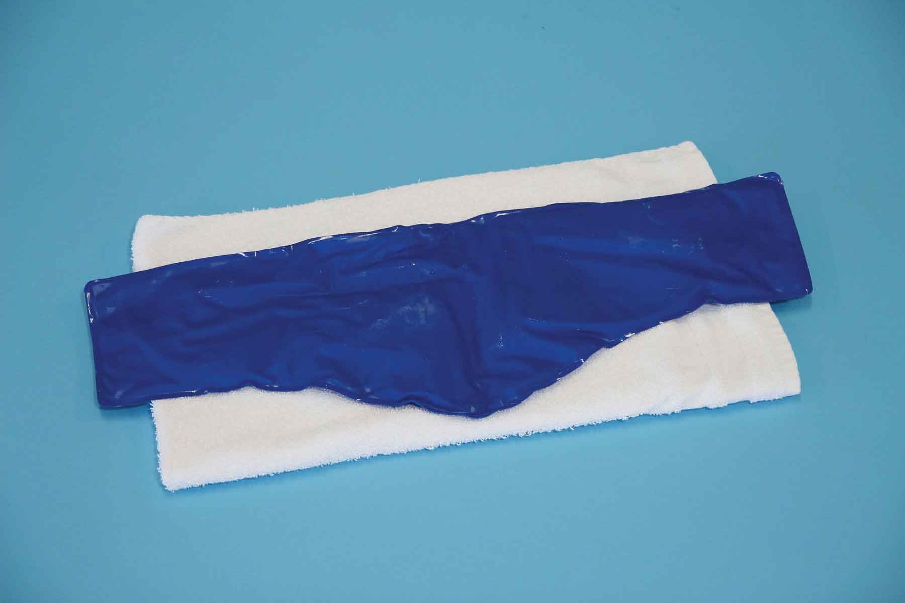
FIGURE 8-1 Cold pack.
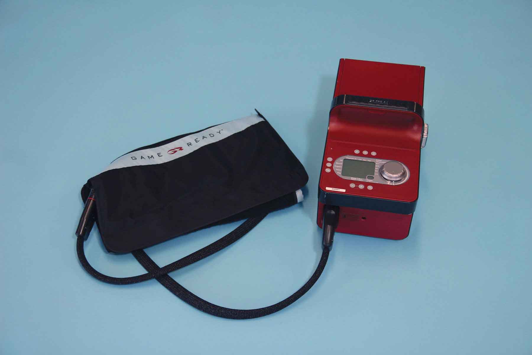
FIGURE 8-2 Pneumatic cold compression unit.
The physiologic effects of local cold application are principally the result of vasoconstriction, reduced metabolic function, and reduced motor and sensory conduction velocities. These effects include the following:
 A decrease in muscle and intraarticular temperature. The decrease in muscle temperature and intraarticular structures occurs because of a decline in local blood flow55,57,60,68,69 and appears to be most noticeable between the temperatures of 40°C and 25°C.70 Temperatures below 25°C, which typically occur after 30 minutes of cryotherapy, actually result in an increase in blood flow (Hunting effect),70 with a consequent unfavorable increase in hemorrhage and an exaggerated acute inflammatory response.63 The reduction in muscle and intraarticular temperature is maintained for several hours after removal of the cooling agent.71 A prolonged application of cold, however, can result in a sympathetically mediated reflex vasodilation in an attempt to rewarm the area, which may actually worsen the swelling.71,72
 Local analgesia.75,83–88 The stages of analgesia achieved by cryotherapy are outlined in Table 8-5.89 It is worth remembering that the timing of the stages depends on the depth of penetration and the depth of the overlying adipose tissue.90 The patient should be advised as to these various stages, especially in light of the fact that the burning or aching phase occurs before the therapeutic phases.
TABLE 8-5 | Stages of Analgesia Induced by Cryotherapy |
Stage | Response | Time after Initiation of Cryotherapy (min) |
1 2 3 4 | Cold sensation Burning or aching Local numbness or analgesia Deep tissue vasodilation without increase in metabolism | 0–3 2–7 5–12 12–15 |
Data from Hocutt JE, Jaffee R, Rylander R, et al. Cryotherapy in ankle sprains. Am J Sports Med. 1982;10:316–319. | ||
 Decreased muscle spasm.23,75,91–93

 A decrease in nerve conduction velocity (NCV).96
Related posts:


Stay updated, free articles. Join our Telegram channel

Full access? Get Clinical Tree


