Techniques of Arthrocentesis
Dennis W. Boulware
 |
A 52-year-old man with rheumatoid arthritis presents with a 36-hour history of acute knee pain with fever after a week of moderate fever and a productive cough. His oral temperature is 39.6°C, and his knee has a large, warm, tense effusion with limited range of motion secondary to pain. His white blood cell count is 24,100 with many immature polymorphonuclear white blood cells. There is concern of septic arthritis, and he requires arthrocentesis for culture and relief.
Arthrocentesis is a frequent part of the evaluation and/or treatment of a patient with a musculoskeletal condition. This chapter focuses on the technique for accessing certain joints with a sterile needle with specific indications for arthrocentesis and treatment recommendations located in the specific chapters regarding that musculoskeletal condition.
Contraindications to arthrocentesis are relative and typically related to the potential for bleeding and/or infection. Caution in performing arthrocentesis should be exercised in the following clinical settings:
Infection of the overlying skin: Passing a sterile needle through an area of skin that is infected or cannot be prepped to retain the needle’s reasonable sterility creates risk of introducing an infection into a joint. Areas of obvious or potential infection must be avoided to preserve the sterility of the joint.
Bacteremia: Performing arthrocentesis in the clinical setting of known bacteremia similarly increases the risk of introducing an infection into the joint. Clinical judgment must be exercised on the relative benefit and risk of performing the arthrocentesis for diagnostic purposes in documenting a polymicrobial infection or therapeutic benefit of the removal of synovial fluid.
Bleeding diathesis: Patients on anticoagulation, with thrombocytopenia, hemophilia, or other causes leading to a bleeding diathesis, are at risk of hemarthrosis from the arthrocentesis. Complications can be avoided by using the smallest needle gauge feasible and providing adequate hemostasis after the procedure. In reality, we are not hesitant to perform venipuncture in these settings with adequate attention to hemostasis postprocedure, so we should have a similar attitude toward arthrocentesis in these settings.
Prosthetic joints: Arthrocentesis of a prosthetic joint can be more challenging because of scarring from the surgical procedure and the risk of infection since the prosthesis can act as a foreign body. Aspiration of the prosthetic joint is possible, but better deferred to the orthopedic surgeon or interventional radiologist under imaging.
Uncooperative patient: Arthrocentesis requires significant cooperation from the patient in positioning and should be performed only on patients who can be fully cooperative.
Clinical Points
Knowledge of local anatomy is essential.
Weigh benefits and risks of arthrocentesis in bacteremia and bleeding diathesis.
Have all needed equipment (syringe, needle, gauge, Band-Aid, etc.) readily accessible to the operator.
Presence of systemic or local infection.
Evidence of local rash overlying injection site.
Consider bleeding diathesis.
Table 27.1 Necessary Equipment | |
|---|---|
|
Equipment
Appropriate equipment (see Table 27.1) should be assembled at the bedside prior to the procedure and easily accessible to the operator during the procedure, without the operator changing position. An assistant is optional and dependent on the operator, but he or she will be helpful if there is concern of patient cooperation or a larger effusion is to be drained requiring changing syringes.
Technique
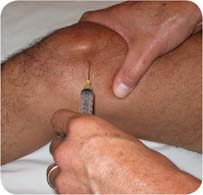
After selecting an appropriate entry site, the entry site can be marked using a ball-point pen with the pen tip retracted. The pen’s aperture can be pressured to the site to leave an impression of the selected entry site before cleansing the area. The selected entry point is cleansed appropriately with an antiseptic solution followed by removal of the antiseptic solution using the alcohol wipes. After cleansing the area, caution should be exercised to avoid contaminating the site by further palpation with an unsterile gloved finger. If further palpation is desired, then a sterile glove can be used or a sterile 4 × 4 gauze can be placed over the area and palpation done over the sterile gauze to preserve the site’s antiseptic condition. Some topical anesthesia is obtained by spraying the site with ethyl chloride until the area “frosts.” Alternatively, a small amount of lidocaine can be injected subcutaneously into the proposed injection area.
When advancing the needle into the joint cavity, the patient experiences discomfort when the needle passes through the skin and again when it crosses the synovium. Less discomfort is experienced when the skin and synovium is crossed quicker as opposed to slowly and deliberately. Once the needle is introduced into the joint cavity, fluid should flow easily into the syringe if using a needle gauge of 20 or larger. If no fluid can be aspirated, or fluid stops flowing, the most common cause is that synovial tissue or solid material (clot, fibrin, cartilage fragments, etc.) within the fluid is obstructing the needle. Rotating the needle or injecting back a small amount of the aspirated fluid into the joint cavity may remove the obstruction. At that point. gentle negative pressure can be placed and the fluid may aspirate. As the total effusion approaches complete drainage, the synovial lining becomes closer to the needle tip and further difficulty is typically experienced or fresh blood now appears in the aspirated fluid. Discomfort by the patient is common at this point, and a decision to continue aspirating at the patient’s discomfort should be weighed by the benefit of removing more fluid at this time. Once sufficient fluid is removed, the needle can be withdrawn and appropriate hemostasis applied to the injection site. Alternatively, if steroids or medication are planned to be injected after aspiration,
the syringe can be separated from the needle that remains in the joint and a syringe with the medication attached to the needle and the medication injected. This situation where an injection follows an aspiration is where a hemostat can be helpful to grasp the hub of the needle while changing syringes. Injecting medication into a joint cavity should not require much pressure on the plunger, although the larger the discrepancy between a large syringe bore and a small-gauge needle, the greater the pressure required. If significant pressure is required, the needle has left the joint space and should be positioned again properly.
the syringe can be separated from the needle that remains in the joint and a syringe with the medication attached to the needle and the medication injected. This situation where an injection follows an aspiration is where a hemostat can be helpful to grasp the hub of the needle while changing syringes. Injecting medication into a joint cavity should not require much pressure on the plunger, although the larger the discrepancy between a large syringe bore and a small-gauge needle, the greater the pressure required. If significant pressure is required, the needle has left the joint space and should be positioned again properly.
Related posts:

Stay updated, free articles. Join our Telegram channel

Full access? Get Clinical Tree


