Systemic Lupus Erythematosus
Michelle A. Petri
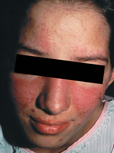 |
A 23-year-old Caucasian woman presents to her primary care doctor with complaints of 9 months of fatigue, pain in muscles including the neck and shoulder area, and red cheeks after sun exposure, lasting for an hour or so. On the physical examination, the cheeks have several pustules. Laboratory data are ordered and show a positive ANA 1:80 (homogeneous pattern), normal complete blood count, normal chemistries, and normal urinalysis. Does she have systemic lupus erythematosus?
Clinical Presentation
Epidemiology
Systemic lupus erythematosus (SLE) is a multisystem autoimmune disease. It occurs predominantly in women, but 10% of patients are men. The onset is predominantly in the 20s and 30s, but it can present in older patients (it is rare before puberty). It is both more common and more severe in African–Americans and Hispanic–Americans than in Caucasians. It is estimated that about 300,000 Americans have SLE.
Pathogenesis
Lupus autoantibodies are present for 5 to 7 years before the clinical onset of SLE occurs. There is a polygenic genetic predisposition, with as many as 100 genes, many affecting inflammatory pathways, such as HLA DR and DQ alleles, interferon, interleukin-6, and the glucocorticoid receptor pathway. Female hormones are another factor in pathogenesis. Men with SLE tend to be hypoandrogenic. Abnormal responses to common viruses, such as Epstein Barr virus, may play an inciting role (1). Environmental precipitants include ultraviolet light, trimethoprim/sulfa, infections, silica, and mercury.
Organ Manifestations
Because SLE is a multisystem disease, multiple presentations are possible. The most common organs involved at presentation are cutaneous and musculoskeletal.
Cutaneous Lupus
In SLE, there can be acute, subacute, and chronic subtypes of cutaneous lupus. Acute cutaneous lupus is a photosensitive maculopapular inflammatory rash. Classically it is called a “malar rash” if on the bridge of the nose and cheeks, but it can also be on the “V” area of the chest or on the forearms. It is usually
raised and lasts for days to months. It must be differentiated from flushes/blushes, acne rosacea (with pustules), seborrhea, solar urticaria (with pruritus), and polymorphous light eruption. In the case presentation, there were pustules and a history of transient rash: both would argue against lupus.
raised and lasts for days to months. It must be differentiated from flushes/blushes, acne rosacea (with pustules), seborrhea, solar urticaria (with pruritus), and polymorphous light eruption. In the case presentation, there were pustules and a history of transient rash: both would argue against lupus.
Subacute cutaneous lupus erythematosus (SCLE) can occur in an annular form (that may be mistaken for a fungal rash or Lyme disease) or a psoriaform rash. SCLE can occur without SLE, and in many cases is caused or aggravated by commonly used medications, including hydrochlorothiazide, terbinafine, statins, calcium-channel blockers, ACE-inhibitors, interferon alpha and beta, and TNF inhibitors (2).
Clinical Points
SLE is both more common and more severe in African–Americans and Hispanic–Americans than in Caucasians.
Most red cheeks are not the lupus malar rash. A lupus rash should be raised and should persist for days to weeks. The precipitating ultraviolet exposure may have been days before (rather than immediate).
SCLE may be caused or worsened by commonly used drugs.
Muscle pain in an SLE patient is usually fibromyalgia (not myositis).
The most frequent anemias in SLE are iron-deficiency and the anemia of chronic disease/inflammation.
The most common chronic cutaneous lupus is discoid lupus. It can occur without SLE. Only about 5% of patients with discoid lupus progress to SLE. Discoid lupus is a scarring rash, usually on the scalp, ears, face, and arms. It can be disfiguring, leading to scarring alopecia, and hypo- and hyperpigmentation.
SLE can also cause cutaneous vasculitis, presenting as palpable purpura or digital gangrene, but this is rare.
Musculoskeletal Lupus
The majority of SLE patients will have inflammatory arthralgias, meaning joint pain with morning stiffness, in the distribution of the small joints of the hands (PIPs and MCPs) and wrists, and, less commonly, large joints. There can be true synovitis of these joints. Erosions are unusual. Instead, SLE patients can develop Jaccoud’s arthropathy, with reducible joint deformation due to tendon and ligament laxity. Myositis can occur in SLE, but it is rare. When a patient with SLE has muscle pain, the usual cause is fibromyalgia.
Lupus Nephritis
Lupus nephritis presents as proteinuria, hematuria, and sometimes red blood cell casts. It is subdivided into mesangial, mesangial proliferative, focal, diffuse proliferative, membranous, and end-stage sclerosis. A renal biopsy is necessary to determine the International Society of Nephrology (ISN) class, which leads to important information on prognosis and treatment. Diffuse proliferative glomerulonephritis (Class IV) is the most likely class to lead to renal failure.
Hematologic Lupus
SLE can affect all cell lines. The most common finding is leukopenia and lymphopenia. Prednisone can cause or worsen lymphopenia. Usually cytopenias from lupus are mild and do not require treatment. Autoimmune hemolytic anemia is usually Coombs positive. The most frequent anemias found in SLE patients, however, are iron-deficiency anemia and the anemia of chronic disease (also called the anemia of chronic inflammation). Thrombocytopenia can occur due to SLE, as well as due to antiphospholipid antibodies.
Serositis
SLE can cause pleurisy, pleural effusions, pericarditis, pericardial effusion, and rarely, ascites.
Neurologic Lupus
Constitutional
Active SLE can lead to fever, weight loss, lymphadenopathy, and splenomegaly. Although fatigue can be part of an acute SLE flare, most chronic fatigue in SLE is not associated with active lupus, but rather with fibromyalgia, deconditioning, depression, hypothyroidism, anemia, and other comorbidity.
Examination
Skin
Acute cutaneous lupus is an erythematosus maculopapular rash on the face, “V” area of the chest, and forearms (i.e., sun exposed areas). Discoid lupus (a deeper rash that can cause scarring) can be found on the scalp, just above the eyebrows, in the ears, and on the palate.
Oral ulcers can be found on the buccal mucosa and the tongue. They can be painful or painless. Nasal ulcers can also occur.
The hair in lupus is both thin and fragile. It tends to break off around the frame of the face (lupus “frizzies”). Circumscribed areas of total hair loss are more likely due to discoid lupus (causing scarring alopecia) or alopecia areata.
SLE patients can have livedo reticularis, a violaceous mottling of the extremities. This can also occur from antiphospholipid antibodies.
Head
SLE patients with secondary Sjögren’s may have parotid enlargement or eye or mouth dryness.
Neck
SLE can cause cervical lymphadenopathy, usually small in size. Thyroid enlargement can occur from autoimmune thyroid disease.
Chest
SLE can cause restrictive lung disease. This can lead to basilar crackles. Lupus pleurisy may cause a pleural rub or pleural effusion.
Heart
Pericarditis can cause a pericardial rub or distant heart sounds, if there is large pericardial effusion. Pulmonary hypertension can cause an accentuated P2. Active lupus causes tachycardia. Heart murmurs are very common in SLE.
Abdomen
Abdominal serositis can cause ascites. Budd-Chiari (from antiphospholipid antibodies) also causes ascites. SLE can cause hepatosplenomegaly.
Extremities
Pedal edema can be a sign of lupus nephritis or pulmonary hypertension. Raynaud’s phenomenon is common in SLE.
Musculoskeletal
Lupus can cause tenderness or true swelling of the PIPs, MCPs, wrists, knees, and ankles (but not the DIP joints). Tenderness in muscles is usually fibromyalgia, not lupus myositis. A proximal myopathy can occur from corticosteroids.
Related posts:

Stay updated, free articles. Join our Telegram channel

Full access? Get Clinical Tree


